
Scientific Session
Advances in Quantitative MRI
Session Topic: Advances in Quantitative MRI
Session Sub-Topic: Quantitative Relaxation Parameter Mapping: Better, Faster, Stronger
Oral
Acquisition, Reconstruction & Analysis
| Wednesday Parallel 5 Live Q&A | Wednesday, 12 August 2020, 14:30 - 15:15 UTC | Moderators: Christian Guenthner & Leigh Johnston |
Session Number: O-64
0887.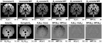 |
Correction of B1 non-uniformity errors in fast macromolecular proton fraction and R1 mapping without B1 maps
Vasily L. Yarnykh1
1Radiology, University of Washington, Seattle, WA, United States
Correction of B1 field non-uniformity is critical for quantitative MRI methods including fast macromolecular proton fraction (MPF) and variable flip angle T1 mapping. However, B1 mapping sequences increase the examination time and are not commonly available in clinics. A new algorithm is presented to enable simultaneous B1 correction in R1=1/T1 and MPF mapping without acquisition of B1 maps. The principle of the algorithm is based on different mathematical dependences of B1-related errors in R1 and MPF allowing extraction of a surrogate B1 map from uncorrected R1 and
MPF maps. The method demonstrated excellent agreement with actual B1 mapping at 3T.
|
|
0888.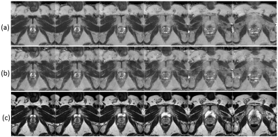 |
A fast T2 mapping protocol for prostate clinical applications using compressed sensing with low-rank and sparsity constraints
Jochen Keupp1, Doneva Mariya1, Jakob Meineke1, and Peter Forthmann1
1Philips Research, Hamburg, Germany
T2w-MRI plays an important role in prostate cancer providing information on the location/grade in diagnosis or surveillance. T2-mapping may provide objective characterization but is hampered by long acquisition time, which has been addressed by dedicated acceleration techniques (e.g. kt-T2 mapping). We investigate further acceleration of T2-mapping by prospective variable sub-sampling in the echo time domain, comparing regular or irregular patterns in combination with compressed sensing using low rank and sparsity constraints, towards a routine clinical T2 mapping protocol with increased volume coverage. Prostate and phantom T2-maps with 24 slices (1×1×3mm3 voxel) were acquired in 5½ minutes with promising map quality.
|
|
 |
0889.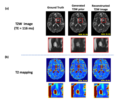 |
Accelerated T2 Mapping by Integrating Two-Stage Learning with Sparse Modeling
Ziyu Meng1,2, Yudu Li2,3, Rong Guo2,3, Yibo Zhao2,3, Tianyao Wang4, Fanyang Yu2,5, Brad Sutton2,5, Yao Li1, and Zhi-Pei Liang2,3
1Institute for Medical Imaging Technology, School of Biomedical Engineering, Shanghai Jiao Tong University, Shanghai, China, 2Beckman Institute of Advanced Science and Technology, University of Illinois at Urbana-Champaign, Urbana, IL, United States, 3Department of Electrical and Computer Engineering, University of Illinois at Urbana-Champaign, Urbana, IL, United States, 4Department of Radiology, The Fifth People's Hospital of Shanghai, Fudan University, Shanghai, China, 5Department of Bioengineering, University of Illinois at Urbana-Champaign, Urbana, IL, United States
We propose a new method to learn the multi-TE image priors for accelerated T2 mapping. The proposed method has the following key features: a) fully leveraging the Human Connectome Project (HCP) database to learn T2-weighted image priors for a single TE, b) transferring the learned single-TE T2-weighted image priors to multi-TE via deep histogram mapping, c) reducing the learning complexity using a tissue-based training strategy, and d) recovering subject-dependent novel features using sparse modeling. The proposed method has been validated using experimental data, producing very encouraging results.
|
|
0890.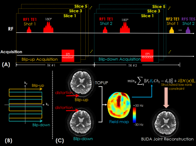 |
T2-BUDA-gSlider: fast T2 mapping with blip-up/down acquisition, generalized SLIce Dithered Enhanced Resolution and subspace reconstruction
Xiaozhi Cao1,2,3, Congyu Liao2,3, Zijing Zhang2,4, Siddharth Srinivasan Iyer2,5, Hongjian He1, Kawin Setsompop2,3,6, Jianhui Zhong1, and Berkin Bilgic2,3,6
1Center for Brain Imaging Science and Technology, Department of Biomedical Engineering, Zhejiang University, Hangzhou, China, 2Athinoula A. Martinos Center for Biomedical Imaging, Massachusetts General Hospital, charlestown, MA, United States, 3Department of Radiology, Harvard Medical School, charlestown, MA, United States, 4State Key Laboratory of Modern Optical Instrumentation, College of Optical Science and Engineering, Zhejiang University, Hangzhou, China, 5Department of Electrical Engineering and Computer Science, Massachusetts Institute of Technology, Cambridge, MA, United States, 6Harvard-MIT Department of Health Sciences and Technology, Cambridge, MA, United States
We propose to combine the gSlider acquisition and blip-up/down acquisition (BUDA) to achieve high-resolution and distortion-free T2 mapping. Firstly, we incorporate Hankel structured low-rank constraint into BUDA reconstruction to recover distortion-free images from blip-up/down shots without navigation. To utilize the similarity among RF-encodings and TEs, we introduce a model-based shuffling-gSlider joint reconstruction to recover high-resolution thin-slice images by gradually eliminating the weak coefficient components during the iterative reconstruction. Finally, the reconstructed images are used to obtain quantitative T2 maps. The proposed method enables distortion-free high-quality whole-brain T2 mapping with 1 mm isotropic resolution within ~1 minute.
|
|
0891.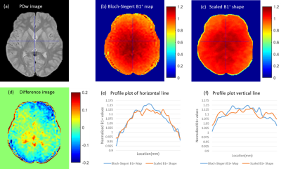 |
Bloch Modelling Enables Robust T2 Mapping using Retrospective Proton Density and T2-weighted Images from Different Vendors and Sites
Gitanjali Chhetri1, Kelly C McPhee1, and Alan H Wilman1
1Biomedical Engineering, University of Alberta, Edmonton, AB, Canada
Differences in pulse sequences between vendors can result in variation in T2 mapping, if not accounted for. We show that Bloch simulation based Indirect and Stimulated Echo Compensation minimizes these differences in T2 maps across different scanners. In contrast, standard exponential fitting results in highly variable T2 values across MR systems even if echo and repetition times are identical. By overcoming errors in T2 quantification through sequence modelling, T2 mapping can be applied in studies across multiple sites and vendors.
|
 |
0892.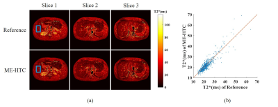 |
Joint Calibrationless Multi-slice Multi-echo Parallel Imaging Reconstruction for Abdominal T2* Mapping
Xiaochuan Wu1,2, Zheyuan Yi1,2,3, Yilong Liu1,2, Fei Chen3, Yanqiu Feng4, and Ed X. Wu1,2
1Laboratory of Biomedical Imaging and Signal Processing, The University of Hong Kong, Hong Kong, China, 2Department of Electrical and Electronic Engineering, The University of Hong Kong, Hong Kong, China, 3Department of Electrical and Electronic Engineering, Southern University of Science and Technology, Shenzhen, China, 4School of Biomedical Engineering, Southern Medical University, Guangzhou, China
T2* mapping in abdominal imaging is challenging due to respiration motions that can result in severe artifacts and affect the accuracy of T2* quantification. Traditional simultaneous autocalibrating and k-space estimation (SAKE) provides a calibrationless parallel imaging approach to reduce the image acquisition time. However, SAKE does not utilize the highly sharable image contents and coil sensitivities among multi-slice multi-echo data. In this study, we proposed a joint calibrationless reconstruction of multi-slice multi-echo images from undersampled MR data for abdominal imaging. Results demonstrated that the resulting T2* maps were in excellent agreement with those from the fully sampled data.
|
0893.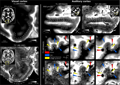 |
7T in-vivo human T2* mapping at 350μm isotropic resolution using ME-GRE with flow artifact mitigation reveals cortical layers & vessels
Omer Faruk Gulban1, Benedikt Poser1, Martin Havlicek1, Federico De Martino1, and Dimo Ivanov1
1Cognitive Neuroscience, Maastricht University, Maastricht, Netherlands
Spatial misencoding of the vascular signal due to flow is an imaging artifact that presents a significant challenge for in vivo MRI at high resolutions (≤0.5mm). Here we propose a method for mitigating this artifact in multi-echo gradient recalled echo (ME-GRE) images at 350μm isotropic resolution by applying 90° rotations to their phase-encoding direction. After applying our method, we demonstrate clearly visible stria of Gennari, intracortical veins, and pial vessels while mitigating the flow artifact. In addition, we report T2* estimates of several human brain tissues (artery, vein, gray/white matter, CSF) in-vivo which are valuable for future hemodynamic signal modeling.
|
|
|
0894.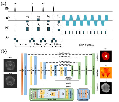 |
Deep Learning-based Single-shot T2* Mapping Using Multiple Overlapping-Echo Detachment Acquisition
Qinqin Yang1, Jian Wu1, Wei Wang1, Jiyang Dong1, Shuhui Cai1, and Congbo Cai1
1Department of Electronics Science, Xiamen University, Xiamen, Fujian, China
Quantitative MRI is of great value to both clinical diagnosis and scientific research. In this study, a novel T2* mapping method, gradient-echo multiple overlapping-echo detachment acquisition (GRE-MOLED) sequence with deep learning-based reconstruction algorithm was proposed. The method is capable of acquiring reliable T2*, M0 and B0 maps simultaneously in a single shot and is robust to B0-inhomogeneities. As a rapid T2* quantitative tool, GRE-MOLED reduces the scan time of T2* mapping to less than 75 ms per slice and has great potential in clinical real-time applications.
|
0895.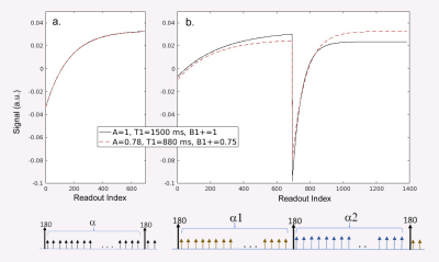 |
Dual Flip-angle IR-FLASH for B1+ Insensitive T1 Mapping: Application to T1 CMR Multitasking.
Fardad Michael Serry1, Sen Ma1,2, Debiao Li1,2, and Anthony G Christodoulou1
1Biomedical Imaging Research Institute, Cedars-Sinai Medical Center, Los Angeles, CA, United States, 2Department of Bioengineering, University of California, Los Angeles, Los Angeles, CA, United States
T1 mapping is important for many diseases, from cancer [1] to cardiovascular disease [2], and more. Many fast T1 mapping protocols rely on an IR-FLASH sequence, especially in the heart. However, the accuracy and repeatability of T1 mapping with IR-FLASH are compromised by B1+ inhomogeneity. Here we present a simple dual-flip-angle (DFA) modification of the IR-FLASH pulse sequence to provide B1+-robust T1 mapping that obviates the need for a separate B1+ scan . We show the improved agreement of DFA-IR-FLASH to IR-TSE in a phantom study as well as its feasibility for in vivo cardiac T1 mapping with MR Multitasking.
|
|
|
0896.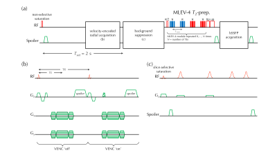 |
Simultaneous measurements of blood flow and blood water T2: a general-purpose sequence for T2-based measurement of whole-organ O2 consumption
Cheng-Chieh Cheng1, Pei-Hsin Wu1, Michael C. Langham1, and Felix W. Wehrli1
1University of Pennsylvania, Philadelphia, PA, United States
A T2-based oximetry method for quantifying whole-organ metabolic rate of oxygen (MRO2): A velocity-encoded acquisition module with golden-angle radial sampling was inserted into a background-suppressed T2-prepared sequence specifically for blood water T2 quantification. Parallel imaging and compressed-sensing techniques were applied to the reconstruction of the sparsely-sampled velocity-encoded k-space data to generate velocity maps. Whole-organ oxygen metabolic rate was estimated by converting T2 to blood oxygenation level via a calibration curve. A pilot study in the superior sagittal sinus showed the method’s ability to estimate whole-brain CMRO2 (136±23 μmol/minute/100g, mean±S.D.) in a single pass.
|
 Watch the Video