
Scientific Session
System Imperfections, Artifacts, and More
Session Topic: System Imperfections, Artifacts, and More
Session Sub-Topic: Measuring & Correcting System Imperfections
Oral
Acquisition, Reconstruction & Analysis
| Tuesday Parallel 3 Live Q&A | Tuesday, 11 August 2020, 15:15 - 16:00 UTC | Moderators: Stephan Orzada & Rudolf Stollberger |
Session Number: O-65
0653.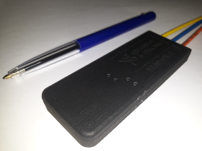 |
Optical Field Probes for MRI
Hans Stærkind1,2, Vincent Oltman Boer1, Kasper Jensen3, Eugene Simon Polzik2, and Esben Thade Petersen1,4
1Danish Research Centre for Magnetic Resonance, Centre for Functional and Diagnostic Imaging and Research, Copenhagen University Hospital Hvidovre, Hvidovre, Denmark, 2Quantop, Niels Bohr Institute, University of Copenhagen, Copenhagen, Denmark, 3School of Physics and Astronomy, University of Nottingham, Nottingham, United Kingdom, 4Department of Health Technology, Technical University of Denmark, Kgs. Lyngby, Denmark
A novel optical field probe is described for the precision measurement of high magnetic fields. The advantages over traditional field probes are two-fold. First, there is no electromagnetic interference with the other parts of the MRI system such as the RF coil. Secondly, the setup allows for a continuous and long readout of the magnetic field during an MRI sequence, where traditional NMR probes typically operate in a pulsed mode.
|
|
 |
0654.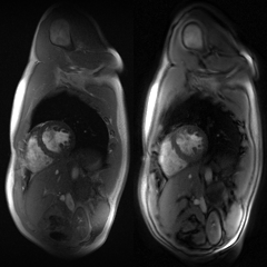 |
Spiral real-time cardiac MR imaging using a GSTF-based pre-emphasis
Philipp Eirich1,2, Tobias Wech1, Julius F. Heidenreich1, Manuel Stich1,3, Nils Petri4, Peter Nordbeck4, Thorsten A. Bley1, and Herbert Köstler1
1Department of Diagnostic and Interventional Radiology, University Hospital Würzburg, Würzburg, Germany, 2Comprehensive Heart Failure Center Würzburg, Würzburg, Germany, 3Siemens Healthcare, Erlangen, Germany, 4Department of Internal Medicine I, University Hospital Würzburg, Würzburg, Germany
An automatic pre-emphasis based on the Gradient System Transfer Function (GSTF) was applied to real-time cardiac MR imaging at 3T to compensate for deviations of spiral k-space trajectories. This yielded cine series with coverage of the whole heart in free-breathing, less than 40 s total scan time and a temporal resolution of 50 ms. The developed framework was compared to a gated Cartesian acquisition in multiple breath-holds, in one healthy volunteer and one patient suffering from cardiac arrhythmia.
|
0655.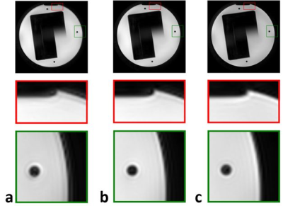 |
Trajectory calculation for spiral imaging based on concurrent reading of the gradient amplifiers’ internal current sensors
Jürgen Rahmer1, Ingo Schmale1, Peter Mazurkewitz1, and Peter Börnert1
1Philips Research, Hamburg, Germany
Spiral sequences sample data during gradient variation and are therefore susceptible to dynamic field deviations caused by eddy currents, timing inaccuracies, or gradient amplifier non-linearities. Linear effects can be corrected using a gradient impulse response function for trajectory calculation. Non-linear effects require a measurement-based approach, e.g. measurement of the gradient fields during imaging using a field camera or an induction-based field measurement. To avoid the need for additional hardware, we propose a hybrid method that combines current measurements using the amplifier-internal current sensors with correction based on the current-to-gradient impulse response function. The approach improves trajectory accuracy in spiral imaging.
|
|
|
0656.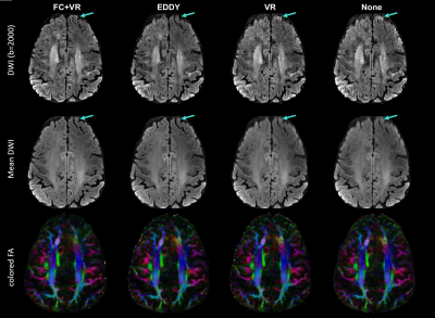 |
Concurrent Field Monitoring in HCP dMRI at 7T: Correction for Eddy Current Induced Signal Blurring and Geometric Distortion.
Ruoyun Emily Ma1, Mehmet Akçakaya1,2, Steen Moeller1, Connor Benson1, Edward Auerbach1, Kâmil Uğurbil1, and Pierre-François Van de Moortele1
1Center for Magnetic Resonance Research, University of Minnesota, Minneapolis, MN, United States, 2Electrical and Computer Engineering, University of Minnesota, Minneapolis, MN, United States
DW-EPI at high diffusion gradients suffers from eddy current induced signal blurring and geometric distortion. In this study, concurrent monitoring of field evolution with NMR probes was implemented for HCP diffusion MRI acquisition at 7T. After image reconstruction with field correction, eddy current induced geometric distortion was largely removed, yielding similar level of correction obtained by data driven approaches such as EDDY. Signal blurring was further reduced than with EDDY. Future efforts will be made to quantify the impact on cortical tractography of this improved DW-EPI reconstruction.
|
0657.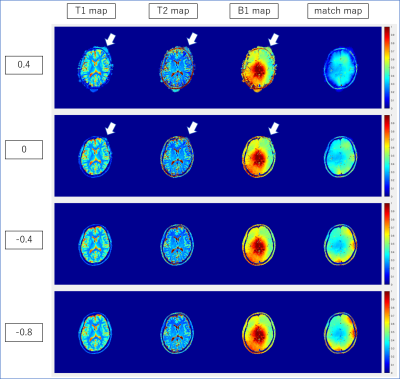 |
A simple method to estimate gradient delay for MRF
Koji Fujimoto1, Martijn A. Cloos2, and Tomohisa Okada1
1Human Brain Research Center, Graduate School of Medicine, Kyoto University, Kyoto, Japan, 2Center for Advanced Imaging Innovation and Research (CAI2R) and Bernard and Irene Schwartz Center for Biomedical Imaging, Department of Radiology, New York University School of Medicine, New York, NY, United States
A self-contained method to estimate the gradient delay for radial based MRF sequences based on the residual error after dictionary matching was presented. Under IRB approval, MRF was performed in two healthy volunteers at 7T. Images were reconstructed with varying degrees of gradient offset in the readout direction. The correlation of the dictionary match (i.e. inner product of the compressed data with the matched dictionary entry) was recorded. The average signal intensity within the head ROI in the “match map” gave the best result, and hence could be an easy and reliable metric for gradient delay correction.
|
|
 |
0658.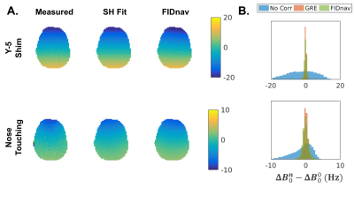 |
Measurement and correction of spatiotemporal B0 fluctuations using an FID-navigated EPI sequence
Tess E. Wallace1,2, Jonathan R. Polimeni2,3, Jason P. Stockmann2,3, W. Scott Hoge2,4, Tobias Kober5,6,7, Simon K. Warfield1,2, and Onur Afacan1,2
1Computational Radiology Laboratory, Boston Children's Hospital, Boston, MA, United States, 2Department of Radiology, Harvard Medical School, Boston, MA, United States, 3Athinoula A. Martinos Center for Biomedical Imaging, Massachusetts General Hospital, Boston, MA, United States, 4Brigham and Women's Hospital, Boston, MA, United States, 5Advanced Clinical Imaging Technology, Siemens Healthcare, Lausanne, Switzerland, 6Radiology, Lausanne University Hospital and University of Lausanne, Lausanne, Switzerland, 7LTS5, École Polytechnique Fédérale de Lausanne (EPFL), Lausanne, Switzerland
Spatiotemporal B0 field fluctuations give rise to dynamic susceptibility-induced distortions in EPI time-series, which reduces signal stability, particularly at higher field strengths. In this work, we propose a novel method for rapid measurement of B0 field changes from FIDnavs embedded in an EPI sequence. We demonstrate the ability of the proposed method to accurately characterize field changes up to second order in controlled phantom and volunteer experiments. Dynamic slice-wise distortion correction using FIDnav field estimates reduced normalized root-mean-square error and improved temporal SNR in volunteers performing deliberate arm motion.
|
0659.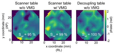 |
Reduction of vibration-induced signal loss by matching mechanical vibrational states: application in high b-value diffusion weighted MRS
Dominik Weidlich1, Mark Zamskiy1, Marcus Maeder2, Stefan Ruschke1, Steffen Marburg2, and Dimitrios C. Karampinos1
1Department of Diagnostic and Interventional Radiology, Technical University of Munich, Munich, Germany, 2Chair of Vibroacoustics of Vehicles and Machines, Technical University of Munich, Munich, Germany
Diffusion gradients are known to yield MR hardware vibrations, which may lead to signal loss in DW measurements especially at high b-values. The present work proposes to mitigate vibration-induced signal loss by introducing a vibration matching gradient (VMG). Laser interferometry was employed to measure the displacements induced by high b-value DW MR spectroscopy, focusing on the quantification of lipids ADC. The measured displacement patterns during the two diffusion gradients were more similar when using the VMG. The application of VMG showed an improvement in lipid ADC quantification in both a water-fat phantom and a volunteer’s tibial bone marrow.
|
|
|
0660.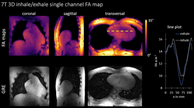 |
Respiration-Resolved 3D Multi-Channel B1 mapping of the body at 7T
Sebastian Dietrich1, Christoph Stefan Aigner1, Juliane Ludwig1, Johannes Mayer1, Simon Schmidt2, Christoph Kolbitsch1,3, Tobias Schaeffter1,3, and Sebastian Schmitter1,2
1Physikalisch-Technische Bundesanstalt (PTB), Braunschweig and Berlin, Germany, 2Medical Physics in Radiology, German Cancer Research Center (DKFZ), Heidelberg, Germany, 3Division of Imaging Sciences and Biomedical Engineering, King's College London, London, United Kingdom A major challenge of ultra-high field MRI is the spatially inhomogeneous transmit radio frequency field that induces spatial contrast variations. In this work we present a novel technique to retrieve channel-wise, motion-resolved absolute 3D FA maps of the human body at 7T. Free breathing 3D scans of the thorax were performed and B1 maps without motion artifacts are obtained for the two motion states inhale and exhale. It allows acquiring B1 libraries that can be used for offline RF pulse calculation including motion-robust RF pulses. |
|
0661.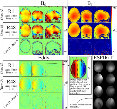 |
PhysiCal: A rapid calibration scan for B0, B1+, coil sensitivity and Eddy current mapping.
Siddharth Srinivasan Iyer1,2, Congyu Liao2, Qing Li3, Mary Katherine Manhard2, Avery Berman2, Berkin Bilgic2, and Kawin Setsompop2
1Electrical Engineering and Computer Science, Massachusetts Institute of Technology, Cambridge, MA, United States, 2Athinoula A. Martinos Center for Biomedical Imaging, Charlestown, MA, United States, 3MR Collaborations, Siemens Healthcare Ltd, Shanghai, China
Calibration scans that acquire coil sensitivity, B0 and B1+ inhomogeneities information play an important role in enabling modern acquisition and reconstruction techniques. This work proposes a unified, rapid calibration sequence termed Physics Calibration (PhysiCal) to obtain accurate B0, Eddy, B1+ and coil sensitivity maps. PhysiCal utilizes a carefully designed mix of full and variable density sampling acquisitions across echoes with synergistic constrained and eigenvalue reconstruction for robust and accurate recovery of whole-brain B0, B1+, Eddy and 32-channel coil sensitivity maps in just 11 seconds at 1 mm x 2 mm x 2mm resolution at 3T.
|
0662. |
Highly 3D accelerated Bloch Siegert B1+ Mapping at 7T
Andreas Lesch1, Christoph Aigner2, and Rudolf Stollberger1
1Institute of Medical Engineering, Graz University of Technology, Graz, Austria, 2Physikalisch-Technische Bundesanstalt (PTB), Braunschweig and Berlin, Berlin-Charlottenburg, Germany
For many applications in MRI fast and accurate $$$B_1^+$$$ -mapping is an important prerequisite to correct for spatially varying RF-field variations. We recently proposed a reconstruction algorithm to reconstruct highly undersampled Bloch Siegert data, which was applied to 3T data. In this work we investigated the applicability of this algorithm to 7T data in terms of accuracy and possible acquisition time. We could successfully show that the proposed algorithm can be applied to 7T data with only slightly increased error compared to 3T. The minimum scan time at 7T is in the order of 45s-1min for a 3D volume.
|
 Watch the Video