
Scientific Session
Diffusion Microstructure Modeling and Validation
Session Topic: Diffusion Microstructure Modeling and Validation
Session Sub-Topic: Microstructure: Validation
Oral
Diffusion
| Wednesday Parallel 4 Live Q&A | Wednesday, 12 August 2020, 13:45 - 14:30 UTC | Moderators: Tim Dyrby |
Session Number: O-67
 |
0722.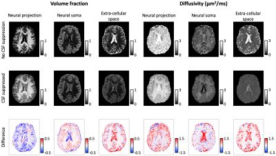 |
High-Resolution Neural Soma Imaging with FLAIR: Eliminating CSF Contamination in Grey Matter
Noemi G Gyori1,2, Iulius Dragonu3, Christopher A Clark2, Daniel C Alexander1, and Enrico Kaden1
1Centre for Medical Image Computing, University College London, London, United Kingdom, 2Great Ormond Street Institute of Child Health, University College London, London, United Kingdom, 3Siemens Healthcare Ltd, Frimley, United Kingdom
In-vivo microstructure imaging in cortical grey matter is limited by low imaging resolution and signal contamination from CSF. In this work, we use FLAIR to eliminate free water signal in the brain, and thus enhance sensitivity to microscopic tissue architecture in the cortex. We present the advantage of CSF suppression in Neural Soma Imaging, a state-of-the-art diffusion technique that focuses on the salient features of grey matter. We show high-resolution maps (1.5 mm isotropic) of neural tissue microstructure and T1- and T2-relaxation times, and demonstrate that neural projection density estimates are significantly higher when the CSF signal is eliminated.
|
|
0723.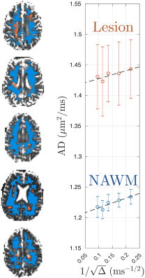 |
Characterizing white matter lesions in multiple sclerosis with time-dependent diffusion MRI reveals the signature of axonal beading
Hong-Hsi Lee1, Dmitry S Novikov1, and Els Fieremans1
1Center for Biomedical Imaging, Department of Radiology, NYU School of Medicine, New York, NY, United States
We observe diffusivity time-dependence along white matter axons in normal-appearing white matter (NAWM) and lesions in 5 relapse remitting multiple sclerosis (MS) patients. The long-time diffusivity along axons is higher in MS lesion than that in NAWM due to persistent demyelination and axonal loss, consistent with previous studies. Further, the axial diffusivity time-dependence is weaker in MS lesions than in NAWM, probably caused by beading due to increased mitochondria in astrocytes/axons in MS lesions. we propose the axial diffusivity time-dependence as a potential specific biomarker for beading, to monitor the progression and treatment response of MS.
|
0724.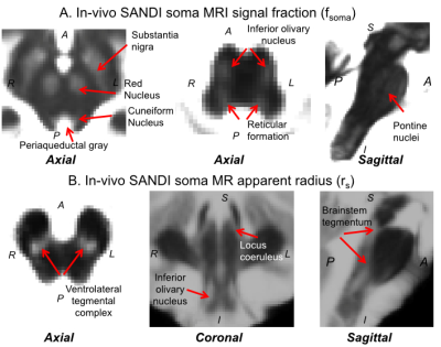 |
Validation and application of soma and neurite density imaging (SANDI) for in-vivo human brainstem nuclei atlasing
Marta Bianciardi1, Maria G. García-Gomar1, Kavita Singh1, Michele Guerreri2, Alejandra Sierra3, Jussi Tohka3, Ali Abdollahzadeh3, Hui Zhang2, and Marco Palombo2
1Department of Radiology, A.A. Martinos Center for Biomedical Imaging, MGH and Harvard Medical School, Boston, MA, United States, 2Centre for Medical Image Computing, Department of Computer Science, University College London, London, United Kingdom, 3A.I. Virtanen Institute for Molecular Sciences, University of Eastern Finland, Kuopio, Finland
Despite the development of detailed in-vivo human cortical atlases, in-vivo brainstem nuclei atlasing is still at its early stages due to reduced gray-white matter MRI-contrast in the brainstem compared to the cortex. Recently, we generated an in-vivo probabilistic atlas of 16 human brainstem nuclei based on multi-contrast 7Tesla MRI. Nevertheless, to further expand the in-vivo brainstem nuclei atlas, there is an unmet need from additional MRI-contrast reflecting brainstem cytoarchitecture. We found that recently developed in-vivo Soma-And-Neurite-Density-Imaging (SANDI) provides original MRI-contrast directly related to ex-vivo brainstem nuclei cytoarchitecture, and can be used to expand the current in-vivo human brainstem nuclei atlas.
|
|
|
0725.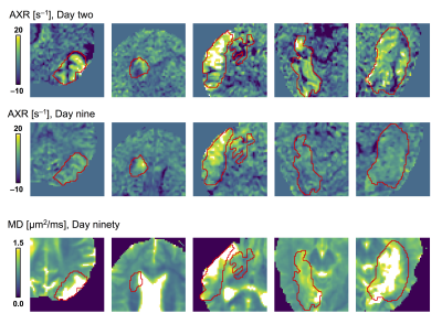 |
Diffusion time dependence and tissue outcome in ischemic stroke
Björn Lampinen1, Jimmy Lätt2, Johan Wasselius3, Danielle van Westen4, and Markus Nilsson4
1Medical Radiation Physics, Lund University, Lund, Sweden, 2Center for Medical Imaging and Physiology, Skåne University Hospital, Lund, Sweden, 3Clinical Sciences Lund, Neurology, Lund University, Lund, Sweden, 4Clinical Sciences Lund, Diagnostic Radiology, Lund University, Lund, Sweden
Many patients with ischemic stroke that would benefit from ‘late’ recanalization go untreated, as current imaging-based predictions of outcome are insufficiently individualized. This study investigated whether diffusion MRI (dMRI), a standard tool in stroke diagnostics, provides additional information through effects of diffusion time dependence. Results showed elevated rates of water exchange within lesions of subacute stroke patients. The absence of such exchange appeared predictive of tissue viability in the chronic stage, even in regions normally considered irreversibly injured. Information on diffusion time dependence may thus improve penumbra definitions and help identifying subjects with favorable outcome of late recanalization.
|
|
0726.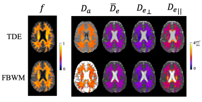 |
Testing white matter tissue modeling with multiple diffusion encoding MRI
Hunter G Moss1,2, Emilie T McKinnon1,2,3, and Jens H Jensen1,2
1Neuroscience, Medical University of South Carolina, Charleston, SC, United States, 2Center for Biomedical Imaging, Medical University of South Carolina, Charleston, SC, United States, 3Neurology, Medical University of South Carolina, Charleston, SC, United States
The validation of white matter (WM) tissue modeling for diffusion MRI is challenging, in part, because some of the predicted microstructural parameters (e.g., compartment-specific diffusivities) cannot be easily measured with independent methods such as histology. Most WM tissue models are designed to utilize single diffusion encoding (SDE) MRI data as provided by conventional diffusion MRI sequences. Since multiple diffusion encoding (MDE) MRI yields more information than SDE, it allows for tissue modeling that requires fewer assumptions. Hence, MDE can be applied to help validate the predictions for all SDE model parameters. Here we give an explicit example of this.
|
0727.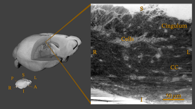 |
Streamline tractography for 3D mapping of axon bundle organization in one MRI voxel using ultra-high resolution synchrotron radiation imaging
Hans Martin Kjer1,2, Mariam Andersson1,2, Yi He2, Marie Louise Elkjaer3, Alexandra Pacureanu4,5, Zsolt Illes3, Bente Pakkenberg6, Anders Bjorholm Dahl1, Vedrana Andersen Dahl1, and Tim B. Dyrby1,2
1DTU Compute, Technical University of Denmark, Kgs. Lyngby, Denmark, 2Danish Research Centre for Magnetic Resonance, Hvidovre, Denmark, 3Department of Neurology, Odense University Hospital, Odense, Denmark, 4X-ray Nanoprobe Group, ID16A, The European Synchrotron, Grenoble, France, 5University College London, London, United Kingdom, 6Research Laboratory for Stereology and Neuroscience, Bispebjerg University Hospital, Copenhagen NV, Denmark
We present an efficient image analysis pipeline that enables us to reveal white matter organization in high-resolution 3D non-MRI structural datasets, in cases where a strict image segmentation is not required nor possible. We apply the method to a synchrotron X-ray holographic tomography scan from a healthy mouse sample, and show the organization of axon bundles in a region covering parts of the corpus callosum and the cingulum. The method has a potential to improve our general understanding of white matter organization and our ability to generate realistic phantoms for validation of microstructure modelling from low-resolution diffusion MRI scans.
|
|
|
0728.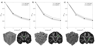 |
Improved contextual fibre growth for generating white matter numerical phantoms with realistic microstructure
Ross Callaghan1, Daniel C Alexander1, Marco Palombo1, and Hui Zhang1
1Department of Computer Science & Centre for Medical Image Computing, University College London, London, United Kingdom
We present an improved version of the ConFiG white matter numerical phantom generator to create realistic white matter microstructure. Building on ConFiG’s novel fibre growth algorithm, the enhancement incorporates a dynamic growth network and global optimisation of fibre positions. Resulting phantoms represent a significant improvement over those from the original ConFiG algorithm, with realistic morphology and an increase in packing density of up to 30%. These improved phantoms result in much more realistic simulated diffusion MRI signals, reducing RMSE to real data by ten times. This improvement demonstrates the potential of ConFiG as a computational model of white matter microstructure.
|
 |
0729.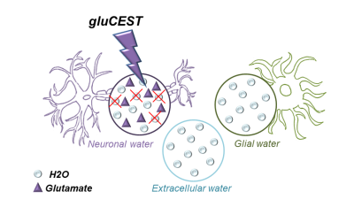 |
Using glutamate-CEST to characterize water diffusion inside neurons: initial results
Mélissa Vincent1,2, Yohann Mathieu-Daudé1,2, Julien Flament1,2, and Julien Valette1,2
1Molecular Imaging Research Center (MIRCen), Commissariat à l'Energie Atomique et aux Energies Alternatives (CEA), Fontenay-aux-Roses, France, 2UMR 9199, Neurodegenerative Diseases Laboratory, Centre National de la Recherche Scientifique (CNRS), Université Paris-Sud, Université Paris-Saclay, Fontenay-aux-Roses, France
It is still unclear how diffusion properties of water differ from one compartment to another (neurons, glial cells, extracellular space…). Here we propose the idea that Chemical Exchange Saturation Transfer of Glutamate (gluCEST) may be used to specifically reduce the contribution of intraneural water to the overall signal attenuation, thus providing enhanced sensitivity to non-neuronal compartments. Acquisitions performed in two rats yields water ADC slightly but significantly higher when gluCEST is performed, supporting the idea that water diffusion is slower inside neurons.
|
|
0730.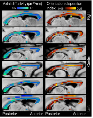 |
Estimating intra-axonal axial diffusivity with diffusion MRI in the presence of fibre orientation dispersion
Amy FD Howard1, Alexandre A Khrapitchev2, Jeroen Mollink1,3, Rogier B Mars1,4, Nicola Sibson2, Jerome Sallet5, Saad Jbabdi1, and Karla L Miller1
1FMRIB Centre, Wellcome Centre for Integrative Neuroimaging, Nuffield Department of Clinical Neurosciences, University of Oxford, Oxford, United Kingdom, 2CR-UK/MRC Oxford Institute for Radiation Oncology, Department of Oncology, University of Oxford, Oxford, United Kingdom, 3Department of Anatomy, Donders Institute for Brain, Cognition and Behaviour, Radboud University Medical Center, Nijmegen, Netherlands, 4Donders Institute for Brain, Cognition and Behaviour, Radboud University Nijmegen, Nijmegen, Netherlands, 5Wellcome Centre for Integrative Neuroimaging, Experimental Psychology, Medical Sciences Division, University of Oxford, Oxford, United Kingdom
Intra-axonal axial diffusivity could be interesting biomarker of disease, yet it is often assumed constant across the white matter. Furthermore, when intra-axonal diffusivity is estimated, few models account for fibre orientation dispersion which (when not explicitly modelled) will greatly affect the estimates of axial diffusion. Here we combine the stick model of intra-axonal diffusion with a simple model of fibre dispersion to simultaneously estimate intra-axonal axial diffusivity and fibre dispersion on a voxel-wise basis in high b-value data. Our results demonstrate considerable variability in the intra-axonal axial diffusivity across the white matter.
|
 Watch the Video