
Scientific Session
Diffusion Microstructure Modeling and Validation
Session Topic: Diffusion Microstructure Modeling and Validation
Session Sub-Topic: Diffusion: Microstructure Modelling
Oral
Diffusion
| Wednesday Parallel 4 Live Q&A | Wednesday, 12 August 2020, 13:45 - 14:30 UTC | Moderators: Maryam Afzali & Marco Palombo |
Session Number: O-70
0712.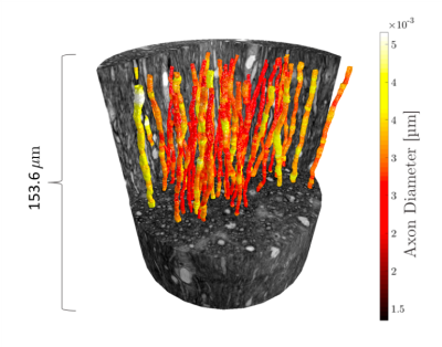 |
The impact of axon orientation dispersion and 3D diameter variations on the transverse apparent diffusion coefficient
Mariam Andersson1,2, Jonathan Rafael-Patino3, Hans Martin Kjer1,2, Vedrana Andersen Dahl1, Alexandra Pacureanu4, Martin Bech5, Anders Bjorholm Dahl1, Jean-Philippe Thiran3,6, and Tim B. Dyrby1,2
1Department of Applied Mathematics and Computer Science, Technical University of Denmark, Kgs. Lyngby, Denmark, 2Danish Research Centre for Magnetic Resonance, Hvidovre, Denmark, 3Ecole Polytechnique Fédérale de Lausanne, Lausanne, Switzerland, 4The European Synchrotron, Grenoble, France, 5Department of Medical Radiation Physics, Clinical Science, Lund University, Lund, Sweden, 6Radiology Department, Centre Hospitalier Universitaire Vaudois and University of Lausanne, Lausanne, Switzerland
We extract the orientation dispersion (OD) and 3D axon diameter variations of long axons (>100 µm) from an ultra-high resolution, synchrotron X-ray nano-holotomography (XNH) scan of the monkey splenium. From this, we discover a relationship between mean axon diameter and along-axon diameter variations. Monte Carlo simulations are then performed on the intra-axonal spaces (IAS) of different substrates which inherit their morphological features from the segmented axons. These simulations show that the OD significantly affects the transverse apparent diffusion coefficient (ADC⟂) of the axon substrate at diffusion times up to 50 ms, while diameter variations do not.
|
|
0713.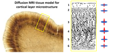 |
Modeling cortical architectonic features by analyzing diffusion MRI data in the cortical reference frame
Alexandru V Avram1,2, Kadharbatcha Saleem1,2, Frank Q Ye3, Cecil Yen4, Michal E Komlosh1,2, and Peter J Basser1
1NICHD, National Institutes of Health, Bethesda, MD, United States, 2The Henry Jackson Foundation, Bethesda, MD, United States, 3NIMH, National Institutes of Health, Bethesda, MD, United States, 4NINDS, National Institutes of Health, Bethesda, MD, United States
We quantified the alignment between the DTI reference frame (DRF) and the cortical reference frame (CRF) throughout the entire cerebral cortex in a macaque brain, and found relatively good correspondence, especially in regions with high curvature such as the gyral walls and the cortical sulci. Based on this correspondence, we analyze cortical diffusion signals in the CRF and construct a simple model of cortical diffusion with distinct radial (columnar) and tangential (sheet-like) diffusion processes in cortical layers. The variation of model parameters with cortical depth reflects architectonic features described in a histologically defined digital macaque brain atlas.
|
|
0714.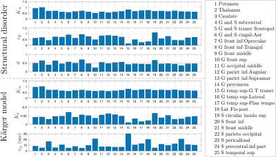 |
In vivo observation and interpretation of time-dependent diffusion in human brain gray matter
Hong-Hsi Lee1, Antonios Papaioannou1, Dmitry S Novikov1, and Els Fieremans1
1Center for Biomedical Imaging, Department of Radiology, NYU School of Medicine, New York, NY, United States
The purpose of this work is to identify the relevant microstructural features for the human brain gray matter. For that, we estimate the diffusivity and kurtosis time-dependence in 25 gray matter sub-regions of 10 healthy subjects, and compare the effects of the structural disorder and exchange (Karger model). The estimated power-law dynamical exponent θ≈1/2 is consistent with the structural disorder picture in a 1-dimensional micro-geometry of randomly positioned restrictions along neurites. In contrast, Karger model yields exchange time much shorter than values in previous studies, and below our shortest diffusion time.
|
|
0715.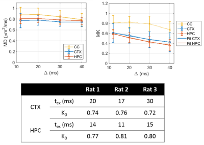 |
Water exchange time between gray matter compartments in vivo
Ileana Ozana Jelescu1 and Dmitry S Novikov2
1Center for Biomedical Imaging, Ecole Polytechnique Fédérale de Lausanne, Lausanne, Switzerland, 2Dept. of Radiology, New York University School of Medicine, New York, NY, United States
In the absence of a myelin sheath, exchange generally is non-negligible over the typical diffusion times of MRI experiments (10 – 100 ms) and should be accounted for in gray matter modeling. Here we use time-dependent kurtosis and the Kärger model (KM) of two slowly exchanging compartments to evaluate water exchange time between intra-neurite and extra-cellular compartments in rat GM in vivo. We report exchange times on the order of 10 – 30 ms. Future work will focus on exploring a broader range of diffusion times to test the asymptotic decay of kurtosis toward zero.
|
|
0716.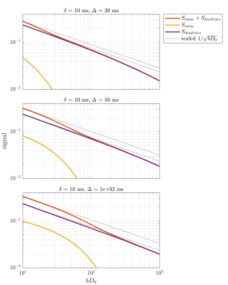 |
Stick power law scaling in neurons withstands realistic curvature and branching
Jonas Lynge Olesen1,2 and Sune Nørhøj Jespersen1,2
1Center of Functionally Integrative Neuroscience (CFIN) and MINDLab, Department of Clinical Medicine, Aarhus University, Aarhus, Denmark, 2Department of physics and Astronomy, Aarhus University, Aarhus, Denmark
A main idea contained in the standard model of diffusion is to model neurons with zero-width sticks. A resulting signature is the prediction that in the large b limit, the isotropically averaged signal scales as $$$1/\sqrt{b}$$$ which has been verified in white matter but not gray matter. This has multiple proposed causes including dendrite curvature and branching. Here, we report on Monte Carlo simulations in 3D reconstructed neurons and find that branching and curvature do not break the power law scaling. On the other hand,the soma is found to limit the regime in which stick scaling is observed.
|
|
 |
0717.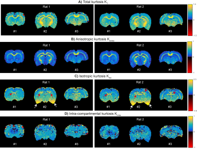 |
Correlation Tensor Imaging - resolving non-Gaussian diffusion sources of in vivo tissues
Rafael Neto Henriques1, Sune Nørhøj Jespersen2,3, and Noam Shemesh1
1Champalimaud Research, Champalimaud Centre for the Unknown, Lisbon, Portugal, 2Center of Functionally Integrative Neuroscience (CFIN) and MINDLab, Clinical Institute, Aarhus University, Aarhus, Denmark, 3Department of Physics and Astronomy, Aarhus University, Aarhus, Denmark
Resolving the different sources of diffusional kurtosis can increase DKI’s specificity towards different microstructural features. Such sources can be resolved using the correlation tensor imaging (CTI) – a novel double diffusion encoding technique that does not rely on common assumptions of time-independent diffusion. Here, a minimal acquisition protocol for CTI is designed and used to characterize the diffusional kurtosis of living rat brains for the first time. We here develop an approach to acquire CTI data in vivo and show that it can robustly decouple inter-compartmental kurtosis sources (anisotropic and isotropic diffusivity variances) from intra-compartmental kurtosis sources.
|
0718. |
A unified framework for analysis of time-dependent diffusion: numerical validation of a restriction-exchange correlation experiment
Markus Nilsson1, Carl-Fredrik Westin2, Jan Brabec1, Samo Lasic3, and Filip Szczepankiewicz1
1Clinical Sciences Lund, Lund University, Lund, Sweden, 2Brigham and Women's hospital, Harvard Medical School, Boston, MA, United States, 3Random Walk Imaging AB, Lund, Sweden
Probing time-dependence with diffusion MRI enables mapping of microstructure features such as cell sizes (restrictions) and membrane permeability (exchange). However, restrictions and exchange have opposite effects on the MR signal, and cannot be distinguished by just varying the diffusion time. We propose a unified framework for analysis of time-dependent diffusion that enables the design of efficient restriction-exchange correlation experiments. A signal representation was developed featuring parameters connected to restricted diffusion and exchange. This connection was validated by numerical simulations.
|
|
0719.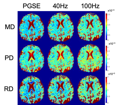 |
Oscillating Gradient Diffusion-Encoding In Human Brain Shows Linear Frequency Correlation in High Amplitude and Slew Rate Head Gradient System
Ek T Tan1,2, Robert Y Shih3, Jhimli Mitra1, Yihe Hua1, Tim Sprenger4, Chitresh Bhushan1, Jennifer A McNab5, Matt A Bernstein6, and Thomas KF Foo1
1GE Research, Niskayuna, NY, United States, 2Radiology and Imaging, Hospital for Special Surgery, New York, NY, United States, 3Walter Reed National Military Medical Center, Bethesda, MD, United States, 4GE Healthcare, Stockholm, Sweden, 5Stanford University, Stanford, CA, United States, 6Radiology, Mayo Clinic, Rochester, MN, United States
High gradient amplitude, high gradient slew rate, and high peripheral nerve stimulation thresholds are required for oscillating gradient spin-echo (OGSE) diffusion imaging on human MRI systems. With 200 mT/m amplitude and 500 T/m/s slew rate, the MAGNUS head gradient coil was used to evaluate OGSE imaging in six healthy subjects at frequencies up to 100 Hz and b=450 s/mm2. Comparisons were made against standard pulsed gradient spin-echo (PGSE) diffusion in-vivo, which show up to 27% increased OGSE diffusivity, excellent linear correlation with frequency, and correlation length scales of 0.8µm in white matter. Diffusivity changes were negligible in an isotropic phantom.
|
|
0720.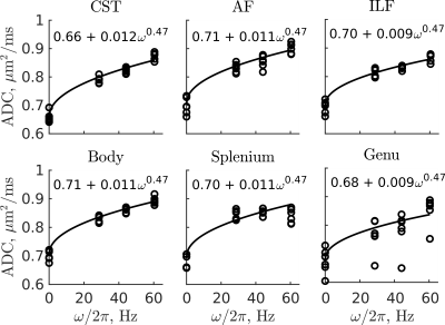 |
Evidence for Short Range Disorder in the in vivo Human Brain using OGSE Diffusion MRI
Aidin Arbabi1, Jason Kai1, Ali R Khan1, and Corey A Baron1
1Robarts Research Institute, University of Western Ontario, London, ON, Canada
Oscillating gradient spin-echo (OGSE) diffusion MRI enables probing of the frequency content of the apparent diffusion coefficient (ADC). A square root dependence of ADC on frequency has been demonstrated in both healthy and globally ischemic rodent brain tissue, which is consistent with short range structural disorder along neurites. In this work, OGSE data was acquired at multiple frequencies to explore the power law scaling of the ADC in the human brain in vivo, where evidence of a square root dependence of ADC on frequency was obtained for the first time in the in vivo human brain.
|
|
0721.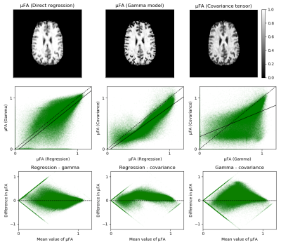 |
Accuracy and precision of microscopy anisotropy estimation using q-space trajectory encoding - a model comparison study
Leevi Kerkelä1, Fabio Nery1, Feng-Lei Zhou2, Geoff J.M. Parker2,3,4, Filip Szczepankiewicz5,6,7, Matt G. Hall1,8, and Chris A. Clark1
1UCL Great Ormond Street Institute of Child Health, University College London, London, United Kingdom, 2Centre for Medical Image Computing, University College London, London, United Kingdom, 3Bioxydyn Limited, Manchester, United Kingdom, 4CRUK and EPSRC Cancer Imaging Centre in Cambridge and Manchester, Manchester, United Kingdom, 5Department of Radiology, Brigham and Women’s Hospital, Boston, MA, United States, 6Harvard Medical School, Boston, MA, United States, 7Medical Radiation Physics, Clinical Sciences Lund, Lund University, Lund, Sweden, 8National Physical Laboratory, Teddington, United Kingdom
Estimation of microscopic fractional anisotropy (μFA) using multidimensional diffusion MRI is a promising novel method for characterising clinically relevant microstructural properties of neural tissue. In this study, three commonly used methods for calculating μFA were compared by imaging a fibre phantom and healthy volunteers. Statistically significant differences were observed in accuracy and precision of the μFA estimates calculated using the covariance tensor model, the gamma distributed diffusivities model, and the direct regression approach. The differences between the methods have to be carefully considered when this promising new metric is applied in characterising microstructural properties of tissue or pathologies.
|
 Watch the Video