
Scientific Session
Diffusion Modeling, Tractography and Applications
| Wednesday Parallel 4 Live Q&A | Wednesday, 12 August 2020, 14:30 - 15:15 UTC | Moderators: Stella Xing |
 |
0858.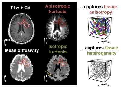 |
B-tensor encoding in gliomas: improved tumor grading by the isotropic kurtosis
Jan Brabec1, Filip Szczepankiewicz2,3,4, Patrik Brynolfsson5, Lampinen Björn1, Faris Durmo4, Anna Rydelius6, Linda Knutsson1,7, Pia Sundgren4,8, and Markus Nilsson4
1Clinical Sciences Lund, Medical Radiation Physics, Lund University, Lund, Sweden, 2Department of Radiology, Brigham and Women's Hospital, Boston, MA, United States, 3Harvard Medical School, Boston, MA, United States, 4Clinical Sciences Lund, Diagnostic Radiology, Lund University, Lund, Sweden, 5Dept. of Translational Medicine, Division of Medical Radiation Physics, Lund University, Lund, Sweden, 6Clinical Sciences Lund, Neurology, Lund University, Lund, Sweden, 7Russell H. Morgan Department of Radiology and Radiological Science, Johns Hopkins University School of Medicine, Baltimore, MD, United States, 8Lund University Bioimaging Center, Lund University, Lund, Sweden
B-tensor encoding enables mapping of the isotropic and anisotropic components of the diffusional kurtosis, which are sensitive to cell eccentricity and variance in cell density, respectively. We measured the kurtosis components in patients with glioma tumors and explored their ability to improve tumor classification. Results showed that the addition of isotropic kurtosis improves the ability to distinguish low- and high-grade gliomas compared with using post-Gd T1w enhancements alone. Also, non-enhancing glioblastomas and oligodendrogliomas could be distinguished based on the within-tumor standard deviation of the isotropic kurtosis.
|
0859.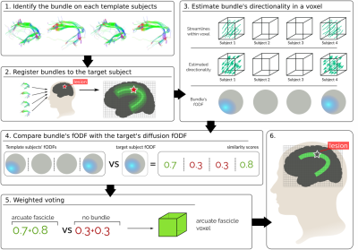 |
Lesion-Robust White-Matter Bundle Identification through Diffusion Driven Label Fusion
Guillermo Gallardo1, Gaston Zanitti2, Samuel Deslauriers-Gauthier3, Matthew Higger4, Sylvain Bouix4, Alfred Anwander5, and Demian Wassermann2
1Neuropsycology, Max Planck Institute for Human Cognitive and Brain Sciences, Leipzig, Germany, 2Parietal, Inria Saclay - Ile de France, PAris, France, 3Athena EPI, Universite Cote d’Azur, Inria, Sophia Antipolis, France, 4Psychiatry Neuroimaging Laboratory, Brigham and Womens Hospital, Harvard Medical School, Boston, MA, United States, 5Max Planck Institute for Human Cognitive and Brain Sciences, Leipzig, Germany White-matter pathologies disrupt the white-matter organization, that manifests as deficits in brain function. When treating such pathologies, it is of great importance to infer which pathways are affected. However, the white-matter lesions hamper the use of tractography to track fiber bundles. In this work, leveraging diffusion imaging, we propose a novel diffusion-driven technique to improve the localization of brain pathways. Aggregating information from few healthy subjects, our technique is able to localize both the affected pathways and the lesion interrupting when tracking is not possible. |
|
 |
0860.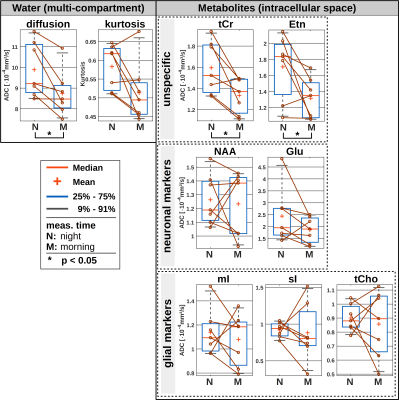 |
Investigation of potential effects of sleep on diffusion characteristics of metabolites and water: initial results
André Döring1, Christian Rummel2, Sandra C. Röthlisberger3, Simone Duss3, Corinne Roth3, Claudio Bassetti3,4, and Roland Kreis1
1Depts. Radiology and Biomedical Research, University of Bern, Bern, Switzerland, 2Support Center for Advanced Neuroimaging, University Institute for Diagnostic and Interventional Neuroradiology, Bern, Switzerland, 3Sleep-Wake-Epilepsy-Center, University Hospital Bern, Bern, Switzerland, 4Dept. of Neurology, University Hospital Inselspital Bern, Bern, Switzerland
Diffusion-weighted MR spectroscopy (DW-MRS) and imaging (DW-MRI) was applied to investigate potential effects of sleep on apparent diffusion coefficients (ADCs) of water and metabolites in human gray matter in 7 healthy subjects. Monitoring the transition from wake to sleep for a period of 4 hours did not reveal any significant alterations, while comparison of night measurements after slight sleep deprivation to morning examinations after a full night’s sleep indicated that ADCs for some metabolites are lower in the morning than before sleep – though these results need corroboration in a larger cohort.
|
|
0861.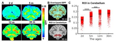 |
Multidimensional Diffusion MRI Assists Myelin-sensitive Bound Pool Fraction in Differentiating Microstructural Maturity of Primate Brains
Yi He1, Henrik Lundell1, Ines Mexia Rodrigues1, Matthew D. Budde2, Mark D. Does3, Maurice Ptito4,5, and Tim Bjørn Dyrby1
1Danish Research Centre for Magnetic Resonance, Centre for Functional and Diagnostic Imaging and Research, Copenhagen University Hospital Hvidovre, Hvidovre, Denmark, 2Neurosurgery, Medical College of Wisconsin, Milwaukee, WI, United States, 3Department of Biomedical Engineering, Vanderbilt University, Nashville, TN, United States, 4School of Optometry, Université de Montréal, Montreal, QC, Canada, 5Department of Nuclear Medicine, University of Southern Denmark, Odense, Denmark
Myelin-sensitive bound pool fraction (BPF) enables the tracking process of myelination in primate brains. The 3D BPF maps demonstrated rapid development of myelination from a 2-day-old brain to a 12-month-old brain and a slower increase from 12 months to 30 months. Even though the process of myelination is slow, multidimensional diffusion MRI indices are indeed helpful in significantly differentiating the microstructural maturity of primate brains. Our findings suggest that both indices, isotropic kurtosis (MKI, associated with cell density variance) and microscopic anisotropy (MKA, correlated with cell eccentricity) are significant imaging markers for microstructural differentiation in the development of primate brains.
|
|
0862.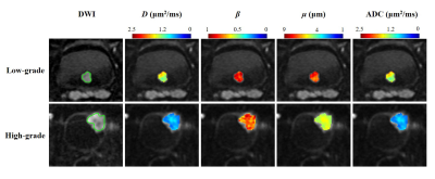 |
Grading bladder urothelial carcinoma using a non-Gaussian fractional order calculus diffusion model
Cui Feng1,2, Yanchun Wang1, Guangyu Dan2,3, Zheng Zhong2,3, M. Muge Karaman2,3, Daoyu Hu1, and Xiaohong Joe Zhou2,3,4,5
1Radiology, Tongji Hospital, Tongji Medical College, Huazhong University of Science and Technology, Wuhan, China, 2Center for MR Research, University of Illinois at Chicago, Chicago, IL, United States, 3Department of Bioengineering, University of Illinois at Chicago, Chicago, IL, United States, 4Department of Radiology, University of Illinois at Chicago, Chicago, IL, United States, 5Department of Neurosurgery, University of Illinois at Chicago, Chicago, IL, United States Diffusion-weighted imaging based on apparent diffusion coefficient (ADC) has been used for bladder urothelial carcinoma grading. However, a considerable overlap between the low- and high-grade bladder urothelial carcinoma has hindered its clinical acceptance. We employed high b-value diffusion imaging with a non-Gaussian fractional-order calculus (FROC) diffusion model for grading bladder urothelial carcinoma. Significant differences were observed in the FROC parameters D, β and μ, between the low- and high-grade urothelial carcinoma. The combination of the FROC parameters provided substantially better performance than ADC. These findings indicate a promising role of FROC parameters for characterizing bladder urothelial carcinoma and beyond. |
|
0863.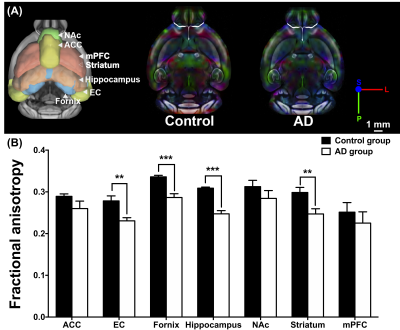 |
Altered Brain and Gut in Alzheimer’s Disease Model Using Diffusion MRI and Intestinal Bacteria Gene Analysis
Yao-Wen Liang1, Ching-Wen Chang1, Ssu-Ju Li1, Ting-Chun Lin1, Hsin-Tzu Lu1, You-Yin Chen1, and Yu-Chun Lo2
1National Yang-Ming University, Taipei, Taiwan, 2Taipei Medical Unversity, Taipei, Taiwan
Microbiota-gut-brain axis, a bidirectional communication, was proposed as an important role in Alzheimer’s disease (AD). However, the correlation between gut microbiota and brain microstructure in AD remained unclear. Triple-transgenic mouse models of AD were used to investigate brain-behavior-gut-microbiome interaction. Diffusion MRI, behavior tasks, and intestinal bacteria gene analysis were applied in this study. The findings implied that the altered brain microstructure and atypical distribution of gut microbiota were associated with the cognitive dysfunction in AD.
|
|
0864.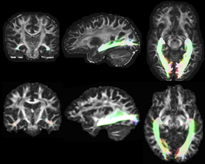 |
Fully-Automated Delineation of the Optic Radiation
Lee Bremner Reid1, Eloy Martínez-Heras2, Magí Andorrà Inglés2, Elisabeth Solana2, Sara Llufriu2, José V Manjón3, and Jurgen Fripp1
1The Australian e-Health Research Centre, CSIRO, Brisbane, Australia, 2Center of Neuroimmunology, Laboratory of Advanced Imaging in Neuroimmunological Diseases, Hospital Clinic Barcelona, Institut d'Investigacions Biomediques August Pi i Sunyer and Universitat de Barcelona, Barcelona, Spain, 3ITACA, Universitat Politècnia de València, Valencia, Spain
The optic radiation (OR), is often severed during temporal lobe resection, resulting in permanent quadrantanopia. To date all published tractography methods that delineate the OR require manual input (e.g. region-of-interest placement and adjustment), or appear to underestimate Meyer’s Loop, limiting their widespread clinical adoption. Here, we present and validate the CONSULT pipeline for OR delineation. This pipeline accepts unprocessed DICOM images as input and produces realistic subject-specific segmentations of the OR, including Meyer’s Loop, without need for any human input. Its validation in 183 datasets demonstrated plausible delineations that are in line with previous dissection studies.
|
0865.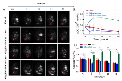 |
Exploring multi-functions of MRI in PDT: imaging-guided tumor therapy, follow-up monitoring and early evaluation of therapeutic efficacy
Xiudong Shi1, Weitao Yang2, Qiong Ma1, and Yuxin Shi1
1Shanghai public health clinical center, Fudan University, Shanghai, China, 2The Institute for Biomedical Engineering & Nano Science, Tongji University, Shanghai, China
Estimating the gross tumor volume by measuring the physical diameters of the tumor with calipers is a common method for evaluating the efficacy of photodynamic therapy (PDT). In this study, the optimal time for determining the efficacy of PDT treatment based on morphological and functional magnetic resonance (MR) imaging techniques is determined.
|
|
0866. |
Contribution of parenchymal water and CSF-like water to changes in brain water diffusivity in post-natal development and aging
Laura D. Reyes1, Amritha Nayak1,2, Alzheimer’s Disease Neuroimaging Initiative *3, and Carlo Pierpaoli1
1NIBIB, National Institutes of Health, Bethesda, MD, United States, 2Henry M. Jackson Foundation, Bethesda, MD, United States, 3Alzheimer’s Disease Neuroimaging Initiative, Los Angeles, CA, United States
In this study, we used a biexponential dual compartment diffusion tensor imaging (DTI) model to characterize different pools of parenchymal water to investigate CSF-like free water and parenchymal diffusivity in childhood, early adulthood, and elderly adulthood, and examine how they change across the lifespan. We identified age-related changes in the contribution of parenchymal and CSF-like free water to age-related changes in overall diffusivity that can be linked to changes in tissue microstructure.
|
 Watch the Video