
Scientific Session
High Resolution fMRI
| Thursday Parallel 4 Live Q&A | Thursday, 13 August 2020, 15:05 - 15:50 UTC | Moderators: Peter Bandettini & Jeroen Siero |
 |
1225.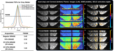 |
Highly Accelerated Sub-Millimeter Resolution 3D GRASE with Controlled T2 Blurring in T2-Weighted FMRI at 7T: Feasibility Study
Suhyung Park1, Salvatore Torrisi2, Jennifer Townsend2, Alexander Beckett2, and David Feinberg1,2
1University of California, Berkeley, Berkeley, CA, United States, 2Advanced MRI Technologies, Sebastopol, CA, United States 3D GRASE is used for cortical layer and columnar fMRI in the absence of signal confounds from draining veins. Its use has been limited by limited slice coverage with blurring. We developed highly accelerated 3D GRASE with controlled T2 blurring by combining compressed sensing with variable flip angles. Compared with current GRASE acquisitions, the proposed method demonstrates that 1) through-plane random encoding with VFA increases the slice coverage with a sharper point spread function, 2) reduced TE from in--plane random encoding provides a high SNR efficiency, and 3) the resulting image sharpness and SNR efficiency lead to increased BOLD activation. |
1226. |
Modelling the Laminar VASO Signal Change in Human V1 at 7T
Atena Akbari1, Saskia Bollmann1, Tonima S Ali 1, and Markus Barth1,2,3
1Centre for Advanced Imaging, The University of Queensland, Brisbane, Australia, 2ARC Training Centre for Innovation in Biomedical Imaging Technology, The University of Queensland, Brisbane, Australia, 3School of Information Technology and Electrical Engineering, The University of Queensland, Brisbane, Australia
In this study, we used the “cortical vascular model” for human V1 at 7T to simulate the laminar VAscular-Space-Occupancy (VASO) signal change. For comparison, we conducted VASO experiments on a group of healthy subjects to measure laminar signal change in V1. Results show a very good agreement between the model prediction and the experimental results once the volume changes of the different vascular compartments (arterioles, capillaries, venules) are taken into account.
|
|
1227. |
Sub-Millimeter Spiral fMRI Combining Magnitude and Phase BOLD Contrast
Lars Kasper1, Maria Engel1, Jakob Heinzle2, Matthias Mueller-Schrader2, Jonas Reber1, Thomas Schmid1, Christoph Barmet1, Bertram Jakob Wilm1, Klaas Enno Stephan2,3,4, and Klaas Paul Pruessmann1
1Institute for Biomedical Engineering, ETH Zurich and University of Zurich, Zurich, Switzerland, 2Translational Neuromodeling Unit, Institute for Biomedical Engineering, University of Zurich and ETH Zurich, Zurich, Switzerland, 3Wellcome Trust Centre for Neuroimaging, University College London, London, United Kingdom, 4Max Planck Institute for Metabolism Research, Cologne, Germany We investigate the spatial specificity of sub-millimeter (0.8mm) single-shot spiral fMRI, and its feasibility for functional phase contrast. Scrutinizing activation patterns of a visual paradigm in 6 subjects, we find that significant contrast changes occur between adjacent voxels, contributing to the evidence of spatial specificity of spiral acquisition as well as gradient echo BOLD contrast, and its possible applications in laminar or columnar fMRI. Furthermore, the vessel-localized nature of the phase activation suggests its suitability for masking macrovascular confound effects. |
|
1228. |
High-resolution line-scanning reveals distinct visual response properties across human cortical layers.
Andrew T. Morgan1, Nils Nothnagel1, Jozien Goense1, and Lars Muckli1
1Institute of Neuroscience & Psychology, University of Glasgow, Glasgow, United Kingdom
Motivated by recent functional line-scanning recordings in rodents, we developed a procedure to record human cortical layers at high spatial (200 μm) and temporal resolution (100 ms). Our technique addresses challenges associated with human line-scanning, such as planning around cortical folding and restrictive SAR limitations. Our results show that line-scanning of human cortical layers corroborates electrophysiological measurements of tuning properties in primary visual cortex. These results demonstrate that line-scanning is a promising technique for investigating local functional circuits in human cortex.
|
|
1229.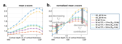 |
Cortical-depth dependence of pure T2-weighted BOLD fMRI with minimal T2’ contamination using Echo-Planar Time-resolved Imaging (EPTI)
Fuyixue Wang1,2, Zijing Dong1,3, Qiyuan Tian1, Jingyuan Chen1, Anna Izabella Blazejewska1, Timothy G. Reese1, Jonathan R. Polimeni1,2, and Kawin Setsompop1,2
1A. A. Martinos Center for Biomedical Imaging, Massachusetts General Hospital, Charlestown, MA, United States, 2Harvard-MIT Health Sciences and Technology, MIT, Cambridge, MA, United States, 33Department of Electrical Engineering and Computer Science, MIT, Cambridge, MA, United States
BOLD fMRI based on T2 contrast has the promise to provide exclusively microvascular specificity, which would optimize the ability of fMRI signals to accurately reflect and localize neuronal activity. However, it is challenging in practice to achieve pure T2 weighting. Here we employ a new highly-efficient acquisition and reconstruction framework based on EPI, Echo-Planar Time-resolved Imaging (EPTI), and extend it to generate blurring- and distortion-free data with purely T2 weighting. We evaluate the technique through a cortical-depth analysis of activation in human visual cortex and demonstrate that it achieves the desired microvascular specificity.
|
|
 |
1230.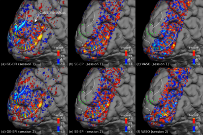 |
Mapping ocular dominance columns in humans using GE-EPI, SE-EPI and SS-SI-VASO at 7 T
Daniel Haenelt1,2, Nikolaus Weiskopf1, Lenka Vaculciakova1,2, Roland Mueller1, Shahin Nasr3,4, Jonathan Polimeni3,4, Roger Tootell3,4, Laurentius Huber5, Martin Sereno6, and Robert Trampel1
1Max Planck Institute for Human Cognitive and Brain Sciences, Leipzig, Germany, 2International Max Planck Research School on Neuroscience of Communication: Function, Structure, and Plasticity, Leipzig, Germany, 3Athinoula A. Martinos Center for Biomedical Imaging, Boston, MA, United States, 4Department of Radiology, Harvard Medical School, Boston, MA, United States, 5Department of Cognitive Neuroscience, Maastricht Brain Imaging Center, Maastricht, Netherlands, 6Department of Psychology, San Diego State University, San Diego, CA, United States
Functional MRI studies classically rely on the use of GE-EPI sequences. However, the GE-based signal is inherently sensitive to large veins, which impairs its use in high-resolution fMRI application. Other BOLD- and CBV-based approaches like SE-EPI and SS-SI-VASO, respectively, promise a higher specificity at the expense of sensitivity. In the present work, we tested if ocular dominance columns (ODCs) can be detected using GE-EPI, SE-EPI and SS-SI-VASO at 7 T. ODCs could be reliably mapped using all three acquisition methods. Furthermore, we could show for the first time ODCs in humans by exploiting the functional CBV response using SS-SI-VASO.
|
1231. |
Mapping directional functional connectivity across brain-wide networks with layer-specific CBV-fMRI
Laurentius Huber1, Emily Finn2, Sean Marrett2, Sriranga Kashyap1, Arman Khojandi2, Rainer Goebel1, Peter Bandettini2, and Benedikt Poser1
1MBIC, Uni Maastricht, Faculty of Psychology and Neuroscience, Maastricht, Netherlands, 2SFIM, NIMH, Bethesda, MD, United States
With recent advances in ultra-high-field MRI hardware and sequence mechanisms, it has become possible to measure CBV-weighted fMRI signal across cortical layers. While initial proof-of-principle layer-fMRI studies in primary brain areas with conventional fMRI task designs are promising, layer-fMRI has not yet realized its full potential to map layer-dependent functional connectivity across large-scale brain networks. In this study, we investigate the applicability of CBV-weighted layer-fMRI to assess functional connectivity during resting-state and naturalistic tasks. We can map common resting-state networks and characterize their internal layer-dependent signatures with respect to directionality and cortical hierarchy.
|
|
|
1232.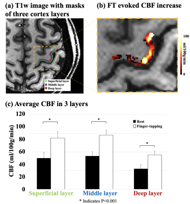 |
In-vivo laminar CBF fMRI using high-resolution pseudo-continuous arterial spin labeling at 7T
Xingfeng Shao1, Kay Jann1,2, Kai Wang1, Fanhua Guo3, Peng Zhang3, and Danny JJ Wang1,2
1Mark & Mary Stevens Neuroimaging and Informatics Institute, Keck School of Medicine, University of Southern California, Los Angeles, CA, United States, 2Department of Neurology, University of Southern California, Los Angeles, CA, United States, 3State Key Laboratory of Brain and Cognitive Science, Beijing MRI Center for Brain Research, Institute of Biophysics, Chinese Academy of Sciences, Beijing, China
In-vivo laminar CBF fMRI was performed by high resolution (0.5×0.5×1.5 mm3) inner-volume GRASE with optimized pCASL labeling at 7T. Activation of finger-tapping task (5 blocks, TA=10 min) was reliably detected in all 4 subjects. Both rest/FT CBF peaks in the middle layers, which corresponds to highest capillary density in cortex layer IV. FT evoked CBF increase shows one peak in middle layer, and a second shoulder in deep layer. The capability to provide quantitative CBF measurements at both baseline and task activation with high specificity to neuronal activities is a unique strength of ASL fMRI compared to other fMRI techniques.
|
1233.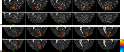 |
On the feasibility of using single-shot perfusion labeling (SSPL) at 7 Tesla for laminar fMRI
Jacco A de Zwart1, Peter van Gelderen1, and Jeff H Duyn1
1Advanced MRI section, LFMI, NINDS, National Institutes of Health, Bethesda, MD, United States
Blood oxygen-level dependent (BOLD) functional MRI (fMRI) based on gradient-echo EPI is the most commonly used fMRI method due to its high sensitivity and robustness. However, large vein contribution negatively affects spatial localization of BOLD activation, of crucial importance for laminar and other high-resolution fMRI applications. Perfusion and blood volume-based methods have been shown to increase spatial accuracy of activation maps. Here we demonstrate feasibility of single-shot perfusion labeling (SSPL) fMRI at up to 1 mm3 resolution, a reference-less perfusion fMRI method twice as efficient as FAIR in which background signal is suppressed, improving temporal stability.
|
|
1234.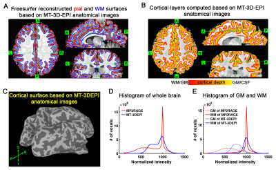 |
A magnetization transfer weighted anatomical reference allows laminar fMRI analysis in native functional image space
Yuhui Chai1, Linqing Li2, Yicun Wang3, Larentius Huber4, Benedikt Poser4, Jeff Duyn3, and Peter Bandettini1,2
1Section on Functional Imaging Methods, Laboratory of Brain and Cognition, NIMH, NIH, Bethesda, MD, United States, 2Functional MRI Core, NIMH, NIH, Bethesda, MD, United States, 3Advanced MRI Section, Laboratory of Functional and Molecular Imaging, NINDS, NIH, Bethesda, MD, United States, 4Maastricht Brain Imaging Center, Faculty of Psychology and Neuroscience, University of Maastricht, Maastricht, Netherlands
In most previous laminar fMRI studies, cortical layers are defined based on an anatomical image that is collected by a different acquisition technique and exhibits different geometric distortion compared to the functional images. We introduce to generate a magnetization transfer (MT) weighted anatomical reference, using identical acquisition design as fMRI measurement. Cortical surface and depth can be reconstructed directly from this MT-weighted anatomical EPI image and all laminar analysis can be performed in the native fMRI image space without the need for distortion correction and registration.
|
 Watch the Video