
Scientific Session
Multimodal fMRI
Session Topic: Multimodal fMRI
Session Sub-Topic: Multimodal Imaging of Brain Function
Oral
fMRI
| Thursday Parallel 4 Live Q&A | Thursday, 13 August 2020, 15:50 - 16:35 UTC | Moderators: Karen Mullinger |
Session Number: O-75
1339.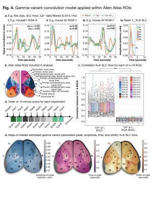 |
Simultaneous fMRI and mesoscopic Ca2+ imaging indicates spontaneous excitatory neural activity accounts for 1/3rd of the variance in BOLD signal
Evelyn MR Lake1, Xinxin Ge2, Xilin Shen1, Peter Herman1, Fahmeed Hyder1, Jessica A Cardin3, Michael J Higley3, Dustin Scheinost1, Xenophon Papademetris1, Michael C Crair2, and R Todd Constable1
1Radiology and Biomedical Imaging, Yale University, New Haven, CT, United States, 2Department of Neurobiology, Yale University, New Haven, CT, United States, 3Department of Neuroscience, Yale University, New Haven, CT, United States
We demonstrate longitudinal simultaneous whole-cortex Ca2+ imaging and fMRI in mice expressing GCaMP in one of five different cell types (excitatory, inhibitory, two interneuron subtypes, and astrocytes). The high SNR of our dual-imaging approach is shown by the indistinguishable Ca2+ responses to hind-paw or visual stimulation measured inside and outside the scanner. We optimize a spatially variable, three-parameter gamma-variant to investigate the transfer function between the BOLD and Ca2+ signals throughout the cortex. This approach is applied in functionally and anatomically defined ROIs. Results show that 1/3rd of the variance in BOLD is accounted for from spontaneous excitatory Ca2+ activity.
|
|
 |
1340.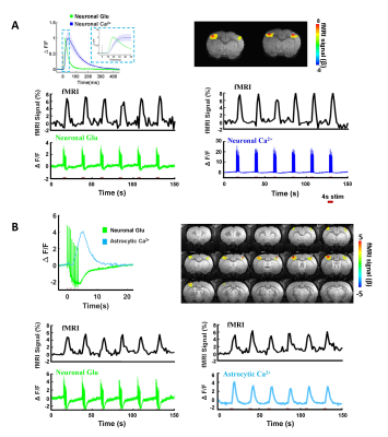 |
Deciphering the contribution of extracellular glutamate and intracellular calcium signaling to the BOLD fMRI signal
Yuanyuan Jiang1, Xuming Chen2, Patricia Pais Roldán2, Bruce Rosen1, and Xin Yu1,2
Video Permission Withheld
1Athinoula A. Martinos Center for Biomedical Imaging, Massachusetts General Hospital, Cambridge, MA, United States, 2Max Planck Institute for Biological Cybernetics, Tuebingen, Germany
We established a multi-modal fMRI platform with two-channel fiber optic recording based on a genetically encoded fluorescent reporter, iGluSnFR, for extracellular glutamate (Glu) sensing and intracellular calcium indicator, GCaMP6f. Different from the intracellular neuronal and astrocytic calcium transients, the Glu signal, peak responses of spikes and baseline drift, show unique correlation features to the BOLD fMRI signal. Here, we applied the multi-modal fMRI platform to decipher the cellular and molecular interaction underlying the BOLD fMRI signal through the neuro-glio-vascular network in animal brains.
|
|
1341.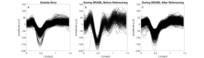 |
Adaptive Virtual Referencing Enables Recording of Extracellular Action Potentials in a 16.4 Tesla Animal Research MRI Scanner
Corey Cruttenden1, Wei Zhu2, Yi Zhang2, Rajesh Rajamani1, Xiao-Hong Zhu2, and Wei Chen2
Video Permission Withheld
1Mechanical Engineering, University of Minnesota, Minneapolis, MN, United States, 2Center for Magnetic Resonance Research, University of Minnesota, Minneapolis, MN, United States
Recording neural signals such as extracellular action potentials during functional magnetic resonance imaging (fMRI) will improve our understanding of neurovascular coupling, which is responsible for the fMRI blood oxygen level dependent (BOLD) signal. Recording electrical neural signals during fMRI is challenging due to interactions between the recording hardware and electromagnetic (EM) fields involved in MRI that introduce noise and artifacts. We developed an adaptive virtual referencing technique to improve the action potential signal quality recorded in the bore of a 16.4T animal scanner during GRASE fMRI. This technique will enable us to further study neurovascular coupling at 16.4T.
|
1342.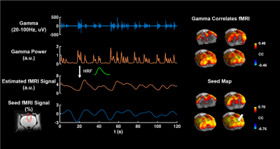 |
Simultaneous whole brain resting state fMRI and full-spectrum electrophysiology in rodents
Wenyu Tu1, Yuncong Ma2, Thomas Neuberger2, and Nanyin Zhang2
1The Huck Institutes of the Life Sciences, Penn State University, University Park, PA, United States, 2Biomedical Engineering, Penn State University, University Park, PA, United States
To elucidate the neural basis of resting state functional network, it is important to continuously record neural activity during rsfMRI. In this study, we developed a platform including animal setup and a signal denoising pipeline to achieve continuous measurement of local field potential (LFP) and neuronal spikes with simultaneous whole-brain rsfMRI in rats.
|
|
1343.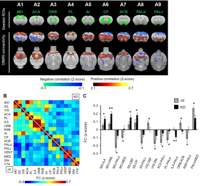 |
Diffusion functional MRI characterizes dynamical brain function in a neuropsychiatric disease model mouse
Yoshifumi Abe1
1Keio University School of Medicine, Tokyo, Japan
We propose an analytical framework to characterize dynamic brain function in neuropsychiatric conditions by taking advantage of the technical aspects of diffusion functional MRI (DfMRI). The pipeline consists of local activity analysis with apparent diffusion coefficient (ADC) data, functional connectivity (FC) analysis with diffusion-weighted data (Sb1800), and ignition-driven mean integration (IDMI) analysis combining both. We illustrated its utility by analyzing model mice with an obsessive–compulsive disorder (OCD)-related behavior. The framework was successful in detecting hyperactivation and biased connectivity across the cortico-striato-thalamic circuitry. The IDMI analysis found unseen local activity-initiated propagation to the global network.
|
|
1344.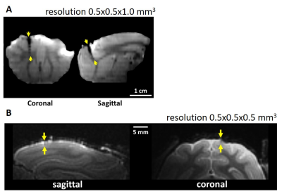 |
Evaluation of an improved microelectrode array for MR-compatibility and MR-simultaneous recording performance in 7T research system
Xiao Yu1,2, Bo-Wei Chen3, Xiaojun Tan1,4, Boyi Qu1,4, Tingting He1,2, Ching-Fu Wang3, Yu-Hao Lan3, You-Yin Chen3, and Hsin-Yi Lai1,2
1Interdisciplinary Institute of Neuroscience and Technology, School of Medicine, Zhejiang University, Hangzhou, China, 2Department of Neurology of the Second Affiliated Hospital, Zhejiang University School of Medicine, Zhejiang University, Hangzhou, China, 3Department of Biomedical Engineering, National Yang Ming University, Taipei, Taiwan, 4College of Biomedical Engineering and Instrument Science, Zhejiang University, Hangzhou, China
Simultaneous recording of electrophysiological signals with functional magnetic resonance imaging (fMRI) can provide a solution for investigation of neurovascular coupling. However, this technique is challenged by 2 aspects, image artifact from electrode and electrophysiological noise from magnetic field. We improved our pervious lab-designed microelectrode array and developed a de-noise method for use of its electrophysiological recording in 7T MRI. The results showed better structural image quality and stable acquisition of spike signals and local field potential. The proposed tool and method has the potential to facilitate simultaneous spike-recording during MR scanning in 7T MRI and further study the neurovascular coupling.
|
|
1345.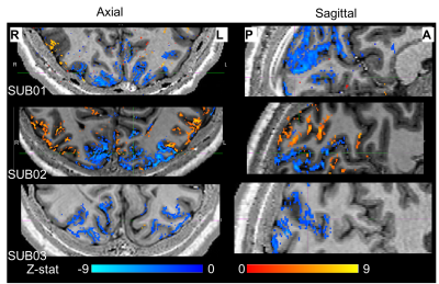 |
Assessing the origin of human alpha oscillations using laminar layer 7T fMRI-EEG
Daniel C. Marsh1, Rodika Sokoliuk2, Kevin M. Aquino1,3, Daisie O. Pakenham1, Ross Wilson2, Rosa Sanchez Panchuelo1, Matthew J Brookes1, Simon Hanslmayr2, Stephen D. Mayhew2, Susan T Francis1, and Karen J Mullinger1,2
1Sir Peter Mansfield Imaging Centre, School of Physics and Astronomy, University of Nottingham, Nottingham, United Kingdom, 2Centre for Human Brain Health, School of Psychology, University of Birmingham, Birmingham, United Kingdom, 3Turner Institute for Brain and Mental Health, Monash University, Melbourne, Australia
EEG alpha (8-13Hz) oscillations occur throughout the cortex but the generating mechanisms are poorly understood. Opinion is divided between alpha being driven by bottom-up, top-down or both these processes. Here we use simultaneous 7T-fMRI-EEG during periods of eyes open/closed to assess the generator of alpha by determining the strongest BOLD-alpha negative layer correlations. We show the feasibility of using high spatial resolution 7T-fMRI with EEG to understand the origin of oscillations. Preliminary analysis shows BOLD-alpha correlations peak in middle layers of V1 (but not in V2/V3) providing suggestion that the alpha oscillations investigated are driven by bottom-up processing.
|
|
1346.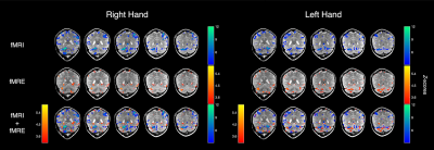 |
Simultaneous fMRI/fMRE reveals BOLD and viscoelastic changes in the cerebellum during motor planning
Patricia S. Lan1, Kevin J. Glaser2, Richard L. Ehman2, and Gary H. Glover3
1Bioengineering, Stanford University, Stanford, CA, United States, 2Radiology, Mayo Clinic, Rochester, MN, United States, 3Radiology, Stanford University, Stanford, CA, United States
In this work, we demonstrate the first fMRE (functional MR elastography) activation in the cerebellum using a motor planning task. A block paradigm of 24s ON (auditory-cued button pressing) and 24s OFF (rest) was used and images were acquired with a single-shot spin-echo EPI MRE sequence. Our results show that tissue stiffness within the cerebellum increases with motor planning. Furthermore, the stiffness and BOLD activation colocalize in the cerebellum but do not match exactly, suggesting that the two modalities may reveal different aspects of the mechanisms for neural activation.
|
|
 |
1347.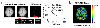 |
Validation of MRI-based Oxygen Extraction Fraction (OEF) Measurement with 15O Positron Emission Tomography
Dengrong Jiang1, Shengwen Deng2, Crystal G. Franklin2, Michael O'Boyle2, Wei Zhang2, Betty L. Heyl2, Li Pan3, Paul A. Jerabek2, Peter T. Fox2, and Hanzhang Lu1
1Department of Radiology, Johns Hopkins University School of Medicine, Baltimore, MD, United States, 2Research Imaging Institute, University of Texas Health Science Center at San Antonio, San Antonio, TX, United States, 3Siemens Healthineers, Baltimore, MD, United States
Cerebral oxygen extraction fraction (OEF) is a potential biomarker in various diseases. The current gold standard to measure OEF is 15O-PET, but its clinical applications are impeded by inherent limitations. To facilitate broader clinical applications of OEF as a disease biomarker, in this work, we compared the whole-brain OEF measurement of a non-invasive MRI technique, T2-relaxation-under-spin-tagging (TRUST), with the gold standard PET measurement, and demonstrated a strong linear correlation and no systematic difference between the two methods.
|
1348.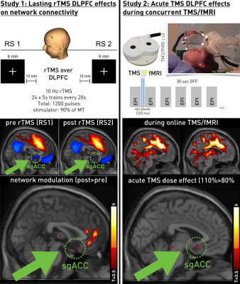 |
Advanced methods for concurrent TMS/fMRI explain target engagement in 10Hz rTMS treatment
Martin Tik1, Michael Woletz1, Anna-Lisa Schuler1, Matic Princic1, Allan Hummer1, and Christian Windischberger1
1Medical University of Vienna, Vienna, Austria
We have established and validated a concurrent TMS/fMRI setup to study target engagement of TMS-treatment during stimulation. The proposed marker for target engagement is a change in anti-correlation of the sgACC to the DLPFC. The direct sgACC effect due to DLPFC stimulation can only be observed by concurrent fMRI. We could show that TMS treatment over the left DLPFC leads to lasting effects in RS connectivity and importantly overlap with acute BOLD response during stimulation. We conclude that concurrent TMS/fMRI can be used to investigate efficacy of treatment and thereby propose a translation into clinical medicine.
|
 Watch the Video