
Scientific Session
High Resolution fMRI
Session Topic: High resolution fMRI
Session Sub-Topic: fMRI Acquisition & Analysis
Oral
fMRI
| Thursday Parallel 4 Live Q&A | Thursday, 13 August 2020, 15:05 - 15:50 UTC | Moderators: Wietske van der Zwaag |
Session Number: O-77
 |
1216.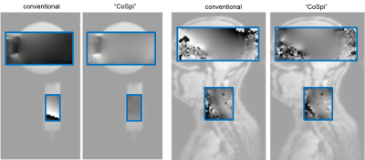 |
A Shim Algorithm to Improve the Field Homogeneity and Image Quality in Cortico-Spinal fMRI
Björn Fricke1 and Jürgen Finsterbusch1
1Department of Systems Neuroscience, University Medical Center Hamburg-Eppendorf, Hamburg, Germany
Cortico-spinal functional MRI covering a brain and a cervical spinal cord volume in the same acquisition, e.g. to investigate the interaction of brain and spinal cord areas, requires a dynamic shim update of the frequency and linear shim terms to obtain a reasonable EPI image quality in both volumes. Unfortunately, the optimum values for static higher-order and volume-specific dynamic linear shim terms cannot be determined with the standard shim algorithms provided by manufacturers. Here, a shim algorithm has been implemented that overcomes this problem and provides a better field homogeneity in the brain and spinal cord volumes.
|
 |
1217.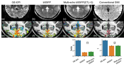 |
Implementing multi-echo balanced SSFP with a sequential phase-encoding order at 7T
Huilou Liang1,2, Ziyi Pan3, Chencan Qian1,2, Kaibao Sun1, Fanhua Guo1,2, Dehe Weng4, Jing An4, Yan Zhuo1,2,5, Hua Guo3, Danny J.J. Wang6, and Rong Xue1,2,7
1State Key Laboratory of Brain and Cognitive Science, Beijing MRI Center for Brain Research, Institute of Biophysics, Chinese Academy of Sciences, Beijing, China, 2University of Chinese Academy of Sciences, Beijing, China, 3Center for Biomedical Imaging Research, Department of Biomedical Engineering, School of Medicine, Tsinghua University, Beijing, China, 4Siemens Shenzhen Magnetic Resonance Ltd, Shenzhen, China, 5CAS Center for Excellence in Brain Science and Intelligence Technology, Chinese Academy of Sciences, Beijing, China, 6Laboratory of FMRI Technology (LOFT), Stevens Neuroimaging and Informatics Institute, University of Southern California, Los Angeles, CA, United States, 7Beijing Institute for Brain Disorders, Beijing, China
In the past decade, passband bSSFP has emerged as an alternative method to the widely-used GE-EPI in fMRI studies at high-fields. Multiline bSSFP with an interleaved phase-encoding order was further proposed to accelerate bSSFP fMRI. However, it intrinsically suffers from high spatial frequency ghosts which blur the image. In this study, we developed a multi-echo bSSFP sequence using a sequential phase-encoding order, combined with the GRAPPA technique for ghost elimination. In vivo experiments demonstrated that this sequence could shorten the imaging time and provide high-quality structural and functional MR images of the human brain at 7T with sub-millimeter resolution.
|
|
1218.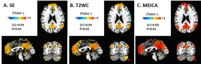 |
Feasibility of high spatial and temporal resolution multi-echo multi-band whole brain resting-state functional MRI on a compact 3T system
Daehun Kang1, Hang Joon Jo1,2, Myung-Ho In1, Erin Gray1, Ek T Tan3,4, Thomas K Foo4, Uten Yarach1, Nolan K Meyer1, Joshua D Trzasko1, John Huston1, Matt A Bernstein1, and Yunhong Shu1
1Mayo Clinic, Rochester, MN, United States, 2Hanyang University, Seoul, Republic of Korea, 3Hospital of Special Surgery, New York, NY, United States, 4GE Global Research, Niskayuna, NY, United States
Multi-echo fMRI has been shown to provide better denoising and result in improved functional analysis compared to single-echo acquisition, but it reduces the temporal resolution and inhibits high-resolution imaging. Multi-band imaging and in-plane acceleration can compensate for the reduced resolution. The high performance gradient on a compact 3T scanner can further reduce the echo-spacing and accelerate the acquisition. Here we demonstrate that high spatial-resolution ME-MB fMRI is achievable with high temporal resolution on the compact 3T. The effectiveness of ME acquisition is evaluated with different artifact reduction strategies in whole brain resting-state fMRI and compared with the standard SE acquisition.
|
|
1219.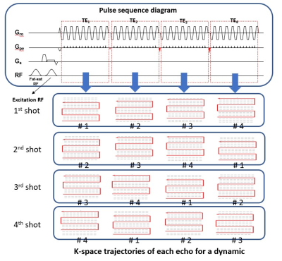 |
Multi-Echo Multi-Segment EPI Based fMRI Using Sliding-Window Acquisition and Multiplexed Sensitivity Encoding (MUSE)
Shihui Chen1, Mei-Lan Chu2, Queenie Chan3, Nan-Kuei Chen4,5, Chun-Jung Juan6, Liyuan Liang1, and Hing-Chiu Chang1
1The University of Hong Kong, Hong Kong, Hong Kong, 2Graduate Institute of Biomedical Electronics and Bioinformatics, National Taiwan University, Taipei, Taiwan, Taipei, Taiwan, 3Philips Healthcare, Hong Kong, China, Hong Kong, Hong Kong, 4Department of Biomedical Engineering, University of Arizona, Tucson, AZ, United States, Tucson, AZ, United States, 5Brain Imaging and Analysis Center, Duke University Medical Center, Durham, NC, United States, Durham, NC, United States, 6Department of Medical Imaging, China Medical University Hsinchu Hospital, Taiwan, Taipei, Taiwan
Multi-echo fMRI (ME-fMRI) has been shown to be useful in differentiating BOLD and non-BOLD signals, therefore improving the sensitivity of fMRI. Parallel imaging with high acceleration factor (e.g., R ≥ 3) is indispensable to achieve reasonable TE interval and desired spatial resolution for ME-fMRI acquisition. However, the reconstructed multi-echo images with high acceleration factor may suffer from underside noise amplification due to SENSE reconstruction. In this work, we further modify multi-echo multi-segment EPI (MEMS-EPI) technique with sliding window acquisition to acquire multi-echo fMRI with high acceleration factor, and then reconstruct highly accelerated multi-echo fMRI images with MUSE algorithm.
|
|
1220.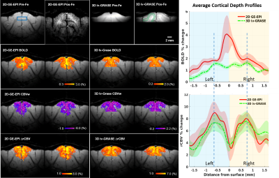 |
Ultrahigh-resolution Laminar fMRI Mapping of Cat Visual Cortex at 9.4T: Comparison of 2D GE-EPI and 3D iv-GRASE Sequences
Wei Zhu1, Djaudat Idiyatullin1, Shinho Cho1, Yi Zhang1, Kâmil Uğurbil1, Xiao-Hong Zhu1, and Wei Chen1
1Center for Magnetic Resonance Research, Department of Radiology, University of Minnesota, Minneapolis, MN, United States
Blood oxygenation-level dependent (BOLD) fMRI studies at the level mesoscopic organizations, such as cortical columns and layers, is challenged by the low signal-to-noise ratio (SNR) and the specificity of different fMRI sequences even at ultrahigh magnetic fields. In this work, we show that when mapping layer-specific activities in the cat visual cortex, ultrahigh-resolution 3D GRASE sequence with inner volume selection achieved similar specificity as at 9.4 Tesla CBV-fMRI using 2D GE-EPI sequence, while attains a higher BOLD sensitivity. Our results indicate that 3D iv-GRASE BOLD is promising for laminar and columnar mapping of brain functions.
|
1221.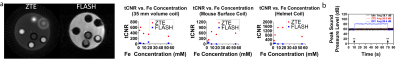 |
iZTE-fMRI
Martin John MacKinnon1, Sheng Song1, Li-Ming Hsu1, Sung-Ho Lee1, G. Allan Johnson2, and Yen-Yu Ian Shih1
1University of North Carolina at Chapel Hill, Chapel Hill, NC, United States, 2Duke University, Durham, NC, United States
In this study we demonstrate how a zero-echo-time (ZTE) technique can overcome several limitations of traditional fMRI experiments. We demonstrate that ZTE fMRI can detect functional activations with positive iron oxide contrast, termed iZTE-fMRI, at an approximate three-fold magnitude increase in tCNR when compared to GRE-techniques in-vivo - with the further potential demonstrated from phantom studies to increase tCNR more significantly under optimal contrast agent dose. We also show that iZTE fMRI experiments can produce functional images with markedly less susceptibility artifacts and acoustic noise than standard GRE techniques.
|
|
|
1222.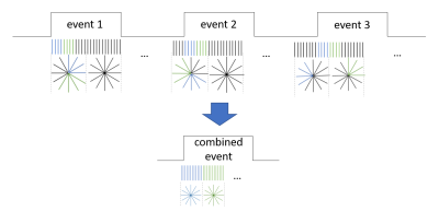 |
Novel resampling approach for 200-ms temporal resolution MB-SWIFT fMRI – application to DBS in rats
Ekaterina Zhurakovskaya1, Lauri Lehto1, Jaakko Paasonen1, Lin Wu2, Sheng Sang2, Jun Ma2, Hanne Laakso1, Tiina Pirttimäki1, Olli Gröhn1, Silvia Mangia2, and Shalom Michaeli2
1University of Eastern Finland, Kuopio, Finland, 2Center for Magnetic Resonance Research, University of Minnesota, Minneapolis, MN, United States
Deep brain stimulation (DBS) is widely used to treat several disorders. Given its minimal sensitivity to electrode-induced artifacts, fMRI with Multi-Band Sweep Imaging with Fourier Transformation (MB-SWIFT) is a powerful tool for identifying the DBS mechanism of action at a network level. However, MB-SWIFT generally suffers from low time resolution, thus limiting the characterization of temporal features. Here, we introduce a novel resampling approach applicable to radial k-space sampling such as used in MB-SWIFT, allowing to track repeating events with 200-ms time resolution. A proof of concept was demonstrated during DBS of the medial septal nucleus in rats.
|
|
1223.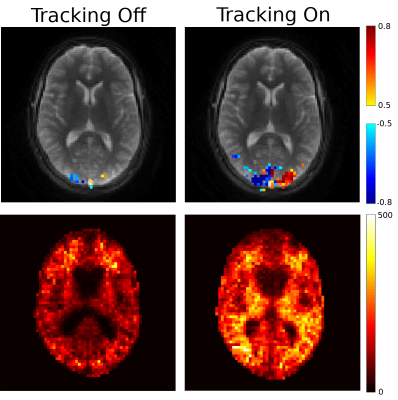 |
Real-Time Respiration Compensation in Oscillating Steady State fMRI
Amos A Cao1 and Douglas Noll2
1University of Michigan, Ann Arbor, MI, United States, 2Biomedical Engineering, University of Michigan, Ann Arbor, MI, United States
Oscillating Steady-State Imaging (OSSI) is a new steady-state sequence for high-SNR fMRI. Respiration-induced B0 changes cause undesirable changes to the OSSI steady-state, resulting in signal artifacts. To address this problem, we present a prospective correction method which utilizes a self-navigating spiral trajectory to measure and correct for B0 changes in real-time. In an initial fMRI proof-of-concept, our real-time correction method increased the number of activated voxels by 454% and increased mean tSNR by 81%. Real-time prospective correction has the potential to outperform retrospective correction methods by directly reducing perturbations to steady-state magnetization during acquisition.
|
|
1224.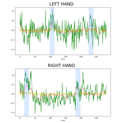 |
A Paradigm Free Regularization Approach to Recover Brain Activations: Validation on Task fMRI
Isa Costantini1, Samuel Deslauriers-Gauthier1, and Rachid Deriche1
1Athena Project-Team, Inria Sophia Antipolis - Méditerranée, Université Côte d’Azur, Biot, France
In this work we propose and validate a Paradigm-Free fMRI (PFFMRI) algorithm that acts directly on the 4-D fMRI image and recover the underlying brain activations without knowledge on the experimental paradigm. PFFMRI is based on the idea that large image variations should be preserved as they occur during brain activation, but small variations should be smoothed to remove noise. Starting from this, we were able to regularize the fMRI image with an anisotropic regularization, thus recovering the location of the brain activations in space and their timing and duration without knowledge of the experimental paradigm.
|
 Watch the Video