
Scientific Session
Interventional
Session Topic: Interventional
Session Sub-Topic: Technology for MRI-Guided Therapy
Oral
Interventional
| Monday Parallel 4 Live Q&A | Monday, 10 August 2020, 13:45 - 14:30 UTC | Moderators: Jan Fritz & Henrik Odéen |
Session Number: O-78
0101.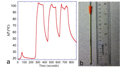 |
Investigation of RF heating risk during MRI-guided cryoablation at 1.5T
Aiming Lu1, Christopher P Favazza1, David A Woodrum1, Joel P Felmlee1, Jacinta Browne1, Brian T Welch1, and Krzysztof R Gorny1
1Radiology, Mayo Clinic, Rochester, MN, United States
Cryoablation with MRI guidance is desirable is a feasible treatment for localized tumors. There is a MRI-conditional cryoablation system and, to the best of our knowledge, no previous report of RF heating/burn incidence. However, since the cryoneedles are metallic and the gas lines have metallic components, potential risks of RF heating/burn exists. In fact, an incidence of skin burn recently occurred in our institution during a MRI-guided liver treatment case. In this work, we demonstrated that RF heating could be significant during MRI-guided cryoablation and showed that several strategies could be potentially used to mitigate the risk.
|
|
0102.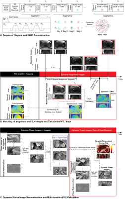 |
Dynamic PRF and T1-based 3D Thermometry in the Liver using a Variable Flip Angle Stack-of-Radial Technique
Le Zhang1, Tess Armstrong1,2, and Holden H. Wu1,2,3
1Radiological Sciences, University of California, Los Angeles, Los Angeles, CA, United States, 2Physics and Biology in Medicine, University of California, Los Angeles, Los Angeles, CA, United States, 3Bioengineering, University of California, Los Angeles, Los Angeles, CA, United States
MR thermometry in the liver is challenged by mismatch between baseline and dynamic images caused by motion, leading to temperature errors. To address motion, previous methods had to compromise spatial coverage to increase temporal resolution. We propose a variable-flip-angle (VFA) 3D stack-of-radial technique for combined proton resonance frequency shift (PRF) and T1-based MR thermometry with volumetric coverage and high spatiotemporal resolution. Accurate VFA T1 calculation is achieved by synthesizing B1+ maps that match the liver position in dynamic images. A multi-baseline approach is used for accurate dynamic PRF measurements. Results from non-heating scans demonstrate reliable liver T1 and PRF measurements.
|
|
0103.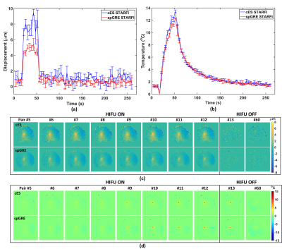 |
Simultaneous MR acoustic radiation force imaging and MR thermometry: comparison of coherent echo-shifted and RF spoiled gradient echo sequence
Yangzi Qiao1, Chao Zou1, Chuanli Cheng1, Changjun Tie1, Xin Liu1, and Hairong Zheng1
1Shenzhen Institutes of Advanced Technology, Chinese Academy of Sciences, Shenzhen, China
Simultaneous MR acoustic radiation force imaging and MR thermometry (STARFI) based on coherent echo-shifted sequence (cES) was proposed and comprehensively compared to RF spoiled gradient echo (spGRE). The calculated displacement of cES STARFI was always larger than the value of spGRE STARFI through both the simulation and experiments, while the accuracy of the temperature monitoring of cES was maintained. The temperature and displacement map acquired during HIFU heating were in good accordance with each other. The cES STARFI can be an alternative for comprehensively monitoring of HIFU treatment with increased displacement sensitivity and time efficiency compared to spGRE STARFI.
|
|
 |
0104.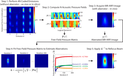 |
Rapid autofocusing of MR-guided focused ultrasound acoustic pressure fields using MR-ARFI with spatially coded emissions
Sumeeth Jonathan1,2, M Anthony Phipps2, Charles F Caskey2, and William A Grissom2
1Biomedical Engineering, Vanderbilt University, Nashville, TN, United States, 2Vanderbilt University Institute of Imaging Science, Vanderbilt University Medical Center, Nashville, TN, United States
Magnetic resonance-guided focused ultrasound (MRgFUS) has many potential neurological applications, but skull-induced aberrations of the acoustic pressure field limit its specificity and safety. MR-acoustic radiation force imaging (MR-ARFI)-based methods have been proposed to refocus the pressure field in situ. However, they take too long for practical in vivo use. We propose a multi-voxel MR-ARFI-based autofocusing method for rapid aberration correction of MRgFUS acoustic pressure fields. We compare our proposed method to the canonical single-voxel MR-ARFI-based refocusing method and demonstrate that as few as two MR-ARFI acquisitions can be used to refocus a programmatically aberrated pressure field.
|
 |
0105.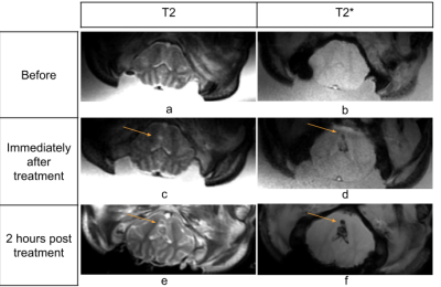 |
MRI assessment and monitoring of cavitation-based ultrasound therapy (histotripsy) for transcranial brain treatment in vivo
Dinank Gupta1, Ning Lu1, Jonathan Sukovich1, Krisanne Litnas2, Aditya Pandey3, Badih Junior Daou3, Timothy Hall1, Zhen Xu1, Scott Peltier2, and Douglas Noll1
1Biomedical Engineering, University of Michigan, Ann Arbor, MI, United States, 2Functional MRI Laboratory, University of Michigan, Ann Arbor, MI, United States, 3Department of Neurosurgery, University of Michigan, Ann Arbor, MI, United States
Transcranial MR guided cavitation-based Focused Ultrasound (FUS) treatment (histotripsy) is performed in vivo for the first time on a pig brain. Transcranial histotripsy is delivered by an MRI compatible FUS transducer array inside a 3T MRI scanner. Real-time MRI monitoring with 2 second temporal resolution is carried with an intra-voxel incoherent motion (IVIM) pulse sequence synchronized with the FUS array. IVIM images show the histotripsy ablation effect at the intended treatment location in real-time, and the ablation zone was confirmed by post-treatment images. This is the first study to show successful in vivo transcranial histotripsy guided by MRI.
|
|
0106.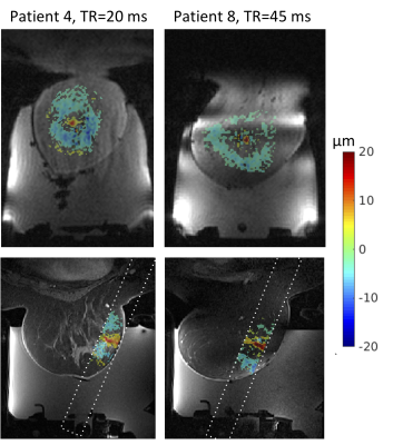 |
Three-dimensional magnetic resonance acoustic radiation force imaging in the breast
Allison Payne1, Lorne Hofstetter2, Henrik Odéen1, Erik Dumont3, Dennis L Parker1, and Jean Palussiere4
1Radiology and Imaging Sciences, University of Utah, Salt Lake City, UT, United States, 2Biomedical Engineering, University of Utah, Salt Lake City, UT, United States, 3Image Guided Therapy, Pessac, France, 4Institut Bergonie, Bordeaux, France
3D MR acoustic radiation force imaging (MR-ARFI) is a useful treatment planning and monitoring tool for magnetic resonance guided focused ultrasound (MRgFUS) treatments in the breast. MR-ARFI displacement is easily visualized in fat, fibroglandular and tumor tissues, allowing for accurate localization of the ultrasound beam and quantitative tissue assessment. The potential formation of standing shear waves in the breast requires careful optimization of the pulse sequence to ensure clear visualization of the radiation force displacement point. This effect is shown in both human breast and phantom data.
|
|
0107.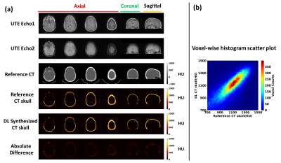 |
Deep learning for improved workflow in MRgFUS treatment planning
Pan Su1,2, Sijia Guo1,3, Florian Maier4, Steven Roys1,3, Himanshu Bhat2, Elias R. Melhem1, Dheeraj Gandhi1, Rao P. Gullapalli1,3, and Jiachen Zhuo1,3
1Department of Diagnostic Radiology and Nuclear Medicine, University of Maryland School of Medicine, Baltimore, MD, United States, 2Siemens Medical Solutions USA Inc, Malvern, PA, United States, 3Center for Metabolic Imaging and Therapeutics (CMIT), University of Maryland Medical Center, Baltimore, MD, United States, 4Siemens Healthcare GmbH, Erlangen, Germany
Transcranial MRI-guided focused ultrasound (tcMRgFUS) is a promising technique to treat multiple diseases. Here we examined the feasibility of leveraging deep-learning to convert MRI dual echo UTE images directly to synthesized CT skull images. We demonstrated that the derived model is capable of not only segmenting the UTE images to generate synthetic CT skull masks that are highly comparable to true CT skull masks, but is also able to reliably predict the CT skull intensities in Hounsfield units. Furthermore, we demonstrated that synthetic CT skull can be reliably used for skull-density-ratio (SDR) determination and predicting target temperature rise in tcMRgFUS.
|
|
0108.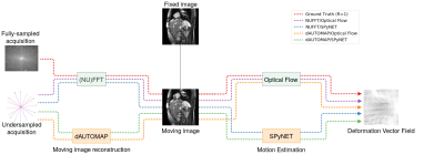 |
Real-time estimation of 2D deformation vector fields from highly undersampled, dynamic k-space for MRI-guided radiotherapy using deep learning
Maarten L Terpstra1,2, Federico d'Agata1,2,3, Bjorn Stemkens1,2, Jan JW Lagendijk1, Cornelis AT van den Berg1,2, and Rob HN Tijssen1,2
1Department of Radiotherapy, Division of Imaging & Oncology, University Medical Center Utrecht, Utrecht, Netherlands, 2Computational Imaging Group for MR diagnostics & therapy, Center for Image Sciences, University Medical Center Utrecht, Utrecht, Netherlands, 3Department of Neurosciences, University of Turin, Turin, Italy
MRI-guided radiotherapy (MRgRT) enables new ways to improve dose delivery to moving tumors and the organs-at-risk (e.g. in abdomen) by steering the radiation beam based on real-time MRI. While state-of-the-art techniques (e.g. compressed sensing) can provide the required acquisition speed, the corresponding reconstruction time is too long for real-time processing. In this work, we investigate the use of multiple deep neural networks for image reconstruction and subsequent motion estimation. We show that a single motion estimation network can estimate high-quality 2D deformation vector fields from aliased images, even for high undersampling factors up to R=25.
|
|
0109.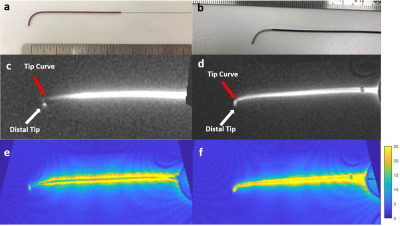 |
A novel active guidewire design with a curved tip geometry for interventional MRI applications under 0.55T.
Korel Dursun Yildirim1,2, Christopher Bruce1, Rajiv Ramasawmy1, Kendall O'Brien1, Adrienne Campbell-Washburn1, Daniel Herzka1, Robert J. Lederman1, and Ozgur Kocaturk1,2,3
1Cardiovascular Branch, Division of Intramural Research, National Heart Lung and Blood Institute, National Institutes of Health, Bethesda, MD, United States, 2Institute of Biomedical Engineering, Bogazici University, Istanbul, Turkey, 3Transmural Systems, Andover, MA, United States
A clinical-grade active MRI 0.035” guidewire design with a curved distal tip geometry and continuous shaft signal ensuring the mechanical and electrical safety, was introduced. Proposed design was tested in-vitro and in-vivo for MRI visibility and mechanical performance, and in-vitro for RF induced heating.
|
0110. |
Dynamic control of RF currents in conductive guidewires with an auxiliary PTx system: First in vivo experience in sheep
Felipe Godinez1, Raphael Tomi-Tricot2, Marylene Delcey3, Gunthard Lykowsky4, Steven E Williams1, Bruno Quesson3, Joseph V Hajnal1, and Shaihan J Malik1
1Biomedical Engineering Department, School of Biomedical Engineering and Imaging Sciences, king's College London, London, United Kingdom, 2Siemens Healthcare Limited, London, United Kingdom, 3Centre de recherche Cardio-Thoracique de Bordeaux, Bordeaux, France, 4RAPID Biomedical GmbH, Rimpar, Germany
This paper presents first in vivo results of a parallel transmit (PTx) system adept at regulating radiofrequency induced guidewire heating, during MRI guided interventions in sheep. The PTx system, which is an add-on to an unmodified conventional 1.5T scanner, regulates heating by operating in modes that couple/decouple the guidewire from the radiofrequency transmit array. With an inserted guidewire decoupling modes allow operation with unrestricted B1+ to safely visualize anatomy, while the coupling mode operated at low radiofrequency power provides safe visualization of the guidewire itself. Temperature measurements at the guidewire tip and in vivo images are shown.
|
 Watch the Video