
Scientific Session
Novel Hardware
Session Topic: Novel Hardware
Session Sub-Topic: New Directions in MR Systems
Oral
Engineering
| Thursday Parallel 5 Live Q&A | Thursday, 13 August 2020, 15:05 - 15:50 UTC | Moderators: Shaoying Huang & Andrew Webb |
Session Number: O-79
1250.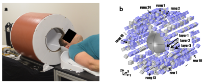 |
A Point-of-Care MRI Scanner for Human Brain Imaging
Clarissa Zimmerman Cooley1,2, Patrick C McDaniel1,3, Jason P Stockmann1,2, Sai Abitha Srinivas1, and Lawrence L Wald1,2,4
1Athinoula A Martinos Center for Biomedical Imaging, Dept. of Radiology, Massachusetts General Hospital, Charlestown, MA, United States, 2Harvard Medical School, Boston, MA, United States, 3Dept. of Electrical Engineering, Massachusetts Institute of Technology, Cambridge, MA, United States, 4Harvard-MIT Division of Health Sciences and Technology, Cambridge, MA, United States
Access to MRI scanners is limited by cost, size, and siting requirements. Specialized low-cost, compact, portable systems could greatly increase accessibility worldwide and enable point-of-care MRI. We present a portable MRI scanner for human brain imaging based on a compact 122kg Halbach cylinder with a built-in readout field. Designing for a built-in encoding field reduces the size of the magnet, the overall system power-consumption, cooling requirements, and acoustic noise. The generalized reconstruction method accounts for non-linearities in the gradient fields. T1 and T2-weighted in vivo images are presented with a resolution of 2x2x7mm.
|
|
 |
1251.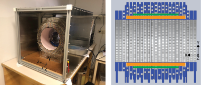 |
Three-dimensional in-vivo human Imaging on a 50 mT low-cost portable Halbach Array
Thomas O'Reilly1, Wouter Teeuwisse1, Bart de Vos2, and Andrew Webb1
1C.J. Gorter Center for High Field MRI, Leiden University Medical Center, Leiden, Netherlands, 2Circuits and Systems, Delft University of Technology, Delft, Netherlands
We show the first 3D in-vivo images acquired from our custom-built 50 mT low-cost Halbach based portable MRI scanner. 3D Images of a knee were acquired with a ~2 mm isotropic resolution using a 3D turbo spin echo sequence in less than 12 minutes. Gradient non-linearity induced image distortions are minimal within the central ~10 cm of the magnet bore length, but require correction beyond this point. These results represent the latest step towards our goal of creating a fully portable MRI scanner targeting pediatric neuroimaging in the developing world for less than 30 000 euros.
|
|
1252.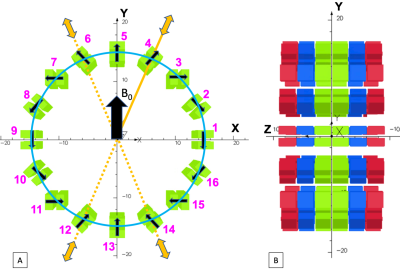 |
Design of a Permanent Magnet for MRI of the Ankle on the International Space Station
Aaron R. Purchase1, Gordon E. Sarty2, Logi Vidarrson3, Keith Wachowicz1, Piotr Liszkowski4,5, Hongwei Sun1, Jonathan C. Sharp1, and Boguslaw Tomanek1,6
1Department of Oncology, University of Alberta, Edmonton, AB, Canada, 2Department of Psychology and Division of Biomedical Engineering, University of Saskatchewan, Saskatoon, SK, Canada, 3LT Imaging, Toronto, ON, Canada, 4AGH University of Science and Technology, Krakow, Poland, 5MRI-Tech Sp. z o.o, Krakow, Poland, 6Institute of Nuclear Physics, Polish Academy of Sciences, Krakow, Poland
MRI is desired for monitoring bone and muscle deterioration in astronauts during long-term spaceflights and on the International Space Station (ISS). However, the magnet is generally too heavy to be transported to the ISS. Therefore, we designed a light (~10kg) magnet that allows transmit array spatial encoding (TRASE) MRI of the ankle on the ISS. The magnet is based on a sparse Halbach geometry with magnetic block-pairs. The positions of the block-pairs were optimized using a genetic algorithm. We intend to manufacture the magnet and test the entire MRI system on a Falcon 20 jet.
|
1253.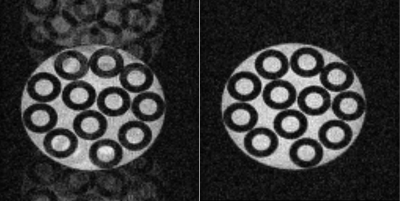 |
First Experiences with a Three-Bore Conduction-Cooled Cryogen-Free Extremity Scanner
Jerome L. Ackerman1,2, Shahin Pourrahimi3, Marcus Donaldson1,2, Nadder Pourrahimi3, Julien Rivoire4, Julien Muller4, Hizami Murad4, Ouri Cohen5, and Isabela Choi1
1Martinos Center, Dept of Radiology, Massachusetts General Hospital, Charlestown, MA, United States, 2Department of Radiology, Harvard Medical School, Boston, MA, United States, 3Superconducting Systems, Inc., Billerica, MA, United States, 4RS2D, Mundolsheim, France, 5Memorial Sloan Kettering Cancer Center, New York, NY, United States
We developed an MRI scanner designed for limb scanning based on a novel conduction-cooled three-bore cryogen-free 1.5T magnet. The two outer bores accommodate the unscanned leg for enhanced patient comfort. We describe the issues relevant to this unique magnet and scanner, including our solution to the field instability problem common in conduction-cooled MRI magnets. By recording the field variation as a function of the phase of the cold head cycle, a compensating waveform may be played out via the numerically-controlled oscillator of the scanner to reduce the periodic field excursion from 25 Hz to about 1-2 Hz.
|
|
| WITHDRAWN | ||
 |
1255.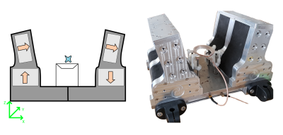 |
Single sided magnet system for relaxometry and diffusion measurement
Dion Thomas1, Petrik Galvosas1, Paul D Teal2, Freya G Harrison3,4, Max Berry3,5, Yu-Chieh Tzeng3, and Sergei Obruchkov6
1School of Chemical and Physical Sciences, Victoria University of Wellington, Wellington, New Zealand, 2School of Engineering and Computer Science, Victoria University of Wellington, Wellington, New Zealand, 3Centre for Translational Physiology, University of Otago, Wellington, New Zealand, 4Department of Surgery and Anaesthesia, University of Otago, Wellington, New Zealand, 5Department of Paediatrics and Child Health, University of Otago, Wellington, New Zealand, 6Robinson Research Institute, Victoria University of Wellington, Wellington, New Zealand We have developed a new permanent magnet based single sided magnetic resonance system, which is suitable for relaxometry and diffusion measurement. Our design generates an external region of homogeneous B0 field, a sweet spot, from which signal can be detected. The magnet has been optimised to have a larger penetration depth and higher B field strength than currently existing systems. We have found the system provides good homogeneity and field strength, making it useful for relaxometry. Additionally, we demonstrate it can be used for diffusion-T2 measurements, which will allow further biomedical applications. |
1256.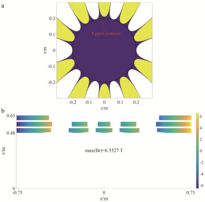 |
A short-bore helium-off 7 T whole-body MRI superconducting magnet design
Yaohui Wang1, Qiuliang Wang1, Jianhua Liu1, Junsheng Cheng1, Zhongbiao Xu2, Zhifeng Chen3, and Feng Liu4
1Institute of Electrical Engineering, Chinese Academy of Sciences, Beijing, China, 2Department of Radiotherapy Cancer Center, Guangdong Provincial People's Hospital & Guangdong Academy of Medical Science, Guangzhou, China, 3School of Biomedical Engineering, Southern Medical University, Guangzhou, China, 4School of Information Technology and Electrical Engineering, The University of Queensland, Brisbane, Australia
7T whole-body MRI system has received comprehensive praises from the worldwide users, but some patients feel clautrophobic when doing the scanning at the long tunnel. This work proposed a ultra-short design scheme for the 7 T MRI magnet, which is nearly a half of the presented commercial system. In addition, the magnet operates at helium-off enviroment, which saves the precious coolant liquid helium and is also conveinent to maintain.
|
|
1257.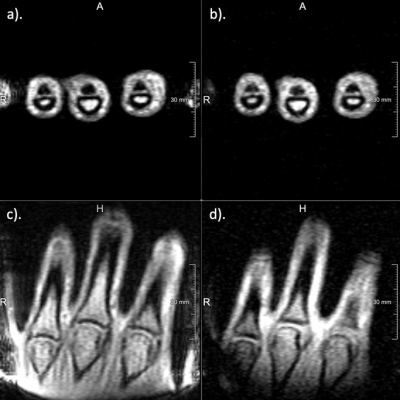 |
In vivo Two-photon Magnetic Resonance Imaging of Human Hand at 1T
Jianshu Chi1, Victor Han1, and Chunlei Liu1,2
1Electrical Engineering and Computer Sciences, University of California, Berkeley, Berkeley, CA, United States, 2Helen Wills Neuroscience Institute, University of California, Berkeley, Berkeley, CA, United States
We demonstrate the first in vivo imaging experiment with a multiphoton excitation technique at 1T. To produce multiphoton excitation, we built a secondary RF coil that produces an RF field, parallel to the $$$B_{0}$$$ field. Designed for kHz frequencies, this coil consists with two layers of traces in a spiral configuration on a printed circuit board (PCB). Adding this low-frequency coil to an Aspect 1T Wrist Scanner, we acquired gradient echo images with two-photon excitation of a human hand in vivo.
|
|
|
1258. |
Improving Homogeneity in Delta Relaxation Enhanced Magnetic Resonance Using Boundary Element Method
Matthew A. McCready1, William B. Handler1, and Blaine A. Chronik1
1Physics and Astronomy, Western University, London, ON, Canada
Delta relaxation enhanced magnetic resonance (dreMR) is a field-shifting quantitative molecular imaging method using activatable MR probes. The dreMR method may be used to produce images with signal proportional to concentration of contrast agent and eliminate signal due to unbound agent. In this work we outline a novel design method for dreMR coils, using an inner layer of windings determined by the boundary element method (BEM). This new design method produces a strong, highly homogeneous field shift which, when coupled with a 0.5T MRI system and activatable MR probes, can reliably image on a larger volume than previous designs.
|
1259.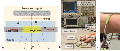 |
Wearable MRS system for blood glucose measurement: Proof of concept
Yongxian Qian1, Bei Zhang1, Shannon Haas1, and Aaron R. Chidakel2
1Radiology, New York University, New York, NY, United States, 2Endocrinology, New York University, New York, NY, United States
This study presents preliminary results supporting a new concept to build a wearable magnetic resonance spectroscopy (MRS) system for noninvasive blood glucose measurement in humans. Computer simulations, hardware buildings and subject studies demonstrated the promising of such a system.
|
 Watch the Video