
Scientific Session
Engineering and Safety of MRI
Session Topic: Engineering and Safety of MRI
Session Sub-Topic: MR Engineering & Safety
Oral
Engineering
| Thursday Parallel 5 Live Q&A | Thursday, 13 August 2020, 14:20 - 15:05 UTC | Moderators: Thomas Denney & Gigi Galiana |
Session Number: O-81
1129.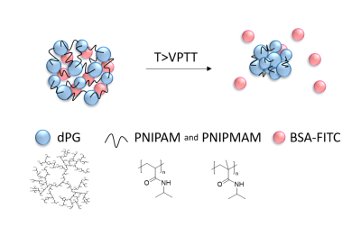 |
Temperature Triggered Release of a Protein from Thermoresponsive Nanogels Using Thermal Magnetic Resonance
Yiyi Ji1, Lukas Winter2, Lucila Navarro3,4, Min-Chi Ku1, João Periquito1, Michal Pham1, Werner Hoffmann2, Loryn E. Theune3, Marcelo Calderón3,5,6, and Thoralf Niendorf1,7
1Berlin Ultrahigh Field Facility (B.U.F.F.), Max Delbrück Center for Molecular Medicine in the Helmholtz Association (MDC), Berlin, Germany, 2Physikalisch-Technische Bundesanstalt (PTB), Berlin, Germany, 3Institute of Chemistry and Biochemistry, Freie Universität Berlin, Berlin, Germany, 4Instituto de Desarrollo Tecnológico para la Industria Química (INTEC), Universidad Nacional del Litoral (UNL) - Consejo Nacional de Investigaciones Científicas y Técnicas (CONICET), Santa Fe, Argentina, 5POLYMAT and Applied Chemistry Department, Faculty of Chemistry, University of the Basque Country UPV/EHU, Donostia-San Sebastián, Spain, 6IKERBASQUE, Basque Foundation for Science, Bilbao, Spain, 7Experimental and Clinical Research Center (ECRC), a joint cooperation between the Charité Medical Faculty and the Max Delbrück Center for Molecular Medicine, Berlin, Germany
Thermal Magnetic Resonance (ThermalMR) adds a thermal dimension to an MR device by exploiting the constructive interference of radiofrequency (RF) waves for temperature intervention. Here, the capacity of ThermalMR is demonstrated in a model system involving the release of a protein from thermoresponsive nanogels. Upon RF heating the nanogels (T=43°C), 29.3% of the protein were released after 6h which is in accordance with the release profile obtained for the reference data derived from a water bath setup. ThermalMR provides an ideal testbed for the study of temperature induced release of drugs, MR probes and other agents from thermoresponsive carriers.
|
|
1130.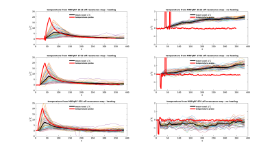 |
Magnetic Resonance Fingerprinting with Quadratic RF Phase for Continuous Temperature Monitoring in Aqueous Tissues
Rasim Boyacioglu1, Megan Poorman2,3, Kathryn Keenan2, and Mark Griswold1
1Radiology, Case Western Reserve University, Cleveland, OH, United States, 2Physical Measurement Laboratory, National Institute of Standards and Technology, Boulder, CO, United States, 3Department of Physics, University of Colorado Boulder, Boulder, CO, United States
Conventional temperature monitoring is based on measurement of off-resonance via gradient-echo phase scans for non-adipose tissue. MRF with quadratic RF phase (MRFqRF) simultaneously quantifies off-resonance, T1, T2, and T2*. For a proof of principle thermometry experiment with MRFqRF, an ex-vivo aqueous sample was heated with laser ablation, temperature was tracked, and multiple continuous MRFqRF scans were obtained with different temporal resolutions. Scanner frequency drifts were removed automatically with Independent Component Analysis. Residual changes in off-resonance predict the temperature change. However, MRFqRF temporal resolution (~10s) needs to be increased further for clinical relevance.
|
|
 |
1131.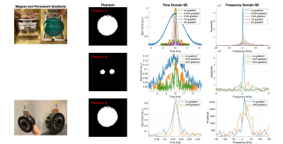 |
Bloch-Optimized Dithered-Ultrasound-Pulse RF for Low-Field Inhomogeneous Permanent Magnet MR Imagers
Irene Kuang1, Nick Arango1, Jason Stockmann2,3, Elfar Adalsteinsson1,4, and Jacob White1
1Electrical Engineering and Computer Science, Massachusetts Institute of Technology, Cambridge, MA, United States, 2A. A. Martinos Center for Biomedical Imaging, Massachusetts General Hospital, Charlestown, MA, United States, 3Harvard Medical School, Boston, MA, United States, 4Institute for Medical Engineering and Science, Massachusetts Institute of Technology, Cambridge, MA, United States
Declining costs of permanent magnets and embedded systems electronics has driven systems engineering of point-of-care diagnostics to the forefront of MR research. We present a low-cost RF signal chain (<$100) for low-field imaging using a Teensy 4.0 microcontroller and STHV800 ultrasound pulser IC. Bloch-optimization simulation of programmable, dithered-pulses enables broadband excitation of the notably inhomogeneous permanent magnets employed in portable, hand-held MR systems.
|
1132.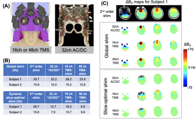 |
Feasibility of using a 3-axis multi-channel TMS coil array for B0 shimming of the brain at 3T
Jason P. Stockmann1,2, Lucia Navarro de Lara1, Larry Wald1,2, and Aapo Nummenmaa1,2
1A. A. Martinos Center, Massachusetts General Hospital, Charlestown, MA, United States, 2Harvard Medical School, Boston, MA, United States
We explore the local field control capability of a 48ch TMS coil array to perform B0 shimming in the brain. The array uses sixteen 3-axis TMS coils built using three independent orthogonal windings that provide added flexibility to orient the resultant magnetic dipole and thus facilitate tailoring the B0 shim field. We propose adapting the TMS coils to carry DC shim currents during EPI fMRI time series acquisitions to reduce artifact levels and improve the utility of TMS-fMRI for studying brain circuits.
|
|
 |
1133.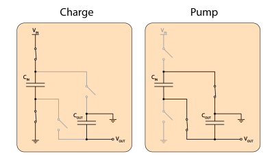 |
In-bore voltage inversion with very low EMI by a switched capacitor converter
Christoph Michael Schildknecht1, David Otto Brunner1, and Klaas Paul Pruessmann1
1ETH Zurich and University of Zurich, Zurich, Switzerland
Complex in-bore electronics (e.g. motion trackers, digitizer etc.) frequently requires various voltage rails. This poses especially a challenge when bi-polar voltages are required because in-bore voltage inversion is typically not possible due to the EMI of such converter. The here presented switched capacitator voltage inverter allows in-bore operation without disturbing the MRI scanner due to the very low EMI. Lab and 3T MRI measurement were performed to verify the EMC compatibility and characterize the device.
|
1134.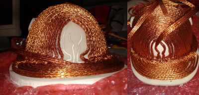 |
Single channel non-linear breast gradient coil for diffusion encoding
Sebastian Littin1, Feng Jia1, Philipp Amrein1, Huijun Yu1, Arthur Magill2, Tristan Kuder2, Mark E. Ladd2, Frederik Laun3, Sebastian Bickelhaupt4, and Maxim Zaitsev1
1Department of Radiology, Medical Physics, University of Freiburg, University Medical Center, Freiburg, Germany, 2Medical Physics in Radiology, German Cancer Research Center, Heidelberg, Germany, 3Department of Radiology, MR Physics, University Medical Center Erlangen, Erlangen, Germany, 4Junior Group Medical Imaging and Radiology – Cancer Prevention, German Cancer Research Center (DKFZ), Heidelberg, Germany
The aim of this project is to design and implement a non-linear single channel breast gradient coil for diffusion encoding. Initial field maps of the prototype implementation are shown. The prototype should allow to generate gradient strengths between 1 and 3.6 [T/m].
|
|
|
1135.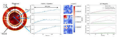 |
Measurement-based safety assessment, prediction and mitigation of RF induced implant heating with parallel transmission: temperature matrix
Berk Silemek1, Lukas Winter1, Frank Seifert1, Harald Pfeiffer1, and Bernd Ittermann1
1Physikalisch-Technische Bundesanstalt (PTB), Braunschweig and Berlin, Germany
A Measurement-based Temperature-Matrix approach is presented that enables a fast, patient and exam specific estimation and mitigation of RF hazards of implants. Various locations in phantom are tested using an 8-channel (300MHz) implant safety testbed. Heating reduction Based on T-Matrix Measurements resulted >3 times heating reduction vs. circularly-polarized mode and >19 times vs. worst-case mode. 2-channel MRI (3T) feasibility experiments using high temperature resolution showed good correlation with transmitted power. In addition, T-matrix-based temperature increase predictions successfully demonstrated. As summary, an easy to implement, cheap, sensor-based method, the T-matrix, to investigate, characterize and mitigate RF heating of implants is introduced.
|
1136.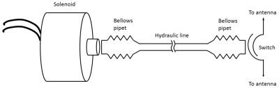 |
A Hydraulically Operated Wireless RF Switch to Control Antenna Tuning in MR-Mediated Radiofrequency Ablation
Jerome L. Ackerman1,2, Erez Nevo3, and Abraham Roth3
1Martinos Center for Biomedical Imaging, Department of Radiology, Massachusetts General Hospital, Charlestown, MA, United States, 2Department of Radiology, Harvard Medical School, Boston, MA, United States, 3Robin Medical, Inc., Baltimore, MD, United States
In magnetic resonance mediated radiofrequency ablation (MR-RFA) the RF energy for the ablation is captured by a wire antenna placed in the scanner bore, and channeled to the ablation needle. There are no external wired connections. The effective length of the antenna is adjusted physically or electrically to be resonant with the scanner RF to maximize energy capture. To suppress heating when desired, the antenna must be detuned. An electronic switch to do so reduces antenna efficiency, but a simple wireless hydraulically activated mechanical switch maintains full antenna efficiency and achieves high on-off ratio.
|
|
|
1137.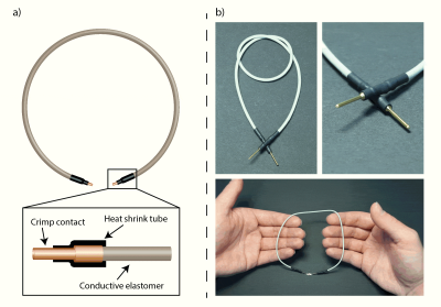 |
Conductive Elastomer for Wearable RF Coils
Andreas Port1, Roger Luechinger1, David Otto Brunner1, and Klaas Paul Pruessmann1
1Institute for Biomedical Engineering, ETH Zurich and University of Zurich, Zurich, Switzerland
Several stretchable conductor concepts have been proposed that rely on a continuous metal phase, rigid or liquid, as conductive path. Conductive elastomers, fundamentally different, form the conductive path through contact between particles such as carbon nano tubes, silver nanowires or silver microparticles. In the present work, we explore the feasibility and performance of MR detection with conductive elastomer coils. Evaluation is performed in terms of Q, SNR and in-vivo imaging. The results indicate that MR receive coils made from conductive elastomer provide good stretchability, adequate electrical performance and promise workflow enhancements as such a coil could even be washed.
|
|
1138.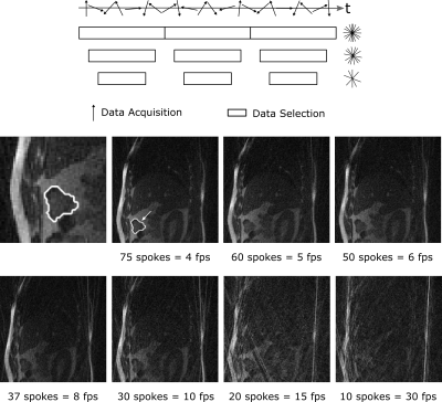 |
Comparison of tumor autosegmentation techniques from an undersampled dynamic radial bSSFP acquisition on a low-field MR-linac
Florian Friedrich1,2, C. Katharina Spindeldreier3, Juliane Hörner-Rieber3, Sebastian Klüter3, Peter Bachert1,2, Mark E. Ladd1, and Benjamin R. Knowles1
1Medical Physics in Radiology, German Cancer Research Center (DKFZ), Heidelberg, Germany, 2Department of Physics and Astronomy, Heidelberg University, Heidelberg, Germany, 3Department of Radiation Oncology,, University Hospital of Heidelberg, Heidelberg, Germany
MR-linac hybrid systems can dynamically image a tumor during radiotherapy to aid in a more precise delivery of the radiation dose. Motion tracking of the target is required and is currently performed by a deformable image registration on Cartesian bSSFP images. This study compares three different tracking methods (convolutional neuronal network, multi-template matching, and deformable image registration) to track a lung tumor in Cartesian images, where the performance of the three methods did not differ significantly. The convolutional neuronal network provided minimal decrease in tracking accuracy in a healthy volunteer when undersampled radial images were used to accelerate image acquisition.
|
 Watch the Video