
Scientific Session
Engineering and Safety of MRI
Session Topic: Engineering and Safety of MRI
Session Sub-Topic: MRI Safety
Oral
MR Safety
| Thursday Parallel 5 Live Q&A | Thursday, 13 August 2020, 14:20 - 15:05 UTC | Moderators: Kiyoko Fujimoto & Emine Saritas |
Session Number: O-82
| 1119. | A 10-year Review of MRI-Related FDA Adverse Event Reports
Jana G Delfino1, Daniel M Krainak1, Stephanie A Flesher1, and Donald L Miller1
1US Food and Drug Administration, Silver Spring, MD, United States We provide a breakdown of the adverse event reports received by FDA during a 10-year period (2008-2017). Reports were manually categorized into eight mutually-exclusive event types. Thermal events were further sub-categorized by probable root cause. Objects that became projectiles were sub-categorized. Adverse events related to MR systems consistent with the known hazards of the MR environment continue to be reported to FDA. Thermal events were the most commonly reported serious injury (59% of analyzed reports). Mechanical events (11%), projectile events (9%), image quality issues (6%), and acoustic events (6%) were also observed.
|
|
 |
1120.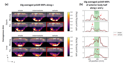 |
Impact of respiration on B1+ field and SAR distribution at 7 T using a novel EM simulation setup
Natalie Schön1, Johannes Petzold1, Frank Seifert1, Christoph Stefan Aigner1, Gregory J. Metzger2, Bernd Ittermann1, and Sebastian Schmitter1,2
1Physikalisch-Technische Bundesanstalt (PTB), Braunschweig and Berlin, Germany, 2Center for Magnetic Resonance Research, University of Minnesota, Minneapolis, MN, United States
At 7T body imaging spatial variations of the transmit magnetic (B1+) and electric (E) fields are observed. Additionally, recent in-vivo studies showed that B1+ patterns vary throughout the respiratory cycle. We present a novel electromagnetic (EM) simulation setup that allows investigating respiration-induced changes of the E- and B1+ fields. Using such simulations, we aim to verify the aforementioned in-vivo results that demonstrated respiration-induced changes of B1+ and corresponding flip angle distributions in the heart. Furthermore, the hitherto neglected, corresponding SAR variations are investigated and we find an up to 100 % change in local SAR throughout the respiratory cycle.
|
1121.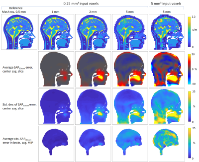 |
Parallel transmit local SAR vs. mesh resolution in EMF simulations of highly detailed anatomical models – a rigorous analysis
Andre Kuehne1, Eva Oberacker2, Helmar Waiczies1, Mostafa Berangi1, Jacek Nadobny3, Pirus Ghadjar3, Peter Wust3, and Thoralf Niendorf1,2,4
1MRI.TOOLS GmbH, Berlin, Germany, 2Max Delbrück Center for Molecular Medicine in the Helmholtz Association, Berlin, Germany, 3Clinic for Radiation Oncology, Charité Universitätsmedizin, Berlin, Germany, Berlin, Germany, 4Experimental and Clinical Research Center (ECRC), joint cooperation between the Charité Medical Faculty and the Max Delbrück Center for Molecular Medicine in the Helmholtz Association, Berlin, Germany
Electromagnetic simulations are an important tool for RF coil and thermal RF applicator development. For rapid design evaluation, fast low mesh resolution simulations would be of benefit, which can however potentially introduce errors in regions of intricate tissue distributions. We rigorously analyze local power deposition errors introduced by using low-resolution meshes in simulations of a highly detailed head model at 297 MHz. Our results indicate, that even at 5mm the introduced error is acceptable. However, artificial current paths are formed in the oronasal cavity, leading to not critical albeit locally elevated power deposition, thus deserving additional attention.
|
|
1122.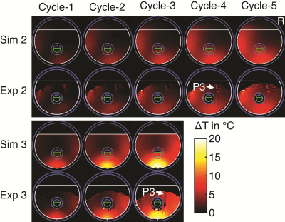 |
Validation of RF induced temperature increase in phantom and in living human tissue: a comparison study
Shubham Gupta1, Keiji Tanaka1, and R. Allen Waggoner1
1Laboratory for Cognitive Brain Mapping, RIKEN Center for Brain Science, Wakoshi, Saitama, Japan
In this study, we compared the temperature increase in a phantom and three human legs that were calculated by the simulations, measured by the MR-thermometry, and the optical thermocouples (phantom only), with good agreement between the modalities. IEC guidelines require simulations of SAR along with validation in phantoms to ensure safety. While it is likely impossible to simulate every possible pulse shape and phase combination in a pTx system with a large number of transmit channels, the results we present here suggest that simulations plus MR-thermometry could provide the verification currently lacking in pTx studies.
|
|
1123.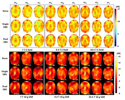 |
Convex Optimized Excitation Control to Reduce RF Heating for DBS Patients at UHF MRI
Youngdae Cho1 and Hyoungsuk Yoo1
1Biomedical Engineering, Hanyang University, Seoul, Republic of Korea
Patients having deep brain stimulation (DBS) can suffer from radio-frequency (RF) heating around the electrode during the MRI scan. Most of previous solutions were conducted based on the birdcage coil; the methods are inappropriate for ultra-high field (UHF) MRI system over 7 T using multi-channel RF coil. Our study introduced an optimized excitation control method by changing input weights of coil elements through convex optimization. Results demonstrated that proposed method effectively reduces RF heating around the electrode as well as acquires MR images of major brain regions with high resolution simultaneously.
|
|
|
1124.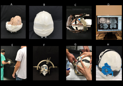 |
Surgical modification of extracranial trajectories of DBS leads can significantly reduce image artifact and RF heating during MRI at 3T
Bhumi Bhusal1, Joshua Rosenow2, Mark Nolt2, Roberto Lopez-Rosado1, Julie Pilitsis3, and Laleh Golestanirad1
1Northwestern University, Chicago, IL, United States, 2Northwestern Medicine, Chicago, IL, United States, 3Albany Medical Center, Albany, NY, United States
Patients with deep brain stimulation (DBS) implants can significantly benefit from MRI, however the interaction between MRI electric fields and DBS leads induces RF currents in the leads that can cause tissue heating and image artifacts. Here we show that modifying the extracranial trajectory of a DBS lead implanted into a cadaver brain significantly reduces both heating in the tissue and the image artifact around electrode contacts during MRI at 3T. Electromagnetic simulations confirm that trajectory modification can reduce induced currents in the lead, which in turn reduces the SAR amplification and distortion of B1 fields around the electrodes.
|
|
1125.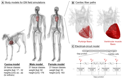 |
Simulation of electromagnetic cardiac stimulation: Validation in dogs and application to human threshold limits for MRI gradient coils
Valerie Klein1,2, Mathias Davids1,2,3, Lothar R. Schad1, Lawrence L. Wald2,3,4, and Bastien Guérin2,3
1Computer Assisted Clinical Medicine, Medical Faculty Mannheim, Heidelberg University, Mannheim, Germany, 2A. A. Martinos Center for Biomedical Imaging, Department of Radiologoy, Massachusetts General Hospital, Charlestown, MA, United States, 3Harvard Medical School, Boston, MA, United States, 4Harvard-MIT Division of Health Sciences and Technology, Cambridge, MA, United States
Lack of detailed data requires a conservative approach in the IEC 60601-2-33 safety limits to prevent cardiac stimulation (CS) by MRI gradient switching. Analogous to our previous peripheral nerve stimulation modeling, we use coupled electromagnetic and electrophysiological simulations to investigate magnetically induced CS in human and canine body models. Our CS simulation pipeline reproduces CS thresholds measured in previous dog experiments. The predicted human CS thresholds are significantly higher than the regulatory safety limits. With further validation, CS simulations could eventually play an important role in determining appropriate MRI safety limits.
|
|
1126.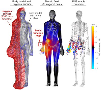 |
Assembly of a PNS predicting “P-matrix” on a Huygens’ surface for rapid PNS assessment of 2D or 3D gradient coil windings
Mathias Davids1,2,3, Bastien Guerin1,2, and Lawrence L Wald1,2,4
1A.A. Martinos Center for Biomedical Imaging, Massachusetts General Hospital, Dept. of Radiology, Charlestown, MA, United States, 2Harvard Medical School, Boston, MA, United States, 3Computer Assisted Clinical Medicine, Medical Faculty Mannheim, Heidelberg University, Mannheim, Germany, 4Harvard-MIT Health Sciences and Technology, Cambridge, MA, United States
Peripheral Nerve Stimulation (PNS) modeling has a potential role for designing and operating therapeutic and diagnostic devices (such as MRI), but is computationally demanding due to the required simulations of EM fields and neural responses. We describe compression of the PNS modeling framework into a single versatile PNS matrix (P-matrix) defined on a Huygens’ surface just outside the subject’s body to allow fast detailed PNS analysis on arbitrary coil windings/formers. This P-matrix can be translated to any coil former within seconds, allowing for rapid PNS assessment or optimization of gradient coil windings with explicit PNS constraints.
|
 |
1127.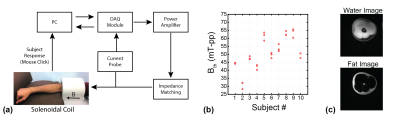 |
Simple Anatomical Measures Correlate with Individual PNS Thresholds for kHz-range Homogeneous Magnetic Fields
Omer Burak Demirel1,2,3,4, Toygan Kilic1,2, Tolga Çukur1,2,5, and Emine Ulku Saritas1,2,5
1Electrical and Electronics Engineering, Bilkent University, Ankara, Turkey, 2National Magnetic Resonance Research Center (UMRAM), Bilkent University, Ankara, Turkey, 3Center for Magnetic Resonance Research, University of Minnesota, Minneapolis, MN, United States, 4Electrical and Computer Engineering, University of Minnesota, Minneapolis, MN, United States, 5Neuroscience Graduate Program, Bilkent University, Ankara, Turkey
This work shows for the first time that fat percentage strongly correlates with peripheral nerve stimulation (PNS) thresholds for kHz-range homogeneous magnetic fields. The correlations get even stronger after taking into account the effects of body part size that is exposed to the magnetic field. These types of magnetic fields are used as excitation field in Magnetic Particle Imaging (MPI). Hence, these results can potentially lead to subject specific threshold prediction, allowing high performance scans within subject specific safety limits.
|
1128.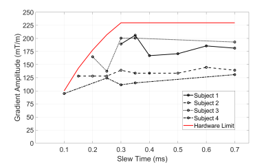 |
Magneto-phosphenes in head-only gradient coils
Colin M McCurdy1, Amgad M Louka1, William B Handler1, and Blaine A Chronik1
1The xMR Labs, Department of Physics and Astronomy, Western University, London, ON, Canada
Magneto-phosphenes are caused by induced potentials in the retina, that result in visual stimulation, appearing as flashing lights. In the MR environment, magneto-phosphenes have been encountered with higher gradient strengths and longer slew times than are typically encountered in MRI. However, in a prototype head-only gradient coil we were able to repeatably induce magneto-phosphenes in four subjects. We then tested the effects of slew times, external light, and eye direction on the subject’s perception of magneto-phosphenes, finding that slew times had little effect but dimming lights and changing eye direction raised thresholds in most cases.
|
 Watch the Video