
Scientific Session
Novel Hardware
Session Topic: Novel Hardware
Session Sub-Topic: Hardware Components
Oral
Engineering
| Thursday Parallel 5 Live Q&A | Thursday, 13 August 2020, 15:05 - 15:50 UTC | Moderators: Aurelien Destruel & Ed Wu |
Session Number: O-83
1260.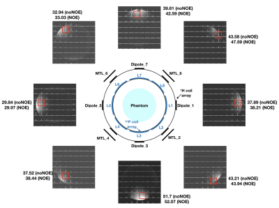 |
A novel interchangeable, double-tuned, twin head coil array design for 1H/23Na MR imaging and 1H/31P MR spectroscopy at 7 T
Chang-Hoon Choi1, Airat Galiamov1, Suk-Min Hong1, Jörg Felder1, Wieland A. Worthoff1, and N. Jon Shah1,2,3,4
1INM-4, Forschungszentrum Juelich, Juelich, Germany, 2INM-11, Forschungszentrum Juelich, Juelich, Germany, 3JARA-BRAIN-Translational Medicine, Aachen, Germany, 4Department of Neurology, RWTH Aachen University, Aachen, Germany
X-nuclei MR offers unique access to important metabolic information in tissues. Multi-tuned coils are required for the X-nuclei measurements, but designing a well-performing coil is challenging. In this study, we present our novel design and performance evaluation of an interchangeable, twin, double-tuned, head coil array for 1H/23Na MR imaging and 1H/31P MR spectroscopy at 7 T. The outer proton array was built using an alternatingly positioned 4-channel dipole antenna and a 4-channel microstrip transmission line array to improve decoupling. The inner 8-channel X-nuclei loop arrays, orthogonal to the 1H, were designed identically to enable conventionally switching between 23Na and 31P.
|
|
1261.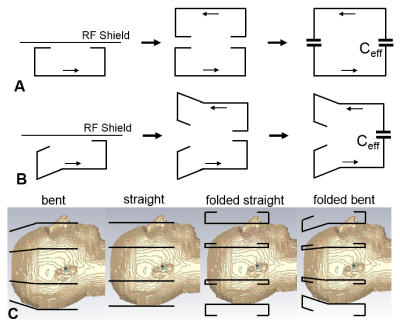 |
Decoupling of Folded Dipole Antenna Elements of a Human Head Array at 9.4T.
Nikolai Avdievich1, Georgiy Solomakha2, Loreen Ruhm1, Anke Henning1,3, and Klaus Scheffler1,4
1Max Planck Institute for Bilogical Cybernetics, Tuebingen, Germany, 2Nanophotonics and Metamaterials, ITMO University, St. Petersburg, Russian Federation, 3Advanced Imaging Research Center, University of Texas Southwestern Medical Center, Dallas, TX, United States, 4Department for Biomedical Magnetic Resonance, University of Tübingen, Tuebingen, Germany
Dipole antennas have been successfully utilized at ultra-high fields (UHF, >7 T) as elements of human body arrays. Usage of dipoles for UHF human head arrays is still under development. In this case, dipoles must be made much shorter, and placed at a relatively large distance to the head. As a result, dipoles are not well loaded and are often purely decoupled. In this work, we developed a novel method of decoupling of adjacent dipole antennas, and used this technique while constructing a novel 9.4 T human head TxRx dipole array coil. The array demonstrates good decoupling and full-brain coverage.
|
|
 |
1262.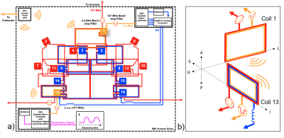 |
Dual-Stream iPRES-W Head Coil Array for MR Imaging, Wireless Respiratory Tracking, and Wireless Localized B0 Shimming
Jonathan Cuthbertson1,2, Trong-Kha Truong1,2, Vani Yadav1,2, Fraser Robb3, Allen Song1,2, and Dean Darnell1,2
1Brain Imaging and Analysis Center, Duke University, Durham, NC, United States, 2Medical Physics Graduate Program, Duke University, Durham, NC, United States, 3GE Healthcare Inc., Aurora, OH, United States
The integrated RF/wireless coil design enables MRI imaging and wireless data transfer with the same coil thereby reducing the number of wired connections in the scanner. Here, we implement this design onto a 48-channel head coil array to enable two independent wireless data streams for two separate applications, specifically, wireless 1) control of the DC currents used for B0 shimming and 2) respiratory tracking with a respiratory belt. In vivo experiments in the brain showed that this coil array significantly reduced B0 inhomogeneities (-41%) and EPI distortions while simultaneously streaming respiratory data from the subject without data loss.
|
1263.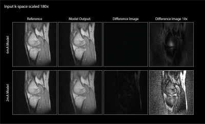 |
Harmonic Balance Modeling of MRI Preamp Impairments
Chris Vassos1, Fraser Robb2, Shreyas Vasanawala3, John Pauly1, and Greig Scott1
1Electrical Engineering, Stanford University, Stanford, CA, United States, 2GE Healthcare, Aurora, OH, United States, 3Radiology, Stanford University, Stanford, CA, United States
A Silicon Germanium alternative to standard HEMT pre-amplifiers is proposed. This is intended to ameliorate the high power consumption associated with current implementations for the wireless use case in which power is a limiting factor. The proposed pre-amp is evaluated for linearity and gain through a behavioral model that is extracted from SPICE simulation to process k-space data. It is found that the non-linearities introduced by the SiGe device begin to have an impact on image quality in high dynamic range cases. This encourages further investigation into SiGe devices as low-power preamplifiers.
|
|
|
1264.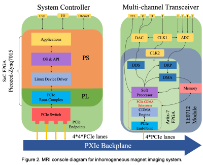 |
An Open-Source Multichannel MRI Console
Guang Yang1, Sergei Obruchkov2, and Robin Dykstra1
1School of Engineering and Computer Science, Victoria University of Wellington, Wellington, New Zealand, 2Robinson Research Institute, Victoria University of Wellington, Wellington, New Zealand This abstracts describes the development of a complete multi-channel MRI console that can be the basis for many MR projects. To support the growing open-source hardware MR community we are making a collection of PXIe modules and IP available to anyone to use and develop applications on. A System Controller Board, a 2-channel Tx and 4-channel Rx transceiver board, an FMC General Purpose Module, PXIe data transfer engine IP, Linux API and device drivers can be accessed online https://github.com/mr-kit/. |
|
1265.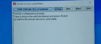 |
Misappropriation of the Scanner Synchronization Trigger for Serial Communication with any UART Device
Andrew Dupuis1, Dominique Franson1, Nicole Seiberlich2, and Mark A Griswold1,3
1Biomedical Engineering, Case Western Reserve University, Cleveland, OH, United States, 2Radiology, University of Michigan, Ann Arbor, MI, United States, 3Department of Radiology, School of Medicine, Case Western Reserve University, Cleveland, OH, United States
Clinical MRI scanners are not designed to allow for easy communication with 3rd party devices, whether for development or clinical purposes. However, most modern scanners provide a synchronization trigger interface that is sequence (and therefore research-user) controllable. We investigated using the synchronization trigger as a serial data output for arbitrary data, and successfully implemented a 192 kilobaud simplex serial interface that can be implemented within any sequence to enable arbitrary data transfer to and control of any external UART device. This opens significant opportunities for 3rd party hardware and software research without manufacturer consent or firmware changes.
|
1266.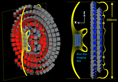 |
Single-sided magnet design for an MR guided lumbar puncture (LP) device
Clarissa Zimmerman Cooley1,2, Patrick C McDaniel1,3, Jason P Stockmann1,2, Farrah J Mateen2,4, and Lawrence L Wald1,2,5
1Athinoula A Martinos Center for Biomedical Imaging, Dept. of Radiology, Massachusetts General Hospital, Charlestown, MA, United States, 2Harvard Medical School, Boston, MA, United States, 3Dept. of Electrical Engineering, Massachusetts Institute of Technology, Cambridge, MA, United States, 4Dept. of Neurology, Massachusetts General Hospital, Boston, MA, United States, 5Harvard-MIT Division of Health Sciences and Technology, Cambridge, MA, United States
Lumbar Punctures (LP) are generally guided by palpation only without visualization of the internal anatomy, leading to repeat attempts and/or avoidance in difficult cases. Image guidance with US and X-ray is possible, but US has poor depth and CSF contrast and radiation from X-ray complicates Point of Care (POC) use. We present a magnet design for a POC MR guided LP device that will couple to a mechanical track for needle insertion. The single-sided magnet is an NdFeB array and achieves a 40mT field and ~50mT/m built-in gradient in the ROI. Simulations of the magnet and imaging procedure are presented.
|
|
1267.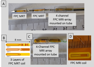 |
A deflectable positionally-localized Virtual Biopsy “Gun”: Construct and initial testing
Ehud J Schmidt1,2, Yue Chen3, Anthony Gunderman3, Junichi Tokuda4, Hassan Elahi5, Ravi T Seethamraju6, Henry R Halperin5, and Akila N Viswanathan2
1Medicine (Cardiology), The Johns Hopkins University, Baltimore, MD, United States, 2Radiation Oncology, Johns Hopkins University, Baltimore, MD, United States, 3Mechanical Engineering, University of Arkansas, Fayetteville, AR, United States, 4Radiology, Brigham and Womens Hospital, Boston, MA, United States, 5Medicine (Cardiology), Johns Hopkins University, Baltimore, MD, United States, 6MRI, Siemens Healthineers, Boston, MA, United States
Evaluating tissue properties prior-to or during therapy, such as locating cancerous and necrotic cells, or characterizing response to radiation or ablation, is conventionally performed by tissue excision, followed by pathologic examination. An alternative is diagnosing tissue in-situ without removing it, as performed using Optical-Coherence-Tomography or Intra-Vascular-UltraSound. We aim to perform tissue definition in soft-tissues not accessed through body-orifices or blood-vessels by combining; (1) Steerable tissue-puncture, (2) MR-Tracking-motion-localization, and (3) imaging along the punctured-holes' walls. Utilization requires rapid high-CNR multiple-contrast MRI. A deflectable virtual-biopsy “gun” for diagnosing cervical-cancer radiation-therapy response was developed. It imaged ~15mm surrounding punctured-holes created for brachytherapy seed-delivery.
|
|
1268.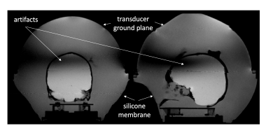 |
Improving Image Quality in Transcranial Magnetic Resonance Guided focused Ultrasound Using a Conductive Screen
J. Rock Hadley1, Henrik Odeen2, Robb Merrill2, Sam Adams2, Viola Rieke2, Allison Payne2, and Dennis Parker2
1Radiology and Imaging Sciences, University of Utah, Salt Lake City, UT, United States, 2University of Utah, Salt Lake City, UT, United States
This work uses an RF screen, placed over the top of a human skull phantom, to reduce image banding artifacts that are common in transcranial transducer MRI. The goals of the study are to improve imaging homogeneity over the region of the brain by changing RF field patterns that cause the artifacts, and to find a solution that doesn’t attenuate or distort the ultrasound properties of the transducer. Hydrophone and focused ultrasound heating studies are performed to measure ultrasound screen transparency and MRI studies are performed to evaluate the effects the screen has on homogeneity and artifact reduction.
|
|
|
1269.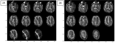 |
Retrospective Electromagnetic interference mitigation in a portable low field MRI system
Sai Abitha Srinivas1, Clarissa Z Cooley1,2, Jason P Stockmann1,2, Patrick C McDaniel1,3, and Lawrence L Wald1,2,4
1Athinoula A Martinos Center for Biomedical Imaging, Charlestown, MA, United States, 2Harvard Medical School, Boston, MA, United States, 3Dept. of Electrical Engineering, Massachusetts Institute of Technology, Cambridge, MA, United States, 4Harvard-MIT Division of Health Sciences and Technology, Cambridge, MA, United States
The performance of a low field Point of Care (POC) MRI system operating outside an RF shielded room is adversely affected by the presence of electromagnetic interference signals, which produce image artifacts, sometimes complicated enough to be confused with image noise. We demonstrate a post-processing interference suppression technique using an external reference coil and dynamically updated transfer function to detect the interference and remove it from the imaging data.
|
 Watch the Video