
Scientific Session
Pediatric Neuro: Fetal to Adolescence
Session Topic: Pediatric Neuro: Fetal to Adolescence
Session Sub-Topic: Pediatric Neuro: Fetal/Newborn/Developmental
Oral
Pediatric
| Monday Parallel 3 Live Q&A | Monday, 10 August 2020, 14:30 - 15:15 UTC | Moderator: Peiying Liu |
Session Number: O-84
 |
0221.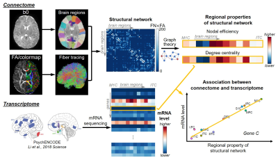 |
Deciphering transcriptomic basis of the human brain structural connectome in the 3rd trimester
Chenying Zhao1,2, Gabriel Santpere3, David Andrijevic3, Minhui Ouyang1, Nenad Sestan3, and Hao Huang1,4
1Department of Radiology, Children's Hospital of Philadelphia, Philadelphia, PA, United States, 2Department of Bioengineering, School of Engineering and Applied Science, University of Pennsylvania, Philadelphia, PA, United States, 3Department of Neuroscience and Kavli Institute for Neuroscience, Yale School of Medicine, New Haven, CT, United States, 4Department of Radiology, Perelman School of Medicine, University of Pennsylvania, Philadelphia, PA, United States
Dramatic development of brain connectome takes place during the 3rd trimester, mediated by transcriptome. Transcriptome is complete set of gene-expressed mRNAs and is heterogeneous across brain regions and dynamic throughout development. The transcriptomic basis of structural connectome in this critical developmental stage is unknown. In this study, we identified transcription genes most significantly correlated to nodal efficiency and degree centrality of macroscale structural connectome based on diffusion MRI of 77 preterm brains and over 60,000 quantified transcriptomes. These identified transcription genes such as MYRF regulating oligodendrocyte differentiation and myelination may shed light into the transcriptomic basis of structural connectome development.
|
 |
0222.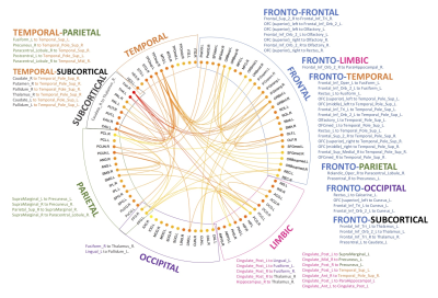 |
THE NEONATAL PRETERM BRAIN: A CONNECTOME ANALYSIS
Joana S. de Almeida1, Djalel-Eddine Meskaldji1,2, Serafeim Loukas1,3, Lara Lordier1, Laura Gui4, François Lazeyras4, and Petra S. Hüppi1
1Department of Women-Children-Teenagers, Hôpitaux Universitaires de Genève, Genève, Switzerland, 2Institute of Mathematics, Ecole Polytechnique Fédérale de Lausanne, Lausanne, Switzerland, 3Institute of Bioengineering, Ecole Polytechnique Fédérale de Lausanne, Lausanne, Switzerland, 4Department of Radiology and Medical Informatics, CIBM, University of Geneva, Genève, Switzerland
Prematurity disrupts brain maturation during a critical period of development, leading to structural brain alterations that might underlie the observed later neurodevelopmental impairments in preterm children. Using diffusion MRI based whole-brain constrained spherical deconvolution tractography, we constructed structural connectomes to study the impact of prematurity on neonatal brain network organization at term-equivalent age. We found that, globally, in comparison to full-term infants, structural networks of very-preterm infants at term showed an increased segregation and decreased capacity to integrate information across brain regions and, in particular, a diminished connectivity strength in subnetworks localized mainly in frontal, limbic and para-limbic regions.
|
|
0223.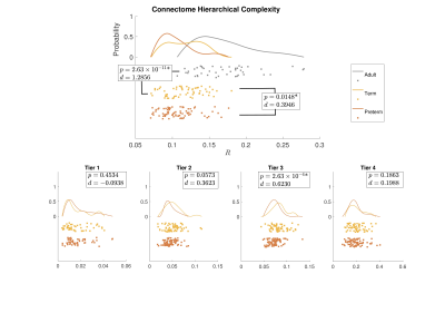 |
Hierarchical complexity of the neonatal brain
Manuel Blesa Cabez1, Paola Galdi1, Simon R Cox1, David Q. Stoye1, Gemma Sullivan1, Gillian J. Lamb1, Alan J Quigley2, Michael J. Thrippleton1, Javier Escudero Rodriguez1, Mark E Bastin1, Keith M Smith1, and James P Boardman1
1University of Edinburgh, Edinburgh, United Kingdom, 2Royal Hospital for Sick Children, Edinburgh, United Kingdom
Preterm birth is associated with long term cognitive deficits and alterations to structural connectivity of developing brain networks. Diversity of connectivity patterns within hierarchically equivalent nodes (hierarchical complexity, HC), is a prominent feature of the adult human connectome. In this work, we show that HC of the structural connectome at birth shares similar properties to HC seen in the adult connectome. Infants born preterm have different HC to infants born at term. In addition, we show that high-level order may be necessary to create structural stability, and this high-level order is resilient to environmental challenges such as preterm birth.
|
|
0224.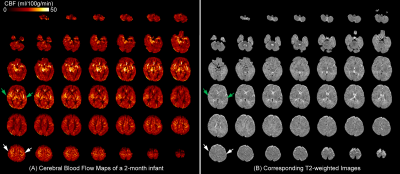 |
High-resolution infant cerebral blood flow map measured with 3D multi-shot, stack-of-spirals pCASL
Minhui Ouyang1, John Detre2, Samantha Linh Lam1, J. Christopher Edgar1,2, and Hao Huang1,2
1Department of Radiology, The Children's Hospital of Philadelphia, Philadelphia, PA, United States, 2Department of Radiology, Perelman School of Medicine, University of Pennsylvania, Philadelphia, PA, United States
During early infancy, dramatic structural and functional maturation of infant brains requires rapid increases of regional cerebral blood flow (rCBF). In this study, we optimized a 3D multi-shot, stack-of-spirals pCASL sequence to obtain high-resolution rCBF maps at isotropic 2.5mm for infants at different maturational stages. Distinctive rCBF distribution patterns at different infant stages of 0-6 months and 7-18 months were revealed. The age-dependent trend lines of rCBF at specific regions were charted. Infant rCBF increases heterogeneously across brain regions, with rCBF increasing faster in visual, prefrontal and parietal cortices than that in precentral and thalamus during this critical period.
|
0225.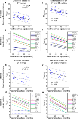 |
Multivariate Evaluation of White Matter Maturation on Neonates and Toddlers by Diffusion Kurtosis Imaging with Mahalanobis Distance
Xianjun Li1, Miaomiao Wang1, Fan Wu1, Qinli Sun1, Heng Liu1, Yuli Zhang1, Mengxuan Li1, Chao Jin1, Congcong Liu1, Xiaocheng Wei1, and Jian Yang1
1Department of Radiology, the First Affiliated Hospital of Xi’an Jiaotong University, Xi'an, China
Mahalanobis distance is a feasible multivariate approach. This work compared performances of Mahalanobis distances based on different combinations of metrics in assessing the white matter maturation on neonates and toddlers. Mahalanobis distance based on the combination of diffusion tensor and kurtosis tensor metrics demonstrated various advantages: stronger correlation with the postmenstrual age and higher developmental speeds could be revealed; distances from the developing to the adult brains and the changes from neonates to toddlers were enlarged. Results in the current work suggest that diffusion kurotsis imaging with the Mahalanobis distance would benefit the characterization of white matter maturation.
|
|
|
0226.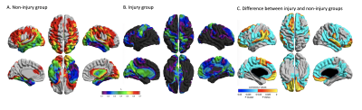 |
Prematurity-related brain injuries disrupt thalamocortical reciprocal growth
Audrey Yin1, Mengting Liu1, Arthur W. Toga1, Duan Xu2, James Barkovich2, and Hosung Kim1
1USC Stevens Neuroimaging and Informatics Institute, Keck School of Medicine of USC, University of Southern California, Los Angeles, CA, United States, 2Department of Radiology and Biomedical Imaging, UCSF School of Medicine, University of California, San Francisco, San Francisco, CA, United States
Prematurity-related injuries often result in aberrant brain maturation, specifically on peri-thalamic white matter. We investigated the effects of these injuries on the intra-thalamic tissue integrity and on thalamocortical connectivity. We found that injuries did not substantially affect thalamic volume or thalamic DTI parameters, but did have a substantial effect on the correlative growth between the thalamus and cortex. This implies that brain injuries disrupt the reciprocal development of the thalamus and cortex, which may indicate abnormal thalamocortical connectivity.
|
0227.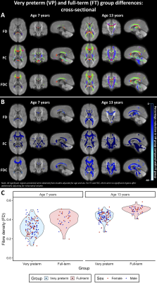 |
Longitudinal development of white matter fibre density and morphology in children born very preterm
Claire E Kelly1,2, Deanne K Thompson1,2,3,4, Sila Genc2,5, Jian Chen2, Joseph YM Yang2,3,6,7, Chris Adamson2, Richard Beare2, Marc L Seal2,3, Jeanie LY Cheong1,8,9, Lex W Doyle1,3,8,9, and Peter J Anderson1,10
1Victorian Infant Brain Study (VIBeS), Murdoch Children's Research Institute, Melbourne, Australia, 2Developmental Imaging, Murdoch Children's Research Institute, Melbourne, Australia, 3Department of Paediatrics, The University of Melbourne, Melbourne, Australia, 4Florey Institute of Neuroscience and Mental Health, Melbourne, Australia, 5Cardiff University Brain Research Imaging Centre (CUBRIC), Cardiff University, Cardiff, United Kingdom, 6Department of Neurosurgery, The Royal Children's Hospital, Melbourne, Australia, 7Neuroscience Research, Murdoch Children's Research Institute, Melbourne, Australia, 8Newborn Research, The Royal Women’s Hospital, Melbourne, Australia, 9Department of Obstetrics and Gynaecology, The University of Melbourne, Melbourne, Australia, 10Turner Institute for Brain and Mental Health, Monash University, Melbourne, Australia
In this long-term follow-up of children following very preterm (VP) birth, we applied fixel-based analysis to study white matter development. At ages 7 and 13 years, VP children had reduced fibre density and cross-section throughout the white matter compared with full-term controls. Longitudinally, VP children had slower macrostructural development of commissural and motor pathways between ages 7 and 13 years. Younger gestational age, smaller birth weight and neonatal brain abnormalities were associated with lower fibre density and cross-section at both ages. Thus, VP birth and concomitant perinatal risk factors are associated with long-term delays and/or disruptions to white matter development.
|
|
0228.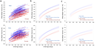 |
Influences of Gender, Physical Growth, and Socioeconomic Characteristics on Early Brain Growth in Children from an LMIC Setting.
Sean Deoni1, Aarti Kumar2, Vishwajeet Kumar2, Madhuri Tiwari2, John Spencer3, and Muriel Bruchhage4
1MNCHD&T, Bill & Melinda Gates Foundation, Seattle, WA, United States, 2CEL, Lucknow, India, 3University of East Anglia, Norwich, United Kingdom, 4Brown University, Providence, RI, United States
Early brain development is influenced by a myriad of environmental and psychosocial exposures that are often amplified in children in low and middle income countries (LMICs). However, few neuroimaging studies have been performed in these settings. Here we report on the first longitudinal neuroimaging study of young children in rural Uttar Pradesh (UP) India, showing the importance of early weight grain and socioeconomic factors on brain growth. We also find significant male-female differences, which may derive from the lesser societal importance of women, including lower education levels, increased malnutrition, and reduced healthcare seeking for girls.
|
|
|
0229.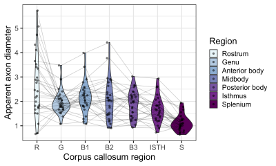 |
Uncovering regional maturation of axon diameter across child and adolescent brain development
Sila Genc1, Erika P Raven1, Mark Drakesmith1, and Derek K Jones1,2
1Cardiff University Brain Research Imaging Centre (CUBRIC), Cardiff University, Cardiff, United Kingdom, 2Mary MacKillop Institute for Health Research, Australian Catholic University, Melbourne, Australia
The maturation of white matter across childhood and adolescence is predominantly driven by the thickening of myelin and increasing axon density. Previous post-mortem studies have suggested that axon count in the corpus callosum reaches adult levels in the early post-natal period, suggesting that the radial growth of axons may be driving the apparent increases in axon density. In this novel application of paediatric microstructural development, we estimate apparent axon diameter using ultra-strong gradient MRI (300 mT/m) for the first time. Our findings reveal age-related maturation of axon diameter in the genu and body of the corpus callosum.
|
0230.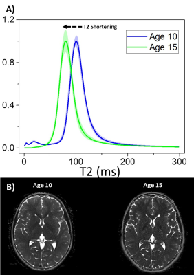 |
Longitudinal Changes of the Extremely Preterm Brain from Age 10 to Age 15: Myelination and Hydration.
Ryan McNaughton1, Hernan Jara2, Xin Zhang1, Mina Botros2, Robert M Joseph2, Asim Z Mian2, Laurie Douglass2, Karl Kuban2, Rebecca C Fry3, and Michael O'Shea3
1Mechanical Engineering, Boston University, Boston, MA, United States, 2Boston University Medical Center, Boston, MA, United States, 3University of North Carolina at Chapel Hill School of Medicine, Chapel Hill, NC, United States
Purpose: To identify new qMRI markers for assessing changes in hydration and myelination during development of the extremely preterm (EP) brain. Methods: Quantitative MR algorithms create maps of the transverse relaxation time (T2) and normalized proton density (PD) for 7 EP born individuals using MR images obtained at age 10 and age 15 years. Results: White and grey matter of the EP brain demonstrate increases in proton density and significant decreases in tissue T2. Conclusion: Decreases in T2 potentially describes the evolution of a more myelin rich environment with age, motivating a new method to assess myelination during brain development.
|
 Watch the Video