
Scientific Session
Pediatric Innovations
Session Topic: Pediatric Innovations
Session Sub-Topic: Pediatric Body & Musculoskeletal
Oral
Pediatric
| Monday Parallel 3 Live Q&A | Monday, 10 August 2020, 13:45 - 14:30 UTC | Moderators: Corin Willers |
Session Number: O-85
 |
0068.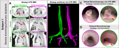 |
Virtual bronchoscopy of neonatal dynamic airway collapse with ultrashort echo-time MRI
Nara S Higano1,2, Alister J Bates1,2,3,4, Erik B Hysinger2,4, Robert J Fleck3,5,6, Andrew D Hahn7, Sean B Fain7,8, Paul S Kingma4,9, and Jason C Woods2,5,6
1Center for Pulmonary Imaging, Cincinnati Children's Hospital, CINCINNATI, OH, United States, 2Pulmonary Medicine, Cincinnati Children's Hospital, CINCINNATI, OH, United States, 3Upper Airway Center, Cincinnati Children's Hospital, CINCINNATI, OH, United States, 4Pediatrics, University of Cincinnati, CINCINNATI, OH, United States, 5Radiology, Cincinnati Children's Hospital, CINCINNATI, OH, United States, 6Pediatrics, University of Cincinnati, Cincinnati, OH, United States, 7Medical Physics, University of Wisconsin - Madison, Madison, WI, United States, 8Radiology, University of Wisconsin - Madison, Madison, WI, United States, 9Neonatology and Pulmonary Biology, Cincinnati Children's Hospital, CINCINNATI, OH, United States
Central airway abnormalities in neonates, e.g. dynamic collapse and stenosis, are serious complications often associated with preterm birth and congenital abnormalities, but have not been extensively studied. These conditions are most often assessed through clinical bronchoscopy, which can be unreliable and poses increased risks to patients. Here, we demonstrate novel visualization of static and dynamic neonatal airway abnormalities on virtual bronchoscopy from high-resolution, retrospectively respiratory-gated ultrashort echo-time MRI, which exhibits good agreement with clinical bronchoscopy. This virtual technique allows for assessment of neonatal airway abnormalities that is readily interpretable to clinicians familiar with clinical bronchoscopy.
|
0069.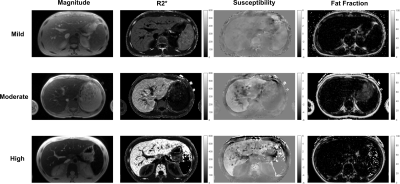 |
Quantitative Susceptibility Mapping using a Multi-spectral ARMA Model for Assessment of Hepatic Iron Overload
Aaryani Tipirneni-Sajja1,2, Ralf Berthold Loeffler2,3, Jane Hankins4, and Claudia Maria Hillenbrand2,3
Video Permission Withheld
1Biomedical Engineering, University of Memphis, Memphis, TN, United States, 2Diagnostic Imaging, St. Jude Children's Research Hospital, Memphis, TN, United States, 3Research Imaging NSW, University of New South Wales, Sydney, Australia, 4Hematology, St. Jude Children's Research Hospital, Memphis, TN, United States
Hepatic iron content (HIC) assessment by R2*-MRI can be confounded by co-existing fibrosis. Instead, quantitative susceptibility mapping (QSM) techniques could be used to assess iron content without being affected by fibrosis. In this study, we demonstrated that the field maps generated from a multi-spectral auto regressive moving average (ARMA) model can be used in conjunction with QSM techniques to measure magnetic susceptibility, as a predictor for HIC.
|
|
0070.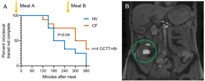 |
Digestive disorders in Cystic Fibrosis: Transit, Motility and MRI Signs of Small Intestinal Bacterial Overgrowth
Neele S Dellschaft1,2, Christabella Ng3, Caroline Hoad1,2, Luca Marciani2,4, Robin Spiller2,4, Penny Gowland1,2, Alan Smyth2,3, and Giles Major2,4
1Sir Peter Mansfield Imaging Centre, University of Nottingham, Nottingham, United Kingdom, 2Nottingham NIHR Biomedical Research Centre, University of Nottingham, Nottingham, United Kingdom, 3Division of Child Health, Obstetrics and Gynaecology, University of Nottingham, Nottingham, United Kingdom, 4Nottingham Digestive Diseases Centre, University of Nottingham, Nottingham, United Kingdom
Cystic Fibrosis (CF) is a genetic disease leading to sticky mucus. We used MRI to characterise the effect of CF on gastrointestinal function, comparing people with CF to matched healthy controls. People with CF had slower orocaecal transit times. No change in gastric emptying rate was apparent but more free water was present in their small bowel with reduced small bowel motility and a reduced gastro-ileal reflex. Some images suggested increased bacterial load in the small bowel. CF colons were larger. These findings are consistent with sticky chyme impeding ileal emptying into the colon, causing obstruction to flow, and constipation.
|
|
 |
0071.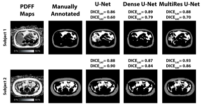 |
Fully Convolutional Networks for Adipose Tissue Segmentation Using Free-Breathing Abdominal MRI in Healthy and Overweight Children
Sevgi Gokce Kafali1,2, Shu-Fu Shih1,2, Xinzhou Li1,2, Tess Armstrong1, Karrie V. Ly3, Shahnaz Ghahremani1, Kara L. Calkins3, and Holden H. Wu1,2
1Radiological Sciences, University of California, Los Angeles, Los Angeles, CA, United States, 2Bioengineering, University of California, Los Angeles, Los Angeles, CA, United States, 3Pediatrics, University of California, Los Angeles, Los Angeles, CA, United States
The volume and fat content of subcutaneous and visceral adipose tissue (SAT and VAT) have strong associations with metabolic diseases in overweight children. Currently, the gold standard to measure the SAT/VAT content is manual annotation, which is time-consuming. Although several studies showed promising results using machine and deep learning to segment SAT and VAT in adults, there is a lack of research on deep learning-based SAT and VAT segmentation in children. Here, we investigated the performance of 3 deep learning network architectures to segment SAT and VAT in children.
|
0072.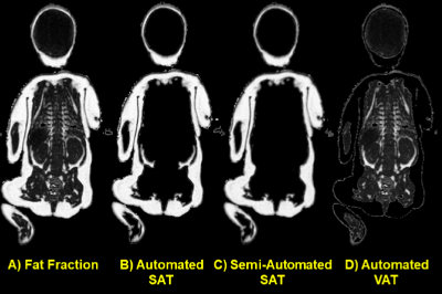 |
Semi-Automated Whole Body Neonatal Regional Fat Quantification Using Dixon Chemical Shift (CSI) MRI at 3.0 Tesla
Jonathan P Dyke1, Kevin Oh1, Amanda Garfinkel2, Alan M Groves3, and Arzu Kovanlikaya1
1Radiology, Weill Cornell Medicine, New York, NY, United States, 2Pediatrics, Weill Cornell Medicine, New York, NY, United States, 3Pediatrics, Mount Sinai School of Medicine, New York, NY, United States
Whole body fat fraction was previously published by our group in term and preterm infants using Dixon CSI MRI. Advanced semi-automated regional fat quantification was performed yielding: subcutaneous adipose tissue(SAT), visceral adipose tissue(VAT), brown adipose tissue (BAT) volumes and hepatic fat fraction (HFF). Whole body VAT volume (cc) was increased in preterm infants (3.4%±1.5%) compared to term (2.4%±0.9%) (p=0.079) when normalized to total body volume (cc). HFF and BAT did not differ between term and preterm infants (p>0.25). CSI MRI allows quantification of regional fat depots in preterm infants which may potentially help optimize nutritional management and monitor growth.
|
|
0073.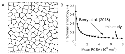 |
Using transverse diffusion to measure changes in muscle fibre number and muscle fibre size during childhood growth in humans
Bart Bolsterlee1,2, Arkiev D'Souza1,3, and Robert D Herbert1,3
1Neuroscience Research Australia, Randwick, Australia, 2Graduate School of Biomedical Engineering, University of New South Wales, Randwick, Australia, 3School of Medical Sciences, University of New South Wales, Randwick, Australia
We used diffusion tensor imaging (DTI) to study macroscopic and microscopic features of skeletal muscles during childhood development (5-17 years). From muscle volume and fibre length measurements, we determined the summed cross-sectional area of all fibres. From measurements of diffusion properties and simulations of restricted diffusion in skeletal muscle, we estimated mean cross-sectional areas of individual fibres. Our findings suggest that human muscles grow both by adding fibres and by increasing fibre cross-sectional areas. DTI-based measurements of skeletal muscle micro- and macrostructure could have important applications in understanding both normal and disordered muscle growth.
|
|
0074.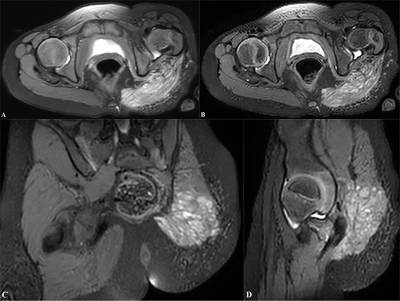 |
3D PDWI Accelerated with Compressed SENSE in Pediatric Joint Imaging: Clinical Feasibility Study
Yupeng Zhu1, Di Hu1, Yanqiu Lv1, Yang Wen1, Huiying Kang1, Xiaomin Duan1, Jiazheng Wang2, Queenie Chan2, and Yun Peng1
1Department of Radiology, Beijing Children’s Hospital, Capital Medical University, National Center for Children's Health, Beijing, China, 2Philips Healthcare, Beijing, China
The use of three dimensional (3D) volumetric acquisition in clinical settings has been limited due to long scan time. However, 3D volumetric acquisition could provide higher spatial resolution and decrease partial volume effects. The introduction of compressed sensing in combination of the parallel imaging technique SENSE allows shortening of scan time and provide comparable overall image quality when compared with standard sequences. The purpose of the study is to determine the feasibility of 3D PDWI accelerated with compressed SENSE(CS) for evaluating the pediatric joint image quality and compared with 2D PDWI.
|
|
0075.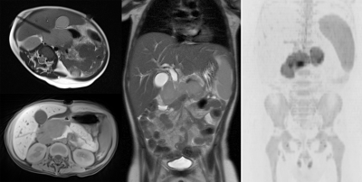 |
MR imaging and MR guided biopsy in initial diagnosis and staging of pediatric malignancies – a one-stop shop approach
Guenther Schneider1, Tobias Woerner1, and Arno Buecker1
1Diagnostic and Interventional Radiology, Saarland University Medical Center, Homburg, Germany
Diagnosis and staging in pediatric malignancies today involves different imaging procedures, from conventional x-ray over ultrasound to MR-, CT- and PET-imaging. We evaluated as if MRI can be used as a comprehensive one-stop shop for diagnosis, staging and biopsy in pediatric malignancies. As a result, when comparing the different imaging modalities, differences between MRI and CT were seen regarding the higher number of small lung lesions detected (<3mm) on CT. Comparable results were seen for abdominal tumors, Hodgkin- and Non-Hodgkin Lymphoma. In Ewing sarcoma MRI showed advantages compared with PET imaging regarding detection of skip lesions and bone marrow metastases.
|
 Watch the Video