
Scientific Session
Pediatric Neuro: Fetal to Adolescence
Session Topic: Pediatric Neuro: Fetal to Adolescence
Session Sub-Topic: Pediatric Neuro: Epilepsy & Brain Injury
Oral
Pediatric
| Monday Parallel 3 Live Q&A | Monday, 10 August 2020, 14:30 - 15:15 UTC | Moderators: Matthew Barkovich & Ashley Harris |
Session Number: O-87
 |
0231.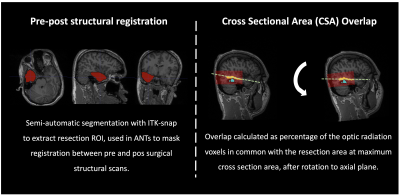 |
Towards clinical implementation of multi-shell diffusion MRI: visual pathway investigation in paediatric epilepsy surgery
Luis Miguel Lacerda1, Jon Clayden1, Sian Handley2, Martin Tisdall3, Enrico Kaden4, Gavin Winston5, Alki Liasis2,6, Helen Cross7, and Chris Clark1
1Developmental Imaging and Biophysics Section, UCL Great Ormond Street Institute of Child Health, London, United Kingdom, 2Clinical and Academic Department of Ophthalmology, Great Ormond Street Hospital for Children NHS Foundation Trust, London, United Kingdom, 3Neurosurgery, Great Ormond Street Hospital for Children NHS Foundation Trust, London, United Kingdom, 4Centre for Medical Image Computing, University College London, London, United Kingdom, 5Department of Clinical and Experimental Epilepsy, UCL Institute of Neurology, London, United Kingdom, 6University of Pittsburgh Medical Centre, Children’s Hospital of Pittsburgh, Pittsburgh, PA, United States, 7Clinical Neurosciences, UCL Great Ormond Street Institute of Child Health, London, United Kingdom
We used multi-shell diffusion imaging to investigate differences in the visual pathways of children undergoing epilepsy surgery and demonstrated its potential for clinical practice. In particular, we compared the traditional Diffusion Tensor Imaging model with the Spherical Mean Technique model and evaluated its potential to produce measures of tissue microstructure not confounded by orientation effects in both a healthy and patient population. Furthermore, we explored the effect of brain surgery and applied Constrained Spherical Deconvolution derived tractography to determine the frequency and influence of the extent and location of resection on the integrity of the visual system after the operation.
|
 |
0232.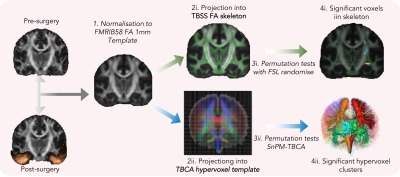 |
Emergence of distinct structural reorganization patterns in children as a result of temporal lobe epilepsy surgery – beyond voxel-based analysis
Luis Miguel Lacerda1, Pedro Luque Laguna2,3, Flavio Dell'acqua2,3, and Chris Clark1
1Developmental Imaging and Biophysics Section, Institute of Child Health, University College London, London, United Kingdom, 2Forensic & Neurodevelopmental Sciences, King's College London, London, United Kingdom, 3Sackler Institute for Translational Neurodevelopment, Institute of Psychiatry, Psychology and Neuroscience, King's College London, London, United Kingdom
We compared Tract-based Spatial Statistics (TBSS) with a recent method which relies on non-statistical parametric mapping in a tractography derived anatomical framework – tract-based cluster analysis (TBCA) - to explore the effect of surgery in Temporal Lobe Epilepsy. In particular, we investigated differences in fractional anisotropy (FA) as an effect of surgery, and if those changes depended on operated hemisphere. We found the same patterns of increased FA on the corona radiata and decreased FA in tracts traversing the temporal lobe with TBSS, whilst TBCA allowed for an increase in anatomical specificity when interpreting differences in this group.
|
0233.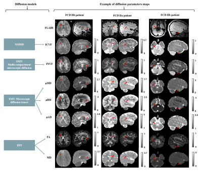 |
MRI profiling of focal cortical dysplasia using advanced diffusion models
Sara Lorio1, Sophie Adler2, Roxana Gunny3, Felice D'Arco3, Enrico Kaden2, Konrad Wagstyl2, Thomas Jacques2,3, Chris Clark2, Helen Cross2, Torsten Baldeweg2, and David W. Carmichael1
1King's College London, LONDON, United Kingdom, 2UCL, London, United Kingdom, 3Great Ormond Street Hospital, London, United Kingdom
Lesion detection and sub-typing for focal cortical dysplasia (FCD), a frequent cause of drug-resistant epilepsy, remain challenging on conventional MRI. New diffusion models such as the spherical mean techniques (SMT) and the neurite orientation dispersion and density imaging (NODDI) provide measurements that are potentially more specific to abnormal tissue microstructure. Quantitative analysis of lesion profiling demonstrated significant changes on NODDI and SMT maps proportional to neurites density, as well on microscopic mean, radial and axial diffusivities. Moreover, signal changes specific to FCD lesions sub-types were observed on those maps, suggesting they can provide features useful for automated lesion detection.
|
|
|
0234.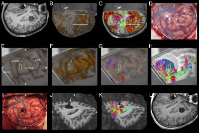 |
One-stage, language-dominant, opercular-insular epilepsy surgery with multimodal structural and functional neuroimaging evaluation
Joseph Yuan-Mou Yang1,2,3,4, Ramshekhar Menon5,6, Sarah Barton2,3,4,5, Simone Mandelstam7,8,9, Rachel Kerr10, Jacquie Wrennall11,12, Catherine Bailey5, Jeremy Freeman2,5, Wirginia Maixner1,2, and A Simon Harvey2,3,5
1Neurosurgery, Royal Children's Hospital, Melbourne, Australia, 2Neuroscience Research group, Murdoch Children's Research Institution, Melbourne, Australia, 3Paediatrics, University of Melbourne, Melbourne, Australia, 4Developmental Imaging group, Murdoch Children's Research Institution, Melbourne, Australia, 5Neurology, Royal Children's Hospital, Melbourne, Australia, 6Neurology, Sree Chitra Tirunal Institute for Medical Sciences and Technology, Thiruvananthapuram, India, 7Medical Imaging, Royal Children's Hospital, Melbourne, Australia, 8Medicine and Radiology, University of Melbourne, Melbourne, Australia, 9The Florey Institute of Neuroscience and Mental Health, Melbourne, Australia, 10Speech pathology, Royal Children's Hospital, Melbourne, Australia, 11Clinical neuropsychology, Royal Children's Hospital, Melbourne, Australia, 12Melbourne School of Psychological Sciences, University of Melbourne, Melbourne, Australia
Language dominant, insular-opercular epilepsies are challenging to manage due complex seizure presentations and proximity to language cortex and associated white matter tracts. Most centres elected for extensive invasive stereo-electroencephalogram recordings and ablative procedures. We demonstrated in our retrospective cohort of 11 patients that focal resections can be undertaken safely and effectively using neuroimaging of seizures, lesions, language fMRI and high order tractography reconstructions based on a multi-fibre white matter modelling technique, and careful microsurgical techniques. This avoids risks associated with invasive procedures. Surgery performed under direct vision is more precise, likely safer, allows tailoring with intraoperative electrocorticography, and provides histopathology.
|
0235.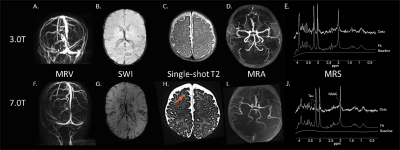 |
Introduction of ultra-high field Magnetic Resonance Imaging in neonates: preparations and feasibility
Evita Wiegers1, Kim Annink2, Niek van der Aa2, Jeroen Dudink2, Thomas Alderliesten2, Floris Groenendaal2, Maarten Lequin1, Floor Jansen3, Koenraad Rhebergen4, Peter Luijten1, Jeroen Hendrikse1, Hans Hoogduin1, Erik Huijing1,
Edwin Versteeg1, Fredy Visser1, Alexander Raaijmakers1, Dennis Klomp1, Manon Benders2, and Jannie Wijnen1
1Department of Radiology, University Medical Center Utrecht, Utrecht, Netherlands, 2Department of Neonatology, University Medical Center Utrecht, Utrecht, Netherlands, 3Department of Paediatric neurology, University Medical Center Utrecht, Utrecht, Netherlands, 4Department of Otorhinolaryngology and Head & Neck Surgery, University Medical Center Utrecht, Utrecht, Netherlands
The aim of this study was to investigate the safety and feasibility of 7T MRI in neonates. RF safety simulations showed that the global and peak specific absorption rates in a baby model do not exceed the specific absorption rates in adult models at 7T. Furthermore an (acoustic) noise damping hood was developed to guarantee hearing protection. In 10 neonates, we show that it is feasible to obtain good quality images at 7T; safety parameters (heart rate, peripheral oxygen saturation, peripheral temperature and comfort scales) were monitored before, during and after the MR scans, no (MRI related) adverse events occurred.
|
|
0236.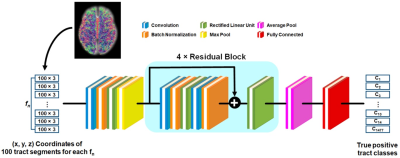 |
Deep convolution neural network-based DWI tractography connectome analysis to predict language improvement after pediatric epilepsy surgery
Jeong-Won Jeong1,2,3,4, Min-Hee Lee1,2, Nolan O'Hara2,4, Eishi Asano1,3,4, and Csaba Juhasz1,2,3,4
Video Permission Withheld
1Pediatrics, Wayne State University, Detroit, MI, United States, 2Translational Imaging Lab, Children's Hospital of Michigan, Detroit, MI, United States, 3Neurology, Wayne State University, Detroit, MI, United States, 4Translational Neuroscience Program, Wayne State University, Detroit, MI, United States
Early surgery helps improve language function in pediatric epilepsy. We investigate if an advanced DWI approach combining deep convolution network-based tract classification with DWI connectome can help early surgery by providing preoperative imaging markers which indicate a high likelihood of postoperative language improvement. Our approach revealed two nodes in preoperative DWI data, including left middle temporal gyrus and left angular gyrus, of which preoperative local efficiency values are not significantly different in patients having postoperative improvement of receptive language, compared with age-matched healthy controls, which can be as effective imaging markers for prediction of the postoperative language improvement.
|
|
0237.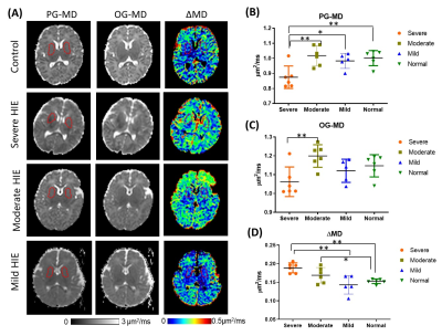 |
Feasibility of oscillating and pulsed gradient diffusion MRI to assess neonatal hypoxia-ischemia on clinical system
Fusheng Gao1, Xiaoxia Shen1, Hongxi Zhang1, Yi Zhang2, Xiaolu Ma1, Jiangyang Zhang3, and Dan Wu2
1Children's Hospital, Zhejiang University School of Medicine, Hangzhou, China, 2Key Laboratory for Biomedical Engineering of Ministry of Education, College of Biomedical Engineering & Instrument Science, Zhejiang University, Hangzhou, China, 3Department of Radiology, New York University School of Medicine, New York, NY, United States
Despite the success of diffusion time (td) dependent diffusion MRI in simulation and preclinical studies, clinical applications of the technique are challenged by difficulties in accessing short td, limited primarily by the clinical gradient system. This study demonstrated the feasibility of td-dependent dMRI using oscillating and pulsed gradients on a 3T clinical system to investigate neonatal hypoxic-ischemic encephalopathy (HIE). Results demonstrated that the td-dependency (ΔMD) increased in the deep gray matter of infants during the first year. In HIE patients, ΔMD increased in the basal ganglia of the severe and moderate HIE, as well as in the penumbra of lesions.
|
|
0238.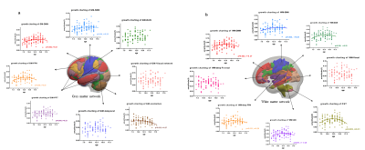 |
Growth Charting of Grey Matter and White Matter Functional Network among Normal Children and Adolescents
Xuan Bu1, Kaili Liang1, Yingxue Gao1, Lu Lu1, Hailong Li1, Lianqing Zhang1, Shi Tang1, Yanlin Wang1, Xinyu Hu1, Qiyong Gong1, Bharat Biswal2, and Xiaoqi Huang1
1Huaxi MR Research Center (HMRRC), Functional and molecular imaging Key Laboratory of Sichuan Province, Department of Radiology, Sichuan University, Chengdu, China, 2Department of Biomedical Engineering, New Jersey Institute of Technology, Newark, NJ, United States
In current study, we used a network growth charting method to map the normative maturational trajectories of major functional network activity in both grey matter and white matter. A quadratic maturation trajectory of functional activity was observed in DMN, SMN and FPN for both grey matter and white matter. The coherent maturational trajectory between these grey matter and white matter network suggests the corresponding refinements of brain network function plays an important role in improvements in higher-order cognitive abilities during normative adolescent development.
|
|
0239.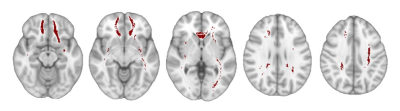 |
Disconnectome-Symptom Mapping in Traumatic Brain Injury: Application to Paediatric Populations
Adam J Shephard1, Jan Novak1, Cathy Catroppa2, Vicki Anderson2, and Amanda G Wood1,3
1School of Life & Health Sciences & Aston Neuroscience Institute, Aston University, Birmingham, United Kingdom, 2Clinical Sciences, Murdoch Children’s Research Institute, Melbourne, Australia, 3School of Psychology, Deakin University, Geelong, Australia
Disconnectome-symptom mapping (DSM) was used to identify relationships between brain and behaviour, by assessing the effect of pathology-intersected white matter tracts on neuropsychological outcomes. This study used DSM to see how IQ, two years post-injury, related to disconnections in the brain, following paediatric traumatic brain injury. For this, two approaches were employed: the BCBtoolkit, designed for use in adults, and a child-analogue. This study found the BCBtoolkit to be less sensitive than the child-analogue, however, in both methods, disconnections in the superior longitudinal fasciculus and external capsule correlated with a reduced IQ when comparing disconnected patients to controls.
|
|
0240.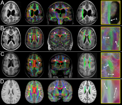 |
White matter tracts organization in patients with polymicrogyria and lissencephaly
Filippo Arrigoni1, Denis Peruzzo1, Simone Mandelstam2,3,4,5, Gabriele Amorosino1, Daniela Redaelli1, Romina Romaniello6, Richard Leventer2,3,4, Renato Borgatti6, Marc Seal2,4, and Joseph Yuan-Mou Yang2,3,4
1Neuroimaging Lab, Scientific Institute, IRCCS E. Medea, Bosisio Parini, Italy, 2Murdoch Children’s Research Institute, Parkville, Australia, 3Royal Children’s Hospital, Parkville, Australia, 4The University of Melbourne, Parkville, Australia, 5The Florey Institute of Neuroscience and Mental Health, Parkville, Australia, 6Scientific Institute, IRCCS E. Medea, Bosisio Parini, Italy
White matter (WM) tracts organization in 42 polymicrogyria (PMG) and 8 lissencephaly (LIS) patients were characterized using the tissue-specific Constrained Spherical Deconvolution modelling technique. Structural appearance of 9 major WM tracts were judged using fiber orientation distribution based direction-encoded color maps and probabilistic algorithm based tractography reconstructions. More abnormal-appearing WM tracts were identified in LIS compared to PMG. Degrees of superior longitudinal fasciculus and cingulum abnormalities were associated with PMG distribution and severity. Thickened superior fronto-occipital fasciculus was demonstrated in three patients. Patterns of WM tracts involvement were related to PMG and LIS distribution and subgroups.
|
 Watch the Video