
Oral - Power Pitch Session
Neurodegeneration 1
| Monday Parallel 2 Live Q&A | Monday, 10 August 2020, 14:30 - 15:15 UTC | Moderators: Sila Genc |
 |
0186.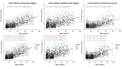 |
Perivascular space imaging across the lifespan
Kirsten M Lynch1, Giuseppe Barisano1, Arthur W Toga1, and Farshid Sepehrband1
1Mark and Mary Stevens Institute for Neuroimaging and Informatics, University of Southern California, Los Angeles, CA, United States
The perivascular space (PVS) is a major component of the glymphatic system and it promotes functional brain clearance. PVS enlargement has been observed in neurological disorders and is considered a biomarker for vascular pathology, however its role in normative development is not well understood. Using a novel technique to segment PVS, we sought to quantify age-related changes in PVS across the lifespan in a large cross-sectional cohort of cognitively normal individuals. We found age was significantly and positively associated with PVS throughout the brain and these results provide a first step towards understanding the typical evolution of brain clearance mechanisms.
|
 |
0187. |
Increased Blood-Brain Interface Water Permeability in the Ageing Brain detected using non-invasive Multiple Echo Time ASL MRI
Yolanda Ohene1, Ian F. Harrison1, David L. Thomas2,3,4, Mark F. Lythgoe 1, and Jack A. Wells 1
1Centre for Advanced Biomedical Imaging, UCL, London, United Kingdom, 2Neuroradiological Academic Unit, UCL Queen Square Institute of Neurology, UCL, London, United Kingdom, 3Dementia Research Centre, UCL Queen Square Institute of Neurology, UCL, London, United Kingdom, 4Wellcome Centre for Humans Neuroimaging, UCL Queen Square Institute of Neurology, UCL, London, United Kingdom
Multi-TE ASL technique detects a significant increase (32%) in blood-brain interface (BBI) permeability in the ageing brain. The change in BBI water permeability is associated with a marked increase (1.9 ± 0.4 fold) in expression of PDGFRβ, an index of pericyte coverage, and changes to aquaporin water channels and their anchoring proteins in the ageing brain. This technique is a promising non-invasive tool to measure age-related changes to the BBI, that may play a mechanistic role in the pathogenesis of neurodegenerative conditions.
|
0188. |
Association of age and sex with cerebral blood flow measured using pseudo-continuous arterial spin labeling imaging
Joseph Alisch1, Nikkita Khattar1, Richard Wonjoong Kim1, Abinand C. Rejimon1, Luis E. Cortina1, Wenshu Qian1, Mustapha Bouhrara1, and Richard G. Spencer1
1NIA, NIH, Baltimore, MD, United States
Cerebral blood flow (CBF) has been shown to decline with age and differs between men and women. However, limited work has been conducted on cognitively unimpaired subjects. Furthermore, most investigations focus on gray matter (GM), with few results reported for white matter (WM), in which CBF is lower and represents a particularly challenging measurement. We investigate associations of age and sex with CBF in GM and WM regions in a cohort of cognitively unimpaired subjects across a wide age range. We find significant correlations between CBF and age, as well as sexual dimorphism of CBF, in critical brain structures.
|
|
0189. |
Dynamic sodium (23Na) MRI for mapping CSF bulk flow in tissue extracellular space for clearance in human brains
Yongxian Qian1, Karthik Lakshmanan1, Yulin Ge1, Yvonne W. Lui1, Thomas Wisniewski2, and Fernando E. Boada1
1Radiology, New York University, New York, NY, United States, 2Neurology, New York University, New York, NY, United States
This study presents preliminary data to demonstrate the potential of dynamic sodium MRI for mapping cerebrospinal fluid (CSF) bulk flow in extracellular space of tissues in whole brain.
|
|
|
0190.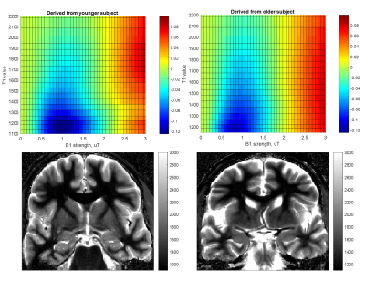 |
Age-dependent variation in CEST signal at low B1 may reflect decline of lipids in older brain tissue
Abigail Cember1,2, Puneet Bagga1, Hari Hariharan1, and Ravinder Reddy1
1Center for Magnetic Resonance and Optical Imaging, University of Pennsylvania, Philadelphia, PA, United States, 2Graduate Group in Biochemistry and Biophysics, University of Pennsylvania, Philadelphia, PA, United States
In investigating the problem of CEST correction methods for low saturation B1, we observed differences in the behavior of the CEST asymmetry signal as a function of age. We believe this incidental observation to be a manifestation of the low saturation power induced NOE reported in other literature. In this case, we hypothesize that the physiological phenomenon underlying the pattern we observe is a decrease in myelin or other lipids in the aging brain. Our T1 maps corroborate literature collected at lower field strength that T1 values increase with age; however, this appears to be an independent, if related, phenomenon.
|
0191.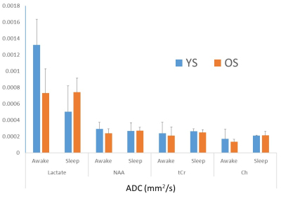 |
Altered lactate dynamics with age in human brain during sleep
Manoj K. Sammi1, Katherine Powers1, Chloe Robinson1, Selda Yildiz1,2, Miranda Lim2,3,4,5,6, Jeffrey J Iliff7,8,9,10,11, and William D Rooney1,2,5,9
1Advanced Imaging Research Center, Oregon Health & Science University, Portland, OR, United States, 2Department of Neurology, Oregon Health & Science University, Portland, OR, United States, 3VA Portland Health Care System, Portland, OR, United States, 4Department of Medicine, Division of Pulmonary and Critical Care Medicine, Oregon Health & Science University, Portland, OR, United States, 5Department of Behavioral Neuroscience, Oregon Health & Science University, Portland, OR, United States, 6Oregon Institute of Occupational Health Sciences, Oregon Health & Science University, Portland, OR, United States, 7Department of Neurology, University of Washington, Seattle, WA, United States, 8Department of Anesthesiology and Perioperative Medicine, Oregon Health & Science University, Portland, OR, United States, 9Knight Cardiovascular Institute, Oregon Health & Science University, Portland, OR, United States, 10VISN 20 Mental Illness Research, Education and Clinical Center (MIRECC), VA Puget Sound Health Care System, Seattle, WA, United States, 11Department of Psychiatry and Behavioral Sciences, University of Washington, Seattle, WA, United States
Lactate dynamics during sleep-awake cycle in human brain are studied non-invasively using single voxel diffusion weighted magnetic resonance spectroscopy (MRS) technique with simultaneous polysomnography (PSG) recordings to characterize sleep stages. Awake lactate apparent diffusion coefficients (ADC) values are large compared to other brain metabolites and may support active transport - Astrocyte-Neuron Lactate Shuttle (ANLS) mechanism. Lactate ADC are reduced in deep sleep stage in young subjects but are unchanged in older subjects. These results may reflect different interstitial fluid exchange activity or changed metabolic state with aging and require further research.
|
|
0192.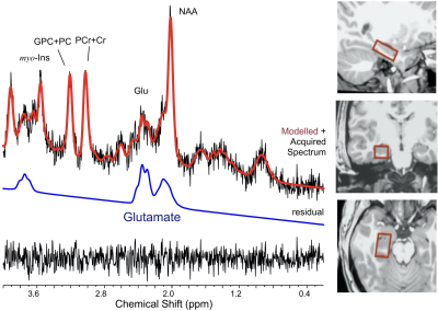 |
Age differences in hippocampal glutamate modulation during associative encoding: A proton functional magnetic resonance spectroscopy study
Chaitali Anand1, Dalal Khatib1, Cheryl Dahle2, Naftali Raz2, and Jeffrey Stanley1
1Psychiatry and Behavioral Neuroscience, Wayne State University, Detroit, MI, United States, 2Psychology, Wayne State University, Institute of Gerontology, Detroit, MI, United States
Memory declines early in normal aging, worsening in dementia. Glutamatergic neurons, abundant in the hippocampus, play a pivotal role in synaptic plasticity underlying formation of associations. We have previously demonstrated with 1H fMRS, significant modulation of hippocampal glutamate during encoding of object-location associations. We observed that the timing of modulation differentiated proficiency in acquiring the associations. Because age-related hippocampal atrophy may be accompanied by glutamatergic dysfunction, age-differences in task-related levels of hippocampal glutamate may provide a marker of age-related memory deficits. Here, we identified age-differences in hippocampal glutamate modulation during associative memory encoding, which may underlie age-related associative memory deficits.
|
|
0193.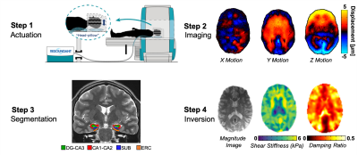 |
Reliable High-Resolution MR Elastography Protocol to Assess Hippocampal Subfield Viscoelasticity in Aging
Peyton L Delgorio1, Lucy V Hiscox1, Ryan T Pohlig1, Faria Sanjana1, Ana M Daugherty2, Hillary Schwarb3, Christopher R Martens1, Matthew DJ McGarry4, and Curtis L Johnson1
1University of Delaware, Newark, DE, United States, 2Wayne State University, Detroit, MI, United States, 3University of Illinois at Urbana-Champaign, Urbana, IL, United States, 4Dartmouth College, Hanover, NH, United States
The goal of this study is to generate the first high-resolution magnetic resonance elastography (MRE) protocol specifically for characterizing viscoelasticity of the hippocampal subfields (HCsf) and analyzing the effects of age on HCsf properties. We demonstrated that the protocol can sensitively and reliably differentiate between HCsf regions. We find that each HCsf decreases in stiffness and increases in damping ratio with age, and that HCsf exhibit differential relationships with age. This protocol shows promise for investigating the HCsf in health and disease.
|
|
0194.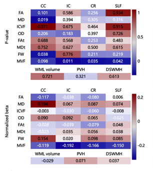 |
Effects of Arterial Stiffness on Cerebral White Matter Integrity in the Elderly
Koji Kamagata1, Christina Andica1, Kazunori Shimada2, Hideyoshi Kaga3, Yuki Someya3,4, Yuya Saito1,5, Toshiaki Akashi1, Akihiko Wada1, Yoshifumi Tamura3,4, Ryuzo Kawamori3,4, Hirotaka Watada3, Hiroyuki Daida2, and Shigeki Aoki1
1Department of Radiology, Juntendo University, Tokyo, Japan, 2Department of Cardiovascular Medicine, Juntendo University, Tokyo, Japan, 3Department of Metabolism & Endocrinology, Juntendo University, Tokyo, Japan, 4Sportology Center, Juntendo University, Tokyo, Japan, 5Department of Radiological Sciences, Tokyo Metropolitan University, Tokyo, Japan
Arterial stiffness has been shown to be associated with structural and functional abnormalities in the brain; however, white matter pathology related to arterial stiffness is poorly understood. In this study, we used white matter (WM) sensitive techniques (diffusion tensor imaging, neurite orientation dispersion and density imaging, free-water imaging, and magnetization transfer-saturation imaging) to better understand the impact of arterial stiffness on the WM microstructure in healthy elderly individuals. Our results suggest that arterial stiffness largely affects the content of cerebral myelin, as reflected by the myelin volume fraction.
|
|
0195.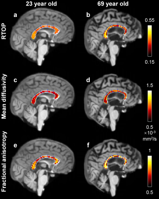 |
Return-to-origin probability from single-shell and multi-shell diffusion MRI data correlates with normal aging
Qiyuan Tian1,2, Qiuyun Fan1,2, Kimberly A. Stephens1, Chanon Ngamsombat1, Maya Polackal1, Brian E. Edlow1, Jennifer A. McNab3, David Salat1, and Susie Y. Huang1,2
1Martinos Center for Biomedical Imaging, Massachusetts General Hospital, Charlestown, MA, United States, 2Department of Radiology, Harvard Medical School, Boston, MA, United States, 3Department of Radiology, Stanford University, Stanford, CA, United States
Return-to-origin probability (RTOP) measures the overall restriction of the microstructural environment and has been used to map microstructural changes related to age and pathology. However, measurement of RTOP requires either specialized acquisition (Cartesian q-space sampling) or processing (q-space gridding or modelling). We show that RTOP from multi-shell data is a weighted summation of the spherical mean signal of each individual shell. We apply our method to a multi-shell dataset of 40 subjects with b-values up to 17,800 s/mm2 and a dataset of 160 subjects from Lifespan Human Connectome Project in Aging and demonstrate its utility in mapping age-related microstructural change.
|
|
0196.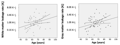 |
Increase in blood-brain barrier disruption during normal aging
Inge Verheggen1, Joost de Jong2, Martin van Boxtel1, Frans Verhey1, Jacobus Jansen2,3, and Walter Backes2
1Department of Psychiatry and Neuropsychology, Maastricht University, Maastricht, Netherlands, 2Department of Radiology and Nuclear Medicine, Maastricht University Medical Center, Maastricht, Netherlands, 3Department of Electrical Engineering, Eindhoven University of Technology, Eindhoven, Netherlands Blood-brain barrier (BBB) disruption is assumed to increase with age, but this has not been demonstrated assessing gadolinium leakage using dynamic contrast-enhanced MRI. We determined BBB leakage rate in healthy middle-aged to elderly individuals (47 - 91 years) combining DCE MRI with pharmacokinetic modeling. Results demonstrated BBB leakage in white and gray matter increased with age. However, this effect was not independent of white matter lesions or cortical thinning, so other physiological changes may influence the age and leakage association.Our study demonstrates that BBB disruption manifests in normal aging, before the emergence of neuropathology. |
|
0197.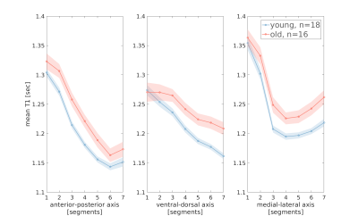 |
Measuring Biological Gradients along the Human Dorsal Striatum in vivo using Quantitative MRI
Elior Drori1, Shir Filo1, and Aviv Mezer1
1The Edmond and Lily Safra Center for Brain Sciences, The Hebrew University of Jerusalem, Jerusalem, Israel
To date there are no in vivo tools for quantifying spatial changes in the microstructure of subcortical gray-matter nuclei. We have developed a quantitative MRI tool, with which we measured variations along the human dorsal striatum, using quantitative T1. We found monotonic gradients along the main axes, consistent with known biological gradients of the striatum. In addition, we found effects of laterality, as well as aging effects. Our method can prove useful for detection and quantification of microstructural irregularities in the striatum in patients suffering from basal ganglia disorders, such as Parkinson’s disease and ADHD.
|
|
0198.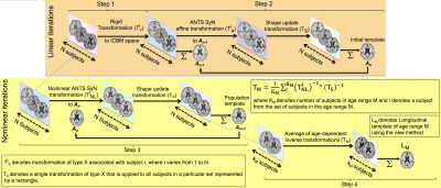 |
Development of a spatio-temporally consistent longitudinal structural template of the older adult brain
Abdur Raquib Ridwan1, Mohammad Rakeen Niaz1, Yingjuan Wu1, Xiaoxiao Qi1, David A. Bennett2, and Konstantinos Arfanakis1,2
1Biomedical Engineering, Illinois Institute of Technology, Chicago, IL, United States, 2Rush Alzheimer's Disease center, Rush University Medical Center, Chicago, IL, United States
One of the major challenges in constructing a longitudinal structural template of the older adult brain is to ensure spatio-temporal consistency. In this work, a new method was introduced to construct a spatio-temporally consistent longitudinal structural template of the older adult brain based on high quality cross-sectional older adult data from a large cohort. The new template was compared to templates generated with previously published methods in terms of spatio-temporal consistency, image quality, and representativeness of age-related brain changes, and was shown to have superior performance.
|
|
0199. |
Diffusion measures and Connectometry in the Human Hippocampal-Subfields Using Super-Resolution HYDI.
Nahla M H Elsaid1,2, Pierrick Coupé3,4, Andrew J Saykin1,2, and Yu-Chien Wu1,2
1Department of Radiology and Imaging Sciences, Indiana University, Indianapolis, IN, United States, 2Indiana Alzheimer Disease Center, Indiana University, Indianapolis, IN, United States, 3LaBRI, UMR 5800, University of Bordeaux, Talence, France, 4LaBRI, UMR 5800, PICTURA, F-33400, CNRS, Talence, France The aging process is known to cause morphological and structural alterations in the human brain. Using a sub-millimeter super-resolution hybrid diffusion imaging (HYDI), we studied the effects of aging on the structural connectivity between the hippocampal subfields as well as between the hippocampus and the cerebral cortex. |
|
0200. |
A multi-center study to investigate the relationship between iron content in deep gray matter nuclei and age
Yan Li1, Sean K. Sethi2,3, Chengyan Wang4, Weibo Chen5, Naying He1, Ewart Mark Haacke3, and Fuhua Yan1
1Department of Radiology, Ruijin Hospital, Shanghai Jiao Tong University School of Medicine, Shanghai, China, 2The MRI Institute for Biomedical Research, Magnetic Resonance Innovations, Inc., Detroit, MI, United States, 3Department of Radiology, Wayne State University, Detroit, MI, United States, 4Human Phenome Institute, Fudan University, Shanghai, China, 5Philips Healthcare, Shanghai, China
To investigate the correlation of iron content in deep gray matter nuclei as a function of age by reconstructed quantitative susceptibility mapping (QSM) using both whole-structural and regional perspectives from three different MRI sites and three different scanners to show that QSM is a robust technology across manufacturers and resolution.
|
 Watch the Video