
Oral - Power Pitch Session
Neurodegeneration 2
| Wednesday Parallel 2 Live Q&A | Wednesday, 12 August 2020, 15:15 - 16:00 UTC | Moderators: Thijs Dhollander & Claire Kelly |
0928.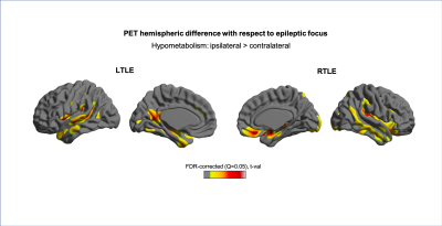 |
Extratemporal cortical morphological changes and hypometabolism revealed in radiological MRI-negative temporal lobe epilepsy
Julia Pia Simon1, Ben A. Duffy1, Yan Li2, Arthur W. Toga1, Wolfgang G. Muhlhofer3, Robert C. Knowlton2, and Hosung Kim1
1University of Southern California, Los Angeles, CA, United States, 2Neurology and UCSF Weill Institute for Neurosciences, San Francisco, CA, United States, 3University of Alabama at Birmingham Epilepsy Center, Birmingham, AL, United States
Radiological MRI-negative temporal lobe epilepsy (TLE) is a common, but challenging subtype for surgical treatment. Compared to MRI-positive cases, these patients often require invasive EEG for localization that may also involve extratemporal regions. Furthermore, these cases entail a lower likelihood of seizure-free surgical outcome. To better understand this important group, we studied cortical surface features of MRI and FDG-PET to relate occult extratemporal damage to epilepsy localization and surgical outcome prediction. Bilateral cortical morphological changes were found. FDG-PET hypometabolism was lateralized in the hemisphere ipsilateral to seizure focus. Extratemporal and bilateral hypometabolism tended to be associated with poor surgical outcome.
|
|
 |
0929.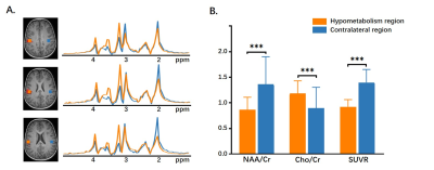 |
Simultaneous 18F-FDG-PET and 1H-MRSI Metabolic Imaging in Epilepsy Patients: A Feasibility Study
Hui Huang1, Miao Zhang2, Rong Guo3,4, Yudu Li3,4, Yibo Zhao3,4, Jialin Hu1, Hongping Meng2, Xinyun Huang2, Xiaozhu Lin2, Wei Liu5, Biao Li2, Bomin Sun5, Yao Li1, Zhi-Pei Liang3,4,
and Jie Luo1
1Institute of Medical Imaging Technology, School of Biomedical Engineering, Shanghai Jiao Tong University, Shanghai, China, 2Department of Nuclear Medicine, Ruijin Hospital, Shanghai Jiao Tong University School of Medicine, Shanghai, China, 3Department of Electrical and Computer Engineering, University of Illinois at Urbana Champaign, Urbana, IL, United States, 4Beckman Institute for Advanced Sciences and Technology, University of Illinois at Urbana Champaign, Urbana, IL, United States, 5Department of Functional Neurosurgery, Ruijin Hospital, Shanghai Jiao Tong University School of Medicine, Shanghai, China
PET and MRSI could provide metabolic information of the epileptogenic zone, which could add value to presurgical planning of epilepsy patients. This study investigated the feasibility of simultaneous high-resolution MRSI and 18F-FDG-PET for whole brain imaging in epilepsy patients, and studied the correlation between metabolic changes found in MRSI and hypometabolism found in FDG-PET. Our experimental results showed a decrease in NAA and an increase in Cho, concomitant with low FDG uptake.
|
0930.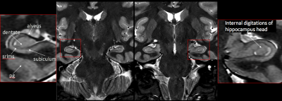 |
Deep Learning Reconstruction Method for Improved Visualization of Hippocampal Anatomical Structures
Patrick Quarterman1, Angela Lignelli2, Marc Lebel3, and Sachin Jambawalikar4
1GE Healthcare, New York, NY, United States, 2Radiology, Columbia University, New York, NY, United States, 3GE Healthcare, Calgary, AB, Canada, 4Columbia University, New York, NY, United States
The purpose of this study was to determine if deep learning reconstruction (DLRecon) method to reduce image noise could lead to improvement in in-vivo anatomical detail of the hippocampus structures without substantial increase in scan/exam time on a clinical 3T system. Evaluation of this new reconstruction technique was performed on a group of 5 volunteers with results indicating that higher resolution scans compared to current seizure protocol was free of imaging noise and led to higher confidence in identifying hippocampal key anatomical structures and temporal lobes.
|
|
0931.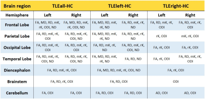 |
White matter microstructure characterisation in left and right temporal lobe epilepsy (TLE) using TBSS
Nicolò Rolandi1, Fulvia Palesi1, Francesco Padelli2, Isabella Giachetti2, Domenico Aquino2, Giuseppe Didato3, Elio Maccagnano3, Paul Summers4, Giancarlo Germani4, Claudia AM Gandini Wheeler-Kingshott1,5,6, and Paolo Vitali4
1Department of Brain and Behavioral Science, University of Pavia, Pavia, Italy, 2Fondazione I.R.C.C.S. Istituto Neurologico Carlo Besta, Milan, Italy, 3Neuroradiology, Fondazione I.R.C.C.S. Istituto Neurologico Carlo Besta, Milan, Italy, 4Neuroradiology Unit, Brain MRI 3T Research Center, IRCCS Mondino Foundation, Pavia, Italy, 5Department of Neuroinflammation, UCL Queen Square Institute of Neurology, Faculty of Brain Sciences, University College London, NMR Research Unit, Queen Square MS Centre, London, United Kingdom, 6Brain MRI 3T Research Center, IRCCS Mondino Foundation, Pavia, Italy
Tract-based spatial statistics investigations of temporal lobe epilepsy (TLE) have been using standard diffusion metrics, without distinguishing patients according to the lateralization of their epileptogenic zone. The aim of this study is to further our knowledge by identifying specific patterns of alteration in left and right TLE patients using diffusion kurtosis imaging and NODDI parameter maps. Our findings demonstrate the presence of specific patterns of white matter alterations, with the left TLE more widely affecting both cerebral and cerebellar regions. These results support the need to consider patients separately, according to the side of their pathology.
|
|
0932.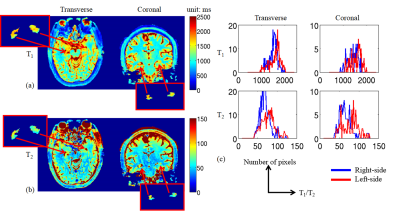 |
Visualization enhancement and quantitative analysis of relaxation time in medial temporal lobe epilepsy based on 3D high-resolution MRF
Xiaozhi Cao1,2,3, Kang Wang4, Congyu Liao2,3, Dengchang Wu4, Qing Li1, Ziyang Chen1, Jun Li1, Huihui Ye1, Hongjian He1, and Jianhui Zhong1
1Center for Brain Imaging Science and Technology, Department of Biomedical Engineering, Zhejiang University, Hangzhou, China, 2Athinoula A. Martinos Center for Biomedical Imaging, Massachusetts General Hospital, charlestown, MA, United States, 3Department of Radiology, Harvard Medical School, charlestown, MA, United States, 4Department of Neurology, the First Affiliated Hospital, School of Medicine, Zhejiang University, Hangzhou, China
We propose to use a 3D MRF technique with multi-axis spiral projection acquisition to achieve 3D high-resolution whole-brain quantitative imaging for patients with MTLE. Isotropic 1-mm resolution relaxivity maps were achieved within 5 minutes. By incorporating Freesurfer’s automatic subcortical segmentation, a whole-brain subcortical segmentation was obtained, enabling feasible and subjective quantitative analysis for each substructure. Additionally, volume information of the substructure was obtained during the process.
|
|
0933.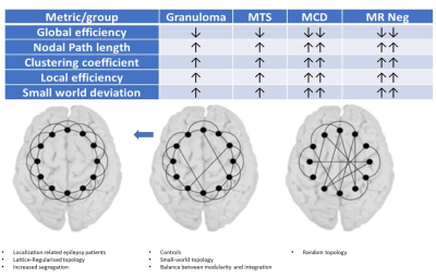 |
CORTICAL THICKNESS COVARIANCE STRUCTURAL NETWORKS IN “FOCAL” EPILEPSY
Karthik Kulanthaivelu1, Kiran Raj V1, Raghavendra Kenchaiah 2, Jitender Saini1, Rose Dawn Bharath1, and Sanjib Sinha2
1Department of Neuroimaging and Interventional Radiology, National Institute of Mental Health and Neurosciences, Bengaluru, India, 2Department of Neurology, National Institute of Mental Health and Neurosciences, Bengaluru, India “Focal” epilepsy is a network aberration. Network characteristics in focal epilepsy due to calcified Neurocysticercal granuloma have not been elucidated. Forty-two patients of focal epilepsy with MRI evidence of either calcified granuloma, malformation of cortical development, mesial temporal sclerosis, or no imaging abnormality were included. Group-level Cortical thickness covariance networks were generated and compared. Focal epilepsy patients (including those with calcific granuloma) had significantly reduced network global efficiency and higher nodal characteristic path length/ Clustering coefficient/ Nodal local efficiency (p<0.05). Networks in focal epilepsy (“ including those due to calcific granuloma”) have higher segregation and lesser integration. |
|
0934.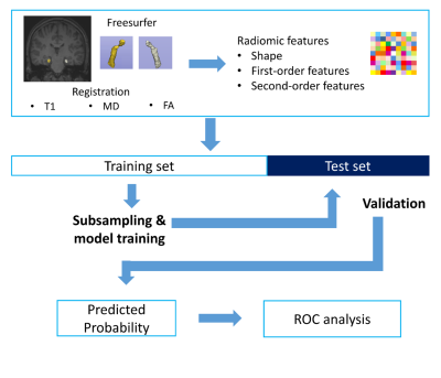 |
Radiomics Features of Hippocampal Regions in Conventional and Diffusion Tensor Imagings can Differentiate Temporal Lobe Epilepsy Patients
Yae Won Park1, Dongmin Choi2, Kyunghwa Han1, Sung Soo Ahn1, Hwiyoung Kim1, and Hyang Woon Lee3
1Yonsei University College of Medicine, Seoul, Republic of Korea, 2Department of Computer Science, Yonsei University, Seoul, Republic of Korea, 3Department of Neurology, Ewha Womans University College of Medicine, Seoul, Republic of Korea
A total of 92 subjects(66 TLE [35 right and 31 left] and 26 healthy controls) were allocated to training(n=66) and test(n=26) sets. Radiomics features (n=558) from the bilateral hippocampi were extracted from T1WI and DTI. Machine learning models were trained. Identical processes were performed to differentiate right TLE from HC and left TLE from HC. The radiomics model in test set showed better performance than hippocampal volume for identifying TLE (AUC 0.82 vs. AUC 0.62, P=0.08). Radiomics models of both subgroups showed better performance than those of hippocampal volume(AUC 0.76 vs. AUC 0.54 [P=0.12] and AUC 0.95 vs 0.68 [P=0.04]).
|
|
0935.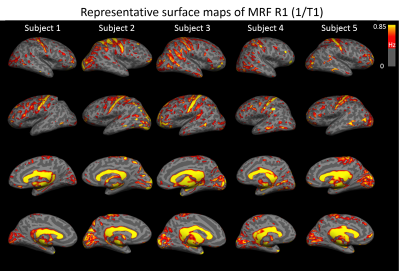 |
Using High-resolution 3D MR Fingerprinting for Characterization of Focal Cortical Dysplasia
Joon Yul Choi1, Rasim Boyacioglu2, Stephen Jones3, Ken Sakaie3, Ingmar Blümcke1,4, Imad Najm1, Mark Griswold2, Dan Ma5, and Zhong Irene Wang1
1Epilepsy Center / Neurological Institute, Cleveland Clinic, Cleveland, OH, United States, 2Radiology, Case Western Reserve University, Cleveland, OH, United States, 3Imaging Institute, Cleveland Clinic, Cleveland, OH, United States, 4Neuropathology, University of Erlangen, Erlangen, Germany, 5Biomedical Engineering, Case Western Reserve University, Cleveland, OH, United States
We investigate in this study quantitative T1 and T2 values as potential biomarkers of tissue properties in epilepsy patients with focal cortical dysplasia (FCD) using a novel high-resolution 3D magnetic resonance fingerprinting (MRF) technique. We first investigated the quantitative T1 and T2 values in various Brodmann areas to verify the sensitivity of MRF in probing tissue properties of the human cortex. We then investigated the MRF T1 and T2 values in different subtypes of FCD lesions, which were higher than their corresponding cortical regions in the controls.
|
|
0936.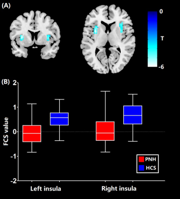 |
Resting-state functional connectivity alterations in periventricular nodular heterotopia related epilepsy
Xinyu Hu1, Wenyu Liu2, Dong Zhou2, Qiyong Gong1, and Xiaoqi Huang1
1Huaxi MR Research Center (HMRRC), West China Hospital of Sichuan University, Chengdu, China, 2Department of Neurology, West China Hospital of Sichuan University, Chengdu, China
We performed the first resting-state fMRI study integrating both whole-brain functional connectivity (FC) and seed-based FC analyses to explore the network-level neural function alterations in patients with periventricular nodular heterotopia (PNH). Our findings (i) identified lower functional connectivity strength (FCS, an index of whole-brain connectivity) in bilateral insula, higher FC in the precuneus and lower FC in the anterior cingulate cortex/medial prefrontal cortex and cerebellum networks in PNH patients and (ii) demonstrated that the significant insular hypoactivation represented the cortical hub of the whole-brain networks in PNH, which might be of clinical significance in predicting disability progression of PNH.
|
|
0937.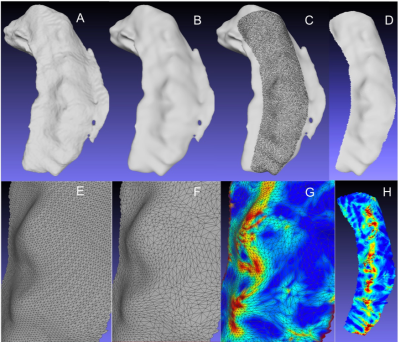 |
Quantifying Hippocampal Dentation in Epilepsy: a comparison of absolute mean curvature versus visual inspection and their memory correlates.
Lawrence Ver Hoef1,2, Mike Zhang3, and Anandh Kilpattu Ramaniharan3
1Neurology, University of Alabama at Birmingham, Birmingham, AL, United States, 2Birmingham VA Medical Center, Birmingham, AL, United States, 3University of Alabama at Birmingham, Birmingham, AL, United States
Hippocampal dentation (HD) is a morphologic feature of the human hippocampus that has been recently described and has been shown to correlate with aspects of verbal and visual memory. It varies dramatically across healthy individuals and can be affected by diseases such as epilepsy. We demonstrate a method to extract ultra-high-resolution surface contours from common MPRAGE images. We also propose a method based on absolute mean curvature to quantify HD and compare that to visual inspection in a cohort of temporal lobe epilepsy patients. Finally, we examine correlations between HD and measures of verbal and visual memory across methods.
|
|
0938.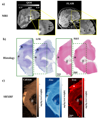 |
Quantitative susceptibility mapping reveals abnormal zinc, calcium and iron levels in focal cortical dysplasia lesions
Sara Lorio1, Po-Wah So1, Jan Sedlacik1, Derek Li2, Emma Dixon3, Sophie Adler3, Harold G. Parkers1, Helen J. Cross3, Torsten Baldeweg3, Thomas Jacques3, Karin Shmueli3, and David Carmichael1,3
1King's College London, London, United Kingdom, 2UCL, London, United Kingdom, 3UCL, LONDON, United Kingdom
We estimated quantitative susceptibility maps (QSM) in 19 children with histologically confirmed focal cortical dysplasia (FCD), a frequent cause of drug-resistant epilepsy. QSM allowed measurement of cortical and sub-cortical layered structure and its alteration in FCD lesions. Moreover, QSM was sensitive to abnormal deposits of calcium, zinc, and iron, which were validated using X-ray fluorescence in brain tissue specimens available following surgical treatment. QSM could provide a non-invasive biomarker of cortical tissue changes in epilepsy and could be used to determine alterations in mineral deposits in different brain disorders.
|
|
0939.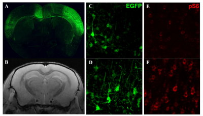 |
Assessing Focal Cortical Dysplasia Using Advanced Diffusion Imaging Sequences
Boyu Zhang1, Shaoping Zhong2, Yuwen Zhang1, Qianfeng Wang1, Jing Ding2, and He Wang1,3
1Institute of Science and Technology for Brain-Inspired Intelligence, Fudan University, Shanghai, China, 2Department of Neurology, Zhongshan Hospital, Fudan University, Shanghai, China, 3Human Phenome Institute, Fudan University, Shanghai, China
Focal cortical dysplasia (FCD) are neurodevelopmental disorders characterized by localized cortical malformation that is highly associated with the drug-resistant epilepsy. In this study, we examined the advanced diffusion MR imaging-neurite orientation dispersion and density imaging (NODDI) in the FCD mice model. The orientation dispersion index (ODI) that represents the dispersion of neurite is significantly higher in the FCD group compared with the control group which are compatible with the pathological observation. Meanwhile, no significant differences are observed in conventional DTI measurements FA and MD indicating that NODDI is more sensitive in detecting FCD lesion.
|
|
0940. |
Significance of Perivascular Spaces in Acute Ischemic Stroke and its Predictions of Epileptogenesis
Nian Yu1,2,3, Benjamin Sinclair4,5, Lina Maria Garcia Posada6, Ben Chen4, Qing Di1, Xingjian Lin1, Qingling Huang7, Scott Kolbe4, Patrick Kwan2,4,5,8, and Meng Law4,6
1Department of Neurology, The Nanjing Brain Hospital Affiliated to Nanjing Medical University, Nanjing, China, 2Department of Neurology, Royal Melbourne Hospital, Melbourne, Australia, 3Department of Radiology, The Nanjing Brain Hospital Affiliated to Nanjing Medical University, Melbourne, China, 4Department of Neuroscience, Monash University, Melbourne, Australia, 5Department of Neurology, Alfred Hospital, Melbourne, Australia, 6Department of Radiology, Alfred Hospital, Melbourne, Australia, 7Department of Radiology, The Nanjing Brain Hospital Affiliated to Nanjing Medical University, Nanjing, China, 8Department of Medicine, University of Melbourne, Melbourne, Australia
Around 10% of patients with stroke go on to develop epilepsy, however, imaging biomarkers for post-stroke epilepsy (PSE) are lacking. Perivascular spaces (PVS) are small interstitial fluid filled spaces lining the blood vessels which have a role in waste clearance in the brain. They have been found to be abnormal in epilepsy, and here we investigate whether they could serve as an early predictor of PSE. We found that the overall number and scores of enlarged PVSs were not associated with PSE, but the inter-hemispheric asymmetry was an independently associated biomarker.
|
 Watch the Video