
Oral - Power Pitch Session
Acquisition & Processing in Neuro
| Tuesday Parallel 2 Live Q&A | Tuesday, 11 August 2020, 14:30 - 15:15 UTC | Moderators: Yuki Kanazawa & Qin Qin |
 |
0532.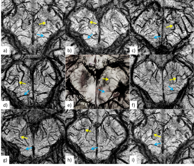 |
Subvoxel Vascular Imaging of the Midbrain Using USPIO-Enhanced MRI
Sagar Buch1, Ying Wang2, Pavan K. Jella1, Min-Gyu Park3, Yongsheng Chen1,4, Jiani Hu1, Yulin Ge5, Kamran Shah1, and E. Mark Haacke1,2
1Department of Radiology, Wayne State University, Detroit, MI, United States, 2Magnetic Resonance Innovations, Inc., Detroit, MI, United States, 3Department of Neurology, Pusan National University School of Medicine, Yangsan, Korea, Republic of, 4Department of Neurology, Wayne State University, Detroit, MI, United States, 5Department of Radiology, New York University School of Medicine, New York, NY, United States
We demonstrate the utility of low dose Ferumoxytol in microvasculature imaging of the midbrain using susceptibility weighted imaging (SWI). Mapping the brain’s vasculature has implications for understanding the etiology of many neurovascular and neurodegenerative diseases such as Parkinson’s disease. By administering this strongly paramagnetic agent, SWI was able to visualize both arteries and veins; and its sensitivity to detect sub-voxel vessels increased tremendously. However, the use of Ferumoxytol exacerbates the signal loss of large vessels, confounding the ability to visualize nearby smaller vessels. Hence, we propose the use of multiple time point SWI to effectively see through the blooming artifacts.
|
 |
0533.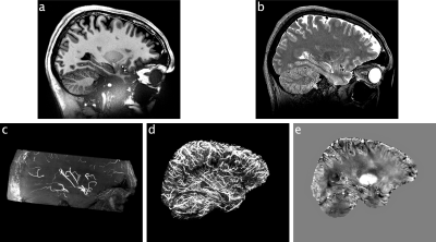 |
The human phantom: Comprehensive ultrahigh resolution whole brain in vivo single subject dataset
Falk Luesebrink1,2, Mattern Hendrik2, Renat Yakupov3, Steffen Oeltze-Jafra1,4, and Oliver Speck2,3,4,5
1Medicine & Digitalization, Otto-von-Guericke University, Magdeburg, Germany, 2Biomedical Magnetic Resonance, Otto-von-Guericke University, Magdeburg, Germany, 3German Center for Neurodegenerative Diseases, Magdeburg, Germany, 4Center for Behavioral Brain Sciences, Magdeburg, Germany, 5Leibniz Institute for Neurobiolgoy, Magdeburg, Germany
Here, we present an extension to our previously published T1-weighted dataset with an ultrahigh isotropic resolution of 250 µm, consisting of multiple additional contrasts. Included are up to 150 µm ToF, an updated 250 µm MPRAGE, 330 µm QSM, up to 450 µm T2-weighted SPACE, 750 µm MPM, 800 µm DTI, one hour continuous rs-fMRI as well as more than 130 MPRAGE volumes collected over 10 years (with varying spatial resolution between 450 µm and 1 mm). All data were acquired on the same 7 T scanner and of the same subject. Basic pre-processing of all data were conducted.
|
0534. |
Low b-value DTI for Analyzing Pseudo-random Flow of CSF
Yoshitaka Bito1, Kuniaki Harada1, Hisaaki Ochi2, and Kohsuke Kudo3
1Healthcare Business Unit, Hitachi, Ltd., Tokyo, Japan, 2Research and Development Group, Hitachi, Ltd., Tokyo, Japan, 3Department of Diagnostic Imaging, Hokkaido University Graduate School of Medicine, Sapporo, Japan
Cerebrospinal fluid (CSF) plays an important role in the clearance system of the brain. Low b-value DTI is reported to be useful for observing the CSF flow; however, the precise flow property observed by low b-value DTI has not been fully investigated. We proposed a mathematical framework of low b-value DTI for analyzing a pseudo-random flow and applied this framework to investigation into CSF. Measured DTI shows high and anisotropic diffusivity, representing large variance of flow velocity, in some segments of CSF. It demonstrates that low b-value DTI can be used for analyzing pseudo-random flow of CSF.
|
|
0535.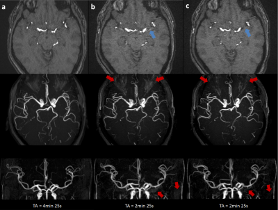 |
3D Flow Compensated Interleaved EPI with Partial Fourier Acquisition: A Feasibility Study for Fast Intracranial TOF-MRA
Wei Liu1 and Kun Zhou1
1Siemens Shenzhen Magnetic Resonance Ltd., Shenzhen, China As commonly used for intracranial vasculature, 3D TOF usually requires long acquisition time. In this study, we implemented a 3D-iEPI sequence with partial flow compensation, combined with partial Fourier acquisition to further reduce the flow artifacts. In specific, each interleave is sequentially acquired twice with alternating readout polarities to reduce the systematic inconsistencies between odd and even echoes. We explored the feasibility of such a sequence for fast intracranial TOF-MRA and demonstrated that the proposed sequence can reduce the acquisition time by approximately a factor of 2 with comparable vasculature depiction to 3D-GRE, which is promising for future applications. |
|
0536.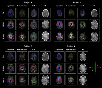 |
Strain Tensor Imaging (STI): Voxelwise assessment of cardiac-induced brain tissue strain at 7T MRI.
Jacob-Jan Sloots1, Alberto De Luca1, Geert Jan Biessels2, and Jaco Zwanenburg1
1Radiology, University Medical Center Utrecht, Utrecht, Netherlands, 2Neurology, University Medical Center Utrecht, Utrecht, Netherlands
The heartbeat induces microvascular blood volume pulsations and subsequent tissue deformations in the brain. Although subtle (typically <1%), these deformations are highly relevant as they accelerate clearance of brain waste products. Moreover, they enable non-invasive assessment of mechanical tissue properties. We developed a sensitive MRI technique with full brain coverage for voxelwise quantification of the cardiac-induced brain tissue strain tensor with 3mm isotropic resolution, based on displacement encoding with stimulated echoes (DENSE). We visualize the strain tensor similar to diffusion tensor imaging. Strain tensor imaging opens a window on brain tissue mechanics and physiological blood volume dynamics in the brain.
|
|
0537. |
Multi b-value Diffusion weighted image Diphase Map (MbDDM) to evaluate cerebrospinal fluid dynamics.
Toshiaki Taoka1,2, Rintaro Ito1,2, Rei Nakamichi2, Toshiki Nakane2, Hisashi Kawai2, and Shinji Naganawa2
1Department of Innovative Biomedical Visualization, Nagoya University, Nagoya, Japan, 2Department of Radiology, Nagoya University, Nagoya, Japan
To visualize the dynamics of cerebrospinal fluid (CSF) motion within the cranium, we evaluated the distribution of the motion-related signal dephasing by CSF on a Multi b-value Diffusion-weighted image Diphase Map (MbDDM). The MbDDM indicated that CSF motion was prominent in areas that included the ventral portion of the posterior fossa, the suprasellar cistern and the Sylvian fissure. Whereas, CSF motion was less in the lateral ventricles and the parietal subarachnoid space, casting doubt on the classical model of CSF dynamics.
|
|
0538.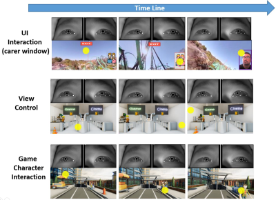 |
Transforming The Experience of Having MRI Using Virtual Reality
Kun Qian1, Tomoki Arichi1, Jonathan Eden2, Sofia Dall'Orso2, Rui Pedro A G Teixeira3, Kawal Rhode3, Mark Neil4, Etienne Burdet2, A David Edwards1, and Jo V Hajnal1
1Centre for the Developing Brain, School of Biomedical Engineering and Imaging Sciences, King's College London, London, United Kingdom, 2Department of Bioengineering, Imperial College London, London, United Kingdom, 3School of Biomedical Engineering and Imaging Sciences, King's College London, London, United Kingdom, 4Department of Physics, Imperial College London, London, United Kingdom Patients undergoing MRI often experience anxiety and sometimes distress prior to and during scanning. We have developed a non-intrusive MR compatible Virtual Reality (VR) system, providing a tailored immersive experience that the user can interact with and control using gaze tracking. Dedicated VR content has been created and tested on adults and children. A key feature is congruency between the VR world and physical sensations during MRI, including VR features corresponding to table motion and scanner noise/vibration. Results suggest the approach has huge clinical potential, and it could represent a platform for conducting a new generation of “natural” fMRI experiments.
|
|
0539.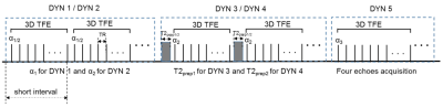 |
Three-Dimensional Simultaneous Quantitative T1-T2-T2* Mapping of Whole Brain (SQUMA): Sequence Design and In-vivo Feasibility
Huiyu Qiao1, Shuo Chen1, Dandan Yang1, Hualu Han1, Zihan Ning1, and Xihai Zhao1
1Tsinghua University School of Medicine, Beijing, China
The feasibility of T1, T2 and T2* in brain imaging and lesion quantification has been proved. However, studies about quantitative imaging seldom quantify T1, T2 and T2* together. This study proposed a three-dimensional (3D) simultaneous quantitative T1-T2-T2* mapping (SQUMA) for the whole brain. SQUMA sequence was composed of five dynamic scans using variable flip angles, variable T2 preparation duration and multi-echo acquisitions. SQUMA sequence showed excellent agreement with reference imaging in measuring T1, T2 and T2* values (R2=0.98, 0.84 and 0.90, respectively) and good to excellent repeatability in in-vivo studies. It is feasible to use SQUMA in clinical applications.
|
|
0540.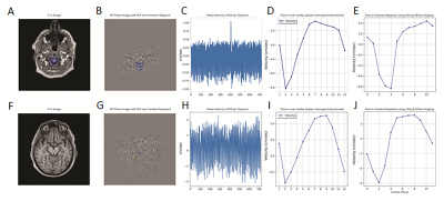 |
Real-Time EPI Phase Contrast Acquisition for Imaging of CSF Dynamics
Petrice Mostardi Cogswell1, Sandeep K Ganji2, Daniel D Borup1, Jeffrey L Gunter1, John Huston III1, and Clifford R Jack Jr1
1Radiology, Mayo Clinic, Rochester, MN, United States, 2Philips Healthcare, Gainesville, FL, United States
CSF flow has been most commonly evaluated using a gated 2D phase contrast (PC) acquisition at the cerebral aqueduct or foramen magnum. However, real-time acquisitions that allow for evaluation of changes in flow with the cardiac and respiratory may provide additional insight into CSF dynamics disorders. In this study we apply a real-time EPI based PC acquisition for imaging of intracranial CSF flow at multiple intracranial locations. Quantitative analyses are validated by comparison with a standard phase contrast acquisition. Frequency spectra analysis demonstrates dominant variations in CSF flow with the cardiac and respiratory cycles.
|
|
0541.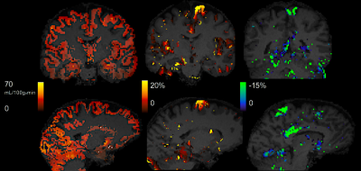 |
Short-term effects of transcranial direct current stimulation (tDCS) on cerebral blood flow measured with ASL MRI
Iris Asllani1,2, Francesco Di Lorenzo3, Katerina Gialopsou3, Joseph G Woods4,5, Marco Bozzali3, and Mara Cercignani3
1Neuroscience, University of Sussex, Brighton, United Kingdom, 2Biomedical Engineering, Rochester Institute of Technologygy, Rochester, NY, United States, 3University of Sussex, Brighton, United Kingdom, 4Radiology, University of California San Diego, San Diego, CA, United States, 5University of Oxford, Oxford, United Kingdom
Short-term effects of transcranial direct current stimulation (tDCS) on CBF were measured using arterial spin labeling (ASL) MRI. Results showed that anodal surface stimulation of the motor region was followed by an increase in gray matter CBF in that region. Conversely, CBF in the stimulated region following a cathodal stimulation decreased. There was no effect of sham stimulation on the CBF of the stimulated area. These findings may help forge a new path toward a better understanding of the neuro-physiological effects of tDCS in humans.
|
|
0542. |
Venous Mapping of Vascular Malformations using Cranial 4D Flow MRI with Improved ‘Virtual Injections’
Laura Eisenmenger1, Grant Steven Roberts2, Michael Loecher3, Leonardo Rivera-Rivera1, Patrick Turski1, Kevin M Johnson1,2, and Oliver Wieben1,2
1Radiology, University of Wisconsin - Madison, Madison, WI, United States, 2Medical Physics, University of Wisconsin - Madison, Madison, WI, United States, 3Radiology, Stanford University, Palo Alto, CA, United States
Endovascular intervention via a venous approach, or trans-venous embolization (TVE), has been increasing employed in the management of intracranial vascular lesions such as arteriovenous malformations (AVMs) and dural arteriovenous fistulas (DAVFs). Current pre-procedural planning is limited by overlapping, complex vascular anatomy and a lack of quantitative hemodynamic feature characterization. Using novel 4D flow MRI methods, high-resolution retrograde venous flow mapping with anatomical detail and dynamic flow fields can provide valuable information prior to TVE. We will present our institutional experience using this method in representative intracranial vascular lesions.
|
|
0543.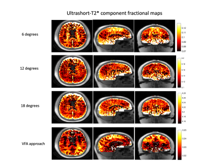 |
Quantification of T1 and relative proton density in the brain ultrashort-T2* component
Nikhil Deveshwar1, Emil Ljungberg2, Misung Han1, and Peder E. Z. Larson1
1Department of Radiology and Biomedical Imaging, University of California, San Francisco, San Francisco, CA, United States, 2Department of Neuroimaging, King’s College London, London, United Kingdom
This study presents a VFA approach for quantification of T1 and relative proton density of the brain ultrashort-T2* component. Measured T1 values corresponding to the ultrashort-T2* component were lower compared to T1 values corresponding to long-T2* components, and did not exhibit gray/white matter differences. Qualitatively, ultrashort-T2* component fraction maps showed better gray/white matter contrast and clearer white matter structure delineation which we expect to be a more accurate representation of relative proton density. These results show that added VFA T1 encoding in characterization of the brain ultrashort-T2* component can more accurately differentiate white matter anatomy.
|
|
0544. |
Feasibility for MR Elastography to Meet Unmet Need in Intracerebral Hemorrhage Surgical Planning
Robert Moskwa1, Dipul Chawla2, Corinne Henak2, Azam Ahmed3, and Walter Block1
1Medical Physics, University of Wisconsin-Madison, Madison, WI, United States, 2Mechanical Engineering, University of Wisconsin-Madison, Madison, WI, United States, 3Neurological Surgery, University of Wisconsin-Madison, Madison, WI, United States
It is hypothesized that the proper surgical approach for intracerebral hemorrhage (ICH) victims should depend on clot rigidity. Neurosurgical experience indicates that brain clot rigidity varies across patients and varies spatially and temporally within each patient. We hypothesize that the wide range of clot rigidity in ICH will allow MR elastography (MRE) techniques to depict the heterogeneity over a wide dynamic range of rigidity. Longitudinal MRE, ultrasound elastography, and mechanical compression testing were performed on large ex-vivo swine blood clots. MR elastography shows promise for characterizing the rigidity of intracerebral hemorrhage as indicated by these ex-vivo tests.
|
|
0545. |
Measurement of Blood-Brain Barrier Permeability in Human Brain using Magnetization Transfer Effect at 7T.
Sultan Zaman Mahmud1,2, Thomas S. Denney1,2, Ronald J. Beyers2, and Adil Bashir1,2
1Department of Electrical and Computer Engineering, Auburn University, Auburn, AL, United States, 2Auburn University MRI Research Center, Auburn, AL, United States
Blood-brain barrier (BBB) plays a very important role in regulating water and nutrients delivery between vascular circulation and central nervous system (CNS). Any disruption in the blood brain barrier may cause the alteration of normal functional activity of the nervous system. The techniques currently available to measure BBB permeability are prone to certain limitations and potential side effects. In this study we demonstrated a non-invasive technique of evaluating BBB permeability using the magnetization transfer (MT) effect on endogenous water labeled by arterial spin labeling (ASL) technique as a perfusion tracer.
|
|
|
0546.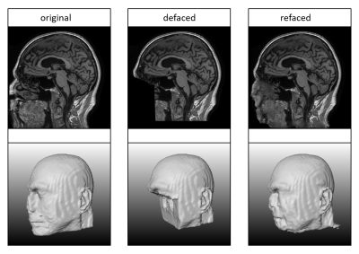 |
Don’t Lose Your Face - Refacing for Improved Morphometry
Till Huelnhagen1,2,3, Mário João Fartaria1,2,3, Ricardo Corredor-Jerez1,2,3, Mazen Fouad A. Wali Mahdi1, Gian Franco Piredda1,2,3, Bénédicte Maréchal1,2,3, Jonas Richiardi1,2, and Tobias Kober1,2,3
1Advanced Clinical Imaging Technology, Siemens Healthcare AG, Lausanne, Switzerland, Lausanne, Switzerland, 2Department of Radiology, Lausanne University Hospital and University of Lausanne, Lausanne, Switzerland, 3LTS5, École Polytechnique Fédérale de Lausanne (EPFL), Lausanne, Switzerland
A growing amount of imaging data is made publicly available. While this is desirable for science and its reproducibility, privacy concerns increase. As the shape of a face can be recovered based on MR images, an increased number of studies remove the face from the data to prevent biometric identification. This defacing can, however, pose a challenge to existing post-processing pipelines e.g. brain volume assessment. This work investigates the impact of regenerating facial structures in defaced images on morphometry in a large cohort using a deep neural network. The results show that refacing can prevent volumetric errors induced by defacing.
|
 Watch the Video