
Oral - Power Pitch Session
Neurovascular Imaging
Session Topic: Neurovascular imaging
Session Sub-Topic: Highlights in Cerebral Lumenography & Vascular Reactivity
Oral - Power Pitch
Neuro
| Thursday Parallel 2 Live Q&A | Thursday, 13 August 2020, 14:20 - 15:05 UTC | Moderators: Henk Mutsaerts |
Session Number: PP-06
 |
1068.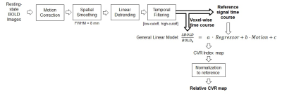 |
Cerebrovascular reactivity mapping using resting-state fMRI: comparison with CO2-inhalation method in 170 controls and 50 Moyamoya patients
Gongkai Liu1, Hanzhang Lu1, Yang Li1, Binu Thomas2, Marco Pinho2, Judy Huang1, Babu G. Welch2, Denise C. Park3, and Peiying Liu1
1Department of Radiology, Johns Hopkins University School of medicine, Baltimore, MD, United States, 2University of Texas Southwestern Medical Center, Dallas, TX, United States, 3The University of Texas at Dallas, Dallas, TX, United States
Cerebral vascular reserve, which indicates the potential of the tissue to receive more blood flow when needed, is desired to evaluate the ischemic risk of brain tissue. However, it is cumbersome to measure vascular reserve using the current methods with Diamox or hypercapnia challenges. Therefore there is a growing interest in using resting-state MRI data to measure cerebrovascular reactivity (CVR). Here, using CO2-inhalation MRI as a gold standard and capitalizing on a large cohort of healthy controls (N=170) and Moyamoya patients (N=50), we sought to identify the optimal strategies for resting-state CVR mapping and establish benchmarks for this new technique.
|
1069.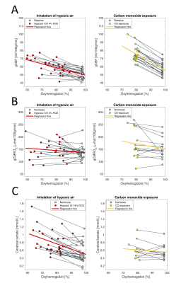 |
Effects from inhalation of hypoxic air and carbon monoxide exposure on human cerebral perfusion, oxygen consumption and lactate production
Mark Bitsch Vestergaard1, Hashmat Ghanizada2, Ulrich Lindberg1, Nanna Arngrim2, Olaf Paulson3, Messoud Ashina2, and Henrik Bo Wiberg Larsson1
1Functional Imaging Unit, Department of Clinical Physiology, Nuclear Medicine and PET, Copenhagen University Hospital Rigshospitalet, Glostrup, Denmark, 2Danish Headache Center, Department of Neurology., Copenhagen University Hospital Rigshospitalet, Glostrup, Denmark, 3Neurobiology Research Unit, Department of Neurology, Copenhagen University Hospital Rigshospitalet, Copenhagen, Denmark
In present study we demonstrate that in healthy humans the cerebral lactate concentration increases during inhalation of hypoxic air but not after exposure to carbon monoxide. This suggests a regulatory mechanism of cerebral glycolytic activity possibly mediated by sensing of arterial oxygen pressure and that the lactate production is not solely a result of hindered oxidative metabolism, at least during non-threatening hypoxic exposure. Phase-contrast mapping and susceptibility-based oximetry were used to acquire global cerebral blood flow and oxygen consumption and MR-spectroscopy was used to measure the lactate concentration in the occipital lope in a total of 51 healthy humans.
|
|
|
1070.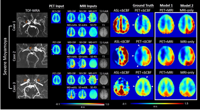 |
Using Deep Learning to Predict PET Cerebrovascular Reserve in Moyamoya Disease from Baseline MRI
David Yen-Ting Chen1,2, Yosuke Ishii1,3, Moss Yize Zhao1, Audrey Peiwen Fan1, and Greg Zaharchuk1
1Radiology, Stanford University, Palo Alto, CA, United States, 2Medical Imaging, Shuang Ho Hospital, Taipei Medical University, New Taipei City, Taiwan, 3Neurosurgery, Tokyo Medical and Dental University, Tokyo, Japan
Cerebrovascular reserve (CVR) is an important hemodynamic parameter for moyamoya disease. Acetazolamide (ACZ) test is often used to measure CVR clinically. However, ACZ is contraindicated in patients with sulfa allergies, severe kidney and liver disease and potentially has severe adverse side effect. Thus, there is a need to assess CVR without pharmacological vasodilation. We utilized a simultaneous [15O]-water PET/MRI dataset to train a convolutional neural network (CNN) to predict CVR. The CNN combined multi-contrast information from baseline perfusion and structural images to predict whole-brain PET-level CVR, with high image quality, quantification accuracy, and diagnostic accuracy for identifying impaired CVR.
|
|
1071.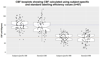 |
Effect of subject-specific labelling efficiency for arterial spin labelling on cerebral blood flow in mild stroke patients
Michael S Stringer1,2, Nithya N Nair3, Una Clancy1,2, Alasadir Morgan1,2, Zahra Shirzadi4,5, Yulu Shi1,2,6, Francesca Chappell1,2, Antoine Vallatos1,2, Maria Valdes Hernandez1,2, Dany Jaime Garcia1,2, Gordon W Blair1,2, Rosalind Brown1,2, Bradley J MacIntosh4,5,
Ian Marshall1,2, Fergus Doubal1,2, Michael J Thrippleton1,2, and Joanna M Wardlaw1,2
1Centre for Clinical Brain Sciences, University of Edinburgh, Edinburgh, United Kingdom, 2UK DRI at the University of Edinburgh, Edinburgh, United Kingdom, 3Centre for Discovery Brain Sciences, University of Edinburgh, Edinburgh, United Kingdom, 4Department of Biomedical Physics, University of Toronto, Toronto, ON, Canada, 5Hurvitz Brain Sciences, Sunnybrook Research Institute, University of Toronto, Toronto, ON, Canada, 6Beijing Tiantan Hospital, Capital Medical University, Beijing, China
Accurate cerebral blood flow (CBF) quantification using arterial spin labelling (ASL) depends on physiological and MR parameters. Labelling efficiency is particularly relevant given it may vary between vascular disease patients. We determined subject-specific labelling efficiency values using phase-contrast MRI scans in a mild stroke cohort. Bland-Altman plots suggested a bias in CBF, with nominal labelling efficiency values underestimating at low and overestimating at high CBF. Using subject-specific, but not nominal, labelling efficiency showed plausible associations between white matter CBF and smoking status, pulse pressure, and age. Subject-specific labelling efficiencies appear to mitigate variance and improve CBF quantification in clinical ASL.
|
|
1072.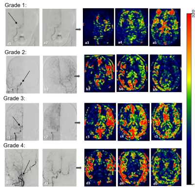 |
Noninvasive Assessment of Cerebral Collaterals with 3D Multi-inversion Time Arterial Spin Labeling in Ischemic Stroke: Comparison with DSA
Hui Wang1, Chuili Kong2, Quanzhi Feng1, Yi Liu1, Yutian Li1, Jinli Li1, Josef Pfeuffer3, Xianchang Zhang4, and Tong Han1
1Radiology, Tianjin Huanhu Hospital, Tianjin, China, 2Radiology, Liaocheng People’s Hospital, Liaocheng, China, 3Siemens Healthcare, Erlangen, Germany, 4MR Collaboration, Siemens Healthcare Ltd, Beijing, China
This study proposed a new method that can directly visualize and assess the collateral status by post-processing multiphase perfusion-weighted images (PWI) generated by multi-inversion time arterial spin labeling (mTI-ASL), and evaluate its performance by comparison with digital subtraction angiography (DSA) in patients with ischemic stroke. Comparison of the results of 28 patients showed that the collateral status assessed by the 3D mTI-ASL grading system was greatly consistent (kappa coefficient k = 0.854) with DSA. This technique is promising for the noninvasive assessment of the collateral status in stroke patients.
|
|
1073.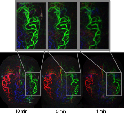 |
High resolution 4D vessel selective angiography in under 5 minutes using a constrained reconstruction
Sophie Schauman1, Thomas W Okell1, and Mark Chiew1
1Wellcome Centre for Integrative Neuroimaging, NDCN, University of Oxford, Oxford, United Kingdom
Arterial spin labeling methods can be used to produce vessel selective angiograms. However, to do this in 3D or 4D is extremely time consuming as many encodings of high spatial (and temporal) resolution images are needed. We propose an optimized acquisition and reconstruction method to create high quality angiogams is five minutes or less. For the acquisition protocol we explore different sampling patterns across encoded images, and for the reconstruction method different ways of constraining the signal temporally.
|
1074.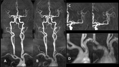 |
Comparison of Spiral and Cartesian k space filling strategies for Time of Flight MR angiography for cervicocerebral arteries – Pilot Study
Ravi Varma Dandu1, Karthick Raj Rajendran2, Rithika Varma Dandu3, Sivakanth Nalubolu4, Kiran Barla1, Narayana Rolla5, and Indrajit Saha6
1Citi Neuro Centre, Hyderabad, India, 2Philips Healthcare, Eindhoven, Netherlands, 3RV College of Engineering, Bengaluru, India, 4Narayana Health City, Bangalore, India, 5Philips Healthcare, Bangalore, India, 6Philips Healthcare, Gurgaon, India
This study compares the performance of Time of Flight MR angiography (ToF-MRA) with spiral k-space filling and ToF-MRA with cartesian filling, for evaluation of the cervicocerebral circulation in 16 healthy volunteers. The imaging protocols were adjusted to give similar coverage and scan times for both techniques. Spiral ToF-MRA showed better visualization of almost all arteries of the cervicocerebral circulation – especially in the small distal intracranial arteries. Artefactual signal drops in segments with slow flow were also fewer with spiral ToF-MRA. Spiral ToF-MRA can potentially evaluate the cervicocerebral arterial system with higher spatial resolution than Cartesian ToF-MRA.
|
|
1075.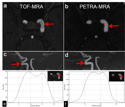 |
Clinical Evaluation of PETRA-MR Angiography in comparison with 3D-TOF-MRA for improved flow dephasing at 3 Tesla
Qing Fu1, Xiao-yong Zhang2, and Ding-xi Liu1
1Department of Radiology, Union Hospital, Tongji Medical College, Huazhong University of Science and Technology, Wuhan, China, 2MR Collaborations, Siemens Healthcare Ltd., Shenzhen, China
This study aimed to demonstrate the image quality and diagnostic performance of subtraction-based pointwise encoding time reduction with radial acquisition (PETRA-MRA) for improved flow dephasing in the intracranial internal carotid artery (ICA) when compared with conventional 3D-TOF-MRA. Our findings showed that image quality and signal homogeneity within the ICA in PETRA-MRA were significantly better than those obtained with TOF-MRA. In conclusion, PETRA-MRA proved to be superior for depicting less flow dephasing artifacts and better image quality in comparison with 3D-TOF-MRA.
|
|
1076.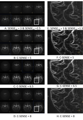 |
Compressed SENSE combined with Keyhole and View-Sharing accelerate Non-contrast enhanced 4D Intracranial MRA based on pCASL
Jilei Zhang1, Weibo Chen1, Jianqing Sun1, Queenie Chan1, Maoxue Wang2, and Bing Zhang2
1Clinical Science, Philips Healthcare, Shanghai, China, 2Drum Tower Hospital, Medical School of Nanjing University, Nanjing, China
The non-contrast enhanced 4D MRA is time-consuming because of the needs to acquire high resolution data at multi-timepoints, which limited its clinical application.In current study, 4D-PACK combined with C-SENSE=6.5 can reduce 23% acquisition time(4 min 42s) for non-contrast enhanced 4D-MRA compared with 4D-PACK with SENSE acceleration, and the excellent acceleration advantage of C-SENSE can improve the clinical application for 4D MRA. the proposed acquisition scheme of 4DMRA with C-SENSE acceleration can be potentially used for evaluating arteriovenous malformation (AVM), arteriovenous fistulas (AVF), moyamoya disease, and stroke patients.
|
|
1077.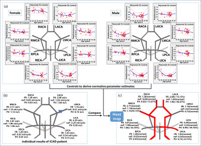 |
Heat Maps of Abnormal Intracranial Hemodynamics in Intracranial Atherosclerotic Disease using 4D Flow MRI
Yue Ma1,2, Maria Aristova1, Sameer Ansari1,3,4, Ann Ragin1, Michael Markl1, and Susanne Schnell5
1Radiology, Northwestern University, Chicago, IL, United States, 2Radiology, Shengjing Hospital of China Medical University, Shenyang, China, 3Neurology, Northwestern University, Chicago, IL, United States, 4Neurosurgery, Northwestern University, Chicago, IL, United States, 5Radiology, University of Greifswald, Chicago, IL, United States
Symptomatic intracranial atherosclerotic disease (ICAD) patients who present with stenosis and hemodynamic abnormalities are at higher risk of recurrent stroke. We propose a methodology that creates patient-specific ‘heat maps’ of abnormal hemodynamic parameters, based on intracranial dual-venc 4D flow MRI. The heat maps were created by detecting and highlighting outlier measurements from 95% confidence interval of normative parameter estimates in healthy controls. Elevated peak velocity (PV) was found in 75% of patients and 58.3% of them with abnormal PV in the uninvolved hemisphere. This novel approach to characterize intracranial hemodynamic impact may allow making patient-specific risk stratification and treatment strategies.
|
|
1078.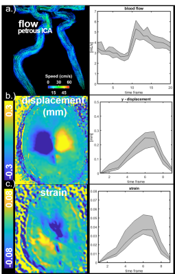 |
Association of Brain Biomechanics and Vascular Dynamics using 4D Flow, MRE and DENSE MRI
Leonardo A Rivera-Rivera1, Grant S Roberts1, Laura B Eisenmenger2, Oliver Wieben1,2, Sterling C Johnson3, and Kevin M Johnson1,2
1Department of Medical Physics, University of Wisconsin - Madison, Madison, WI, United States, 2Department of Radiology, University of Wisconsin - Madison, Madison, WI, United States, 3Department of Medicine, University of Wisconsin - Madison, Madison, WI, United States
The coupling of brain biomechanics and hemodynamics is complex as it includes arterial pressure pulsations, venous and CSF flow, and tissue compliance. Experimental evidence has demonstrated alterations of each the multiple compartments in disease; however, the relationships and coupling between brain biomechanics (e.g. strain and stiffness) and vascular flow dynamics is not well characterized. This study investigates the relationships between brain blood flow, stiffness, and strain using a multi-scale brain imaging platform that includes 4D flow, MRE, and DENSE MRI. Results suggest strong correlations between blood flow, strain, and stiffness and age-related changes in these parameters.
|
|
1079.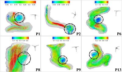 |
Three Dimensional Vortex Identification and Characterization in Small Intracranial Aneurysms based on Sub-millimetric 4D Flow MRI at 7 Tesla
Ang Zhou1, Sean Moen2, Bharathi Jagadeesan2,3,4, and Pierre-Francois Van de Moortele1
1Center for Magnetic Resonance Research, University of Minnesota, Minneapolis, MN, United States, 2Department of Neurosurgery, University of Minnesota, Minneapolis, MN, United States, 3Department of Radiology, University of Minnesota, Minneapolis, MN, United States, 4Department of Neurology, University of Minnesota, Minneapolis, MN, United States
Asymptomatic small intracranial aneurysms affect about 1 in 50 people and are often considered at a low risk of rupture. There are no effective hemodynamic parameters accurately predicting the evolution of small aneurysms. Three dimensional vortex motion is observed in aneurysms which reflects the hemodynamic environment and potentially impact the development of small aneurysms. We propose an approach to describe the three dimensional main vortex motion as a whole inside small aneurysms based on 4D Flow MRI at 7 Tesla. This approach defines the high vortex motion region and gives the direction of the main vortex motion and its center.
|
|
1080.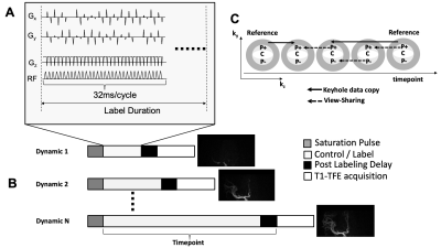 |
Vessel-selective 4D-MRA Using Superselective pCASL Combined with CENTRA-Keyhole (4D-S-PACK) for Intracranial Dural Arteriovenous Fistulas
Osamu Togao1, Akio Hiwatashi2, Makoto Obara3, Michael Helle4, Kazufumi Kikuchi1, Daichi Momosaka1, Yoshitomo Kikuchi1, Tatsuhiro Wada5, Hiroo Murazaki5, and Marc Van Cauteren3
1Department of Clinical Radiology, Graduate School of Medical Sciences, Kyushu University, Fukuoka, Japan, 2Department of Molecular Imaging & Diagnosis, Graduate School of Medical Sciences, Kyushu University, Fukuoka, Japan, 3Philips Japan, Tokyo, Japan, 4Philips Research, Hamburg, Germany, 5Division of Radiology, Department of Medical Technology, Kyushu University Hospital, Fukuoka, Japan
In this study, we demonstrated the ability of 4D-S-PACK (4D-MRA based on superselective pCASL with CENTRA-keyhole and view-sharing) to visualize intracranial DAVFs. 4D-S-PACK enables a time-resolved and vessel-selective angiography within 5 minutes without a use of contrast agents. It was shown that good vessel selectivity for the internal and external carotid arteries was achieved with 4D-S-PACK. 4D-S-PACK enabled accurate identification of feeding arteries. Although the superselective labelling in 4D-S-PACK caused a slight reduction in CNR, compared to full labelling in 4D-PACK, this was acceptable since visualization was well preserved. 4D-PACK can be a non-invasive clinical tool for assessing intracranial DAVFs.
|
 Watch the Video