
Oral - Power Pitch Session
Machine Learning, Imaging Optimization, and Cancer
Session Topic: Machine Learning, Imaging Optimization, and Cancer
Session Sub-Topic: Body Trunk Cancer
Oral - Power Pitch
Body
| Wednesday Parallel 3 Live Q&A | Wednesday, 12 August 2020, 14:30 - 15:15 UTC | Moderators: Oliver Gurney-Champion & Tom Scheenen |
Session Number: PP-10
0829.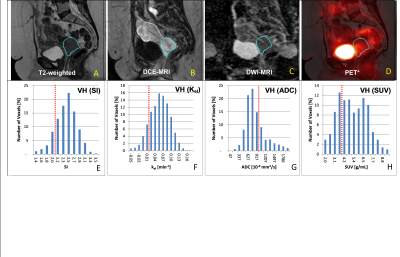 |
Early Prediction of Treatment Response and Mortality in Advanced Cervical Cancer: Temporal Changes of Functional MRI and 18FDG PET/CT Radiomics
Murat Alp Oztek1,2, Stephen R Bowen2, Savannah C Partridge1, Daniel S Hippe1, William T. Yuh1, Aaron S Nelson3, Simon S Lo2, Elaine Y Lee4, Eric Leung5, John C Grecula6, Matthew Harkenrider7, Michael V Knopp6, Wei Wu1,
and Nina A Mayr2
Video Permission Withheld
1Radiology, University of Washington, Seattle, WA, United States, 2Radiation Oncology, University of Washington, Seattle, WA, United States, 3MIM Software, Beachwood, OH, United States, 4Radiology, The University of Hong Kong, Hong Kong, Hong Kong, 5Sunnybrook Health Sciences Centre, University of Toronto, Toronto, ON, Canada, 6Ohio State University, Columbus, OH, United States, 7Loyola University, Chicago, IL, United States
DCE, ADC and 18FDG PET/CT radiomics parameters, obtained simultaneously before, early during and midway during ongoing radiation/chemotherapy correlate with tumor response and particularly mortality, and can serve as early predictors of treatment outcome in advanced cervical cancer. Longitudinal development of functional heterogeneity may be a sensitive measure reflecting responsiveness of individual tumors to a specific cytotoxic treatment regimen. Particularly the persistence of skewness of the dynamic contrast enhancement within the tumor volume predicted cancer mortality. Functional radiomics assessment may help address the unmet need for a patient- and treatment-specific early indicator of tumor responsiveness and survival.
|
|
0830.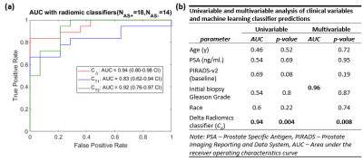 |
Delta Radiomic Features from serial bi-parametric MRI are associated with biopsy upgrading of prostate cancer patients on Active Surveillance
Rakesh Shiradkar1, Ruyuan Zuo1, Amr Mahran2, Lin Li1, Britt Conroy2,3, Lee Ponsky2,3, Sree Harsha Tirumani2,3, and Anant Madabhushi1
1Case Western Reserve University, Cleveland, OH, United States, 2UH Cleveland Medical Center, Cleveland, OH, United States, 3Case Western Reserve University School of Medicine, Cleveland, OH, United States
Serial MRI allows for non-invasive monitoring of prostate cancer patients on Active Surveillance (AS). However, repeat biopsies continue to be defacto standard for AS monitoring due to limitations of MRI. In this study, we sought to compute delta changes in radiomic features on serial bi-parametric MRI and evaluate their associations with biopsy upgrading. We observed that delta radiomic features that quantify underlying spatial, gradient based heterogeneity were associated with biopsy upgrading. On univariable and multivariable analysis with routine clinical variables, we observed that none of the clinical variables were significant while delta radiomic classifier predictions were significant and independently predictive.
|
|
0831.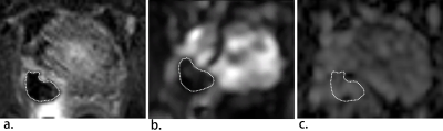 |
T2*-weighted MRI as a non-contrast enhanced method for assessment of focal laser ablation zone extent in prostate cancer thermotherapy
Chongpeng Sun1,2, Shiyang Wang1,3, Aritrick Chatterjee1, Milica Medved1, Scott Eggener4, Gregory S Karczmar1, and Aytekin Oto1
1Radiology, University of Chicago, Chicago, IL, United States, 2Radiology, The First Affiliated Hospital of Guangzhou Medical University, Guangzhou, China, 3Medical Physics, University of Missouri, Columbia, MO, United States, 4Urology, University of Chicago, Chicago, IL, United States
This study evaluated the utility of T2*-weighted (T2*W) MRI for intra-operative identification of ablation zone extent during focal laser ablation (FLA) of prostate cancer. Ablation zone ROI sizes and contrast-to-background ratio (CBR) were calculated on T2*W and apparent diffusion coefficient (ADC) maps and compared to those in the reference standard subtracted contrast-enhanced T1-weighted (sceT1W) images. CBRs on T2*W (TE=32,63 ms) did not differ significantly from those in sceT1W, and ROI sizes in T2*W (TE=63 ms) and sceT1W were well-correlated and differed by only 15%. Therefore, T2*W MRI with long TE visualizes post-procedure ablation zone comparably to contrast-enhanced T1W MRI.
|
|
 |
0832.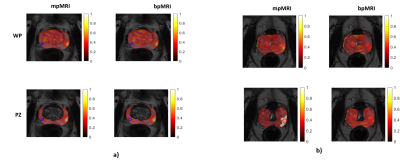 |
Added Value of DCE in Machine Learning-based Tumor Probability Maps for Predicting Clinically Significant Cancer Foci in Pre-biopsy MR images
Gabriel Addio Nketiah1, Léo Pallas2, Adrian L Breto 3, Radka Stoyanova3, Mattijs Elschot 1, and Tone F. Bathen1,4
1Circulation and Medical Imaging, Norwegian University of Science and Technology, Trondheim, Norway, 2Department of Digital Science, CPE Lyon, Lyon, Norway, 3Sylvester Comprehensive Cancer Center, University of Miami, Miami, FL, United States, 4Clinic of Radiology and Nuclear Medicine, St. Olavs University Hospital, Trondheim, Norway
The utility of multiparametric (mp) versus biparametric (bp) MRI protocol in prostate cancer diagnosis has been compared in several large studies, but mainly using manual qualitative evaluation. In this study we employed machine learning models to investigate the added value of DCE (i.e. mpMRI) in predicting significant cancer foci in pre-biopsy MR images. Whereas both protocols had comparable results in the whole prostate and transition zone analyses, we found mpMRI model to be more useful in the peripheral zone, where significant differences (p < 0.05) were found for all performance measures i.e. area under the curve, accuracy, sensitivity and specificity.
|
|
0833.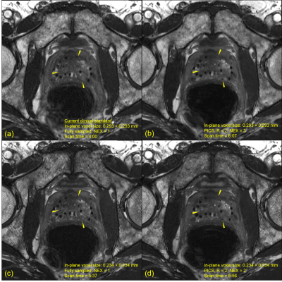 |
Parallel imaging compressed sensing in fully balanced SSFP for prostate brachytherapy MRI without an endorectal coil: a prospective study
Jeremiah W Sanders1, Steven J Frank2, Aradhana M Venkatesan3, Tharakeswara K Bathala3, Chad Tang2, Rajat J Kudchadker4, Teresa L Bruno2, Mark D Pagel5, and Jingfei Ma1
Video Permission Withheld
1Imaging Physics, University of Texas MD Anderson Cancer Center, Houston, TX, United States, 2Radiation Oncology, University of Texas MD Anderson Cancer Center, Houston, TX, United States, 3Diagnostic Radiology, University of Texas MD Anderson Cancer Center, Houston, TX, United States, 4Radiation Physics, University of Texas MD Anderson Cancer Center, Houston, TX, United States, 5Cancer Systems Imaging, University of Texas MD Anderson Cancer Center, Houston, TX, United States
Parallel imaging and compressed sensing (PICS) techniques have demonstrated the ability to accelerate MRI acquisitions in a number of clinical MRI protocols. For postimplant prostate brachytherapy MRI, an endorectal coil (ERC) is currently used to achieve images of sufficient signal-to-noise ratio (SNR) for postimplant quality assessment. Previously we retrospectively demonstrated the feasibility of using PICS to accelerate postimplant prostate brachytherapy MRI. In this work, we prospectively demonstrate that combining PICS with fully balanced steady-state free precession MRI enables high resolution and high SNR images of the prostate without an ERC.
|
|
0834.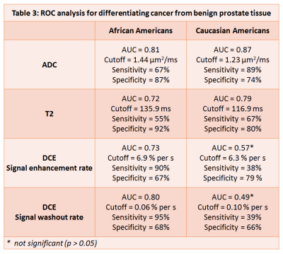 |
Quantitative multi-parametric MRI of the prostate reveals differences between ethnicities
Aritrick Chatterjee1, Xiaobing Fan1, Ambereen Yousuf1, Tatjana Antic2, Gregory Karczmar1, and Aytekin Oto1
1Department of Radiology, University of Chicago, Chicago, IL, United States, 2Department of Pathology, University of Chicago, Chicago, IL, United States
This study investigates whether quantitative MRI of the prostate reveals differences between ethnicities that can affect diagnosis. This study shows that the different ethnicities, specifically AAs and CAs have different quantitative MRI values that affects the utility of MRI in the diagnosis of PCa. Different thresholds are needed for PCa diagnosis for different ethnicities. Despite more high grade lesions in AA, the ADC and T2 for lesions in AA were nominally higher than in CA. DCE-MRI significantly improves differentiation of PCa from benign tissue in AA, due to significantly higher cancer signal enhancement rate in AA compared to CA.
|
 |
0835. |
Amide Proton Transfer Imaging of Rectal Cancer: Baseline values and Feasibility for Predicting Tumor Pathological Features
Lan Zhang1, Xin Li1, Ping Han1, and Fan Yang1
1Radiology, Union Hospital, Tongji Medical College,Huazhong University of Science and Technology, Wuhan, China
We speculate that APT value may be a useful biomarker for assessing rectal pathological characteristics, which could have a potential impact on the clinical therapeutic strategies for patients. We have established a baseline for the APT values in rectal cancer, which was shown significantly correlated with pathologic features (eg. tumor grade and Ki-67 index).
|
0836. |
3D Amide proton transfer weighted imaging in predicting Ki-67 proliferation state of rectal adenocarcinoma
Ling Li1, Yingjie Mei2, Zhaoxian Yan3, Jieping Feng3, Bo Liu3, and Xian Liu3
1Guangzhou University of Chinese Medicine; The second Affiliated hospital of Guangzhou University of Chinese Medicine;, Guangzhou, China, 2Clinical Science,Philips Healthcare;, Shanghai, China, 3Radiology, The second Affiliated hospital of Guangzhou University of Chinese Medicine, Guangzhou, China
The Accurate preoperative staging and grading are significant on the treatment of protocol selection rectal adenocarcinoma. As a new molecular MR imaging technique, APT imaging could provide information that correlates with tumor cell proliferation. In this study, the capability of APT in differentiating WHO grades and pathologic stages of rectal adenocarcinoma was investigated and compared with DWI. The results show that APT value have a significantly positive correlation with the stage and grade of the rectal adenocarcinoma, and the prognosis factor Ki67.
|
|
0837. |
CT and MRI based multi-modal and multi-parametric radiomics pre-operative prediction of perineural invasion in patients with rectal cancer
Xiangchun Liu1, Yu Fu1, Yan Guo2, Kan He1, Qi Yang1, and Huimao Zhang1
1The First Hospital of Jilin University, changchun, China, 2GE Healthcare, beijing, China
Perineural invasion (PNI), defined by tumor invasion of nervous structures and nerve sheaths, which is thought an independent predictor of outcome in rectal cancer. However, for a radiologist, neither MRI nor CT can reliably evaluate PNI. Radiomics is an emerging and effective method for quantitative analysis and prediction using big data of medical imaging. Therefore, this study aims to develop and validate a radiomics prediction model based on MRI and CT for the preoperative prediction of PNI in rectal cancer. The results indicated that excellent diagnostic performance can be yielded with such multi-modal radiomics.
|
|
0838.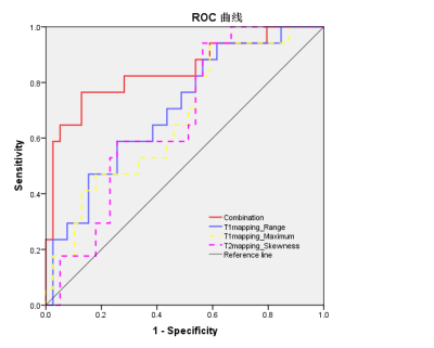 |
Synthetic magnetic resonance imaging-derived histogram metrics for prediction of lymph node metastasis in rectal cancer
Li Zhao1, Hongmei Zhang1, Lizhi Xie 2, and Xinming Zhao1
1Diagnostic Radiology, Cancer Hospital, Chinese Academy of Medical Sciences and Peking Union Medical College, Beijing, China, 2MR Research, GE Healthcare, Beijing, China
The aim of this study was to evaluate the feasibility of quantitative synthetic magnetic resonance imaging (SyMRI)-derived histogram of the primary tumor for predicting the regional lymph node (LN) metastasis in patients with rectal cancer (RC). Our study indicated that histogram parameters of primary tumor on T1 mapping and T2 mapping were associated with regional LN status in RC. Moreover, the combination of the quantitative SyMRI parameters, pathological extramural venous invasion (EMVI), and maximum tumor diameter may significantly improve the predictive performance of LN metastasis.
|
|
0839.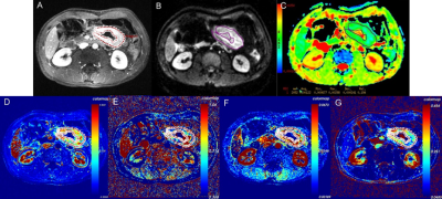 |
Value of quantitative DCE and DW-MRI in predicting extramural venous invasion in locally advanced gastric cancer and the prognostic significance
Yongjian Zhu1, Ying Li1, Jun Jiang1, Yutao Zhou1, Liming Jiang1, Liyan Xue2, and Lizhi Xie3
1Department of Imaging Diagnosis, National Cancer Center/National Clinical Research Center for Cancer/Cancer Hospital, Chinese Academy of Medical Sciences and Peking Union Medical College, Beijing, China, 2Department of Pathology, National Cancer Center/National Clinical Research Center for Cancer/Cancer Hospital, Chinese Academy of Medical Sciences and Peking Union Medical College, Beijing, China, 3GE healthcare, China, Beijing, China
Extramural vascular invasion (EMVI) has been found as an independent risk factor for recurrence and distant metastasis in patients with gastric cancer. Dynamic contrast-enhanced magnetic resonance imaging (DCE-MRI) and diffusion weighted imaging (DWI) has been applied in diagnosis of different cancers. In this study, we research into the value of DCE-MRI parameters and ADC in predicting EMVI and the prognostic significant. It was found that Ktrans, Ve and ADC are independent predictors of pathological EMVI in LAGC, and MRI-predicted EMVI (mrEMVI) confirmed to be a poor prognosis predictor in terms of 2-year recurrence-free survival (RFS).
|
|
0840.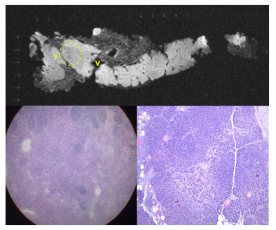 |
Ex Vivo MRI for Direct Radiologic-Histologic Correlation in the Pancreas: Protocol Development with Cadaver Specimens
Alexandra W. Acher1, TJ Colgan1, Yuxin Zhang1, Krisztian Kovacs2, Jitka Starekova1, Victoria Rendell3, Daniel E. Abbott3, Erin Brooks2, Rashmi Agni2, Emily Winslow4, and Scott B. Reeder5
1Department of Radiology, University of Wisconsin, Madison, WI, United States, 2Department of Pathology, University of Wisconsin, Madison, WI, United States, 3Department of Surgery, University of Wisconsin, Madison, WI, United States, 4Department of Surgery, Georgetown University, Washington DC, MD, United States, 5Department of Radiology and Department of Medical Physics, University of Wisconsin, Madison, WI, United States
The purpose of this study was to develop a protocol for use of a previously validated radiologic-histologic correlation device to evaluate the pancreas with ex vivo MRI. Precise radiologic-histologic correlation of pancreatic anatomy was achieved in cadaveric pancreas specimens. The final protocol will be applied to co-localize pancreas cancer margins radiologically and histologically, as well as nodal burden in pancreaticoduodenectomy specimens.
|
|
0841.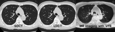 |
Pulmonary MR Imaging with Ultra-Short TE: Capability of Nodule Detection and Lung RADS Classification for Lung Cancer Screening
Yoshiharu Ohno1,2, Masao Yui3, Daisuke Takenaka4, Yoshimori Kassai3, Kazuhiro Murayama1, and Takeshi Yoshikawa2
1Radiology, Fujita Health University School of Medicine, Toyoake, Japan, 2Radiology, Kobe University Graduate School of Medicine, Kobe, Japan, 3Canon Medical Systems Corporation, Otawara, Japan, 4Diagnostic Radiology, Hyogo Cancer Center, Akashi, Japan
No report has been found to compare the capability of pulmonary MR imaging with UTE for nodule detection and Lung-RADS classification as compared with low-dose CT (LDCT) and standard-dose CT (SDCT). We hypothesized that pulmonary MR imaging with UTE has a similar potential to detect pulmonary nodules and evaluate Lung-RADS classification as well as LDCT and SDCT. The purpose of this study was to compare the capability of pulmonary MR imaging with UTE for lung nodule detection and evaluation of Lung-RADS classification with LDCT and SDCT.
|
|
0842.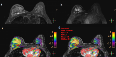 |
A Pilot Evaluation of Amide Proton Transfer-Weighted (APTw) MR Imaging in Characteristics and Diagnosis of Premenopausal Breast Tumors
Nan Zhang1, Qingwei Song1, Lina Zhang1, Ailian Liu1, Haonan Zhang1, Yu Song1, Liangjie Lin2, Jiazheng Wang2, and Zhiwei Shen2
1The First Affiliated Hospital of Dalian Medical University, Dalian, China, 2Philips Healthcare, Beijing, China
Amide proton transfer (APT) imaging is based on the chemical exchange between free bulk water protons and the amide protons (-NH) of endogenous mobile proteins and peptides in tissue. Previous studies have shown that APT-weighted (APTw) MRI could noninvasively identify and differentiate tumors in brain, head and neck etc. This study aims to explore the feasibility of APTw-MRI in characteristics and discrimination of premenopausal malignant and benign breast tumors.The results show that the APTw MR imaging efficiency of diagnosis of premenopausal breast tumors were 0.904.
|
 Watch the Video