
Oral - Power Pitch Session
Hepatobiliary & Pancreas
Session Topic: Hepatobiliary & Pancreas
Session Sub-Topic: Pancreaticohepatobiliary
Oral - Power Pitch
Body
| Monday Parallel 3 Live Q&A | Monday, 10 August 2020, 15:15 - 16:00 UTC | Moderators: Manil Chouhan & Vicente Martinez Sanjuan |
Session Number: PP-11
 |
0325.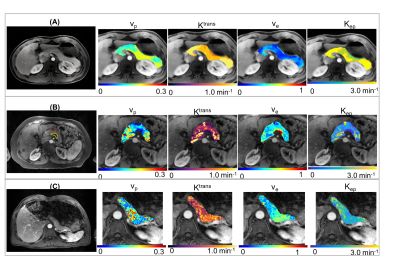 |
Six-Dimensional Quantitative DCE MR Multitasking in the Characterization of Pancreatic Ductal Adenocarcinoma Versus Chronic Pancreatitis
Nan Wang1,2, Srinivas Gaddam3, Lixia Wang1,4, Yibin Xie1, Zhaoyang Fan1, Simon Lo3, Stephen Pandol3, Anthony G Christodoulou1, and Debiao Li1
1Biomedical Imaging Research Institute, Cedars-Sinai Medical Center, Los Angeles, CA, United States, 2Bioengineering, University of California, Los Angeles, Los Angeles, CA, United States, 3Division of Digestive and Liver Diseases, Cedars-Sinai Medical Center, Los Angeles, CA, United States, 4Chaoyang Hospital, Beijing, China
The differentiation of pancreatic ductal adenocarcinoma (PDAC) and chronic pancreatitis (CP) is crucial to the diagnosis and prognosis of PDAC. DCE MRI serves as a promising imaging tool, but still faces several technical challenges. In this work, we evaluated the characterization of PDAC versus CP using the proposed Multitasking DCE technique, which enables free-breathing acquisition, 3D whole-abdomen coverage, high temporal resolution at 500ms, and dynamic T1 mapping throughout all DCE phases. In vivo studies on 16 healthy volunteers, 14 PDAC patients, and 8 CP patients demonstrated the capability of Multitasking DCE in differentiating normal pancreas, PDAC, and CP.
|
0326.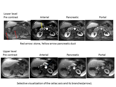 |
Free Breathing Dynamic Contrast Enhanced MR Imaging of the Hepatopancreatobiliary lesions with improved 3D Stack-of-Stars k-space trajectory
Takayuki Masui1, Motoyuki Katayama1, Yuji Iwadate2, Naoyuki Takei2, Mitsuharu Miyoshi2, Masako Sasaki1, Takahiro Yamada1, Ty Cashen3, Sagar Mandava4, and Kang Wang5
1Radiology, Seirei Hamamatsu General Hospital, Hamamatsu, Japan, 2Global MR Applications and Workflow, GE Healthcare, Hino, Japan, 3Global MR Applications and Workflow, GE Healthcare, Madison, WI, United States, 4GE Healthcare, Tucson, AZ, United States, 5GE Healthcare, Waukesha, WI, United States
The feasibility of dynamic Gd-contrast study for evaluation of hepatopancreatobiliary lesions under free breathing was demonstrated. Superb image quality with high temporal resolutions could be obtained using a stack-of-stars k-space trajectory with golden angle ordering a CG-SENSE algorithm that supports parallel imaging and soft-gating for accelerated motion robust imaging. Selective recognition of vasculatures and lesions in the liver and pancreas can be made with this technique, which may be equivalent to fast breath-hold dynamic contrast image in young and old aged population.
|
|
0327.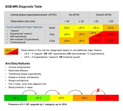 |
Modifying LI-RADS on gadoxetate disodium-enhanced MRI in a prospective cohort: toward improving simplicity and sensitivity
Hanyu Jiang1,2, Bin Song1, and Mustafa Shadi Rifaat Bashir2
1Department of Radiology, West China Hospital, Sichuan University, Chengdu, China, 2Department of Radiology, Duke University Medical Center, Durham, NC, United States
We aimed to develop a modified Liver Imaging Reporting and Data System (mLI-RADS) with comparisons against original LI-RADS version 2018 (v2018) for diagnosing hepatocellular carcinoma (HCC) on gadoxetate disodium-enhanced magnetic resonance imaging (EOB-MRI). 1002 hepatic observations in 272 consecutive at-risk patients were prospectively included. Ancillary features were assessed based on inter-rater agreement, prevalence, diagnostic accuracy, and added value to the major features. Compared with the original LI-RADS v2018, mLI-RADS demonstrated superior simplicity, sensitivity and accuracy without substantial loss of specificity; hence should be the preferred diagnostic criteria for HCC in high-risk patients on EOB-MRI.
|
|
0328.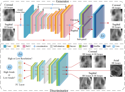 |
Super-resolution Generative Adversarial Network for improving malignancy characterization of hepatocellular carcinoma
Wu Zhou1, Yunling Li1, Hui Huang1, Yaoqin Xie2, Lijuan Zhang2, and Guangyi Wang3
1School of Medical Information Engineering, Guangzhou University of Chinese Medicine, Guangzhou, China, 2Shenzhen Institutes of Advanced Technology, Chinese Academy of Sciences, Shenzhen, China, 3Department of Radiology, Guangdong General Hospital, Guangzhou, China
Deep feature derived from data-driven learning has consistently shown to outperform conventional texture features for lesion characterization. However, due to the slice thickness of medical imaging, through-plane has worse resolution than in-plane resolution. Therefore, the performance of deep feature extracted from the through plane slices may be worse, and their contributions to the final characterization may also be very limited. We propose an end-to-end super-resolution and self-attention framework based on generative adversarial network (GAN), in which the through-plane slices with low resolution are enhanced by learning the in-plane slices with high resolution to improve the performance of lesion characterization.
|
|
|
0329.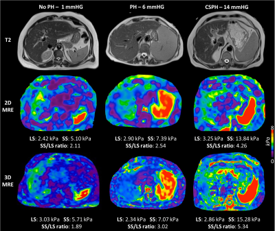 |
Utility of magnetic resonance elastography and ultrasound shear wave elastography for assessment of portal hypertension
Paul Kennedy1,2, Octavia Bane1,2, Stefanie Hectors1,2,3, Daniel Stocker1,2, Bradley D Bolster Jr. 4, Scott Friedman5, Thomas Schiano6, Isabel M Fiel7, Swan Thung7, Aaron Fischman2, and Bachir Taouli1,2
1BioMedical Engineering and Imaging Institute, Icahn School of Medicine at Mount Sinai, New York, NY, United States, 2Department of Diagnostic, Molecular and Interventional Radiology, Icahn School of Medicine at Mount Sinai, New York, NY, United States, 3Department of Radiology, Weill Cornell Medicine, New York, NY, United States, 4Siemens Medical Solutions USA, Inc., Salt Lake City, UT, United States, 5Division of Liver Diseases, Icahn School of Medicine at Mount Sinai, New York, NY, United States, 6Department of Medicine, Icahn School of Medicine at Mount Sinai, New York, NY, United States, 7Department of Pathology, Icahn School of Medicine at Mount Sinai, New York, NY, United States
In this study we investigate the ability of MR elastography (MRE) and ultrasound shear wave elastography (SWE) to assess portal hypertension (PH) severity in patients with liver disease and hepatic venous pressure gradient (HVPG) measurement. 3D MRE spleen stiffness correlated with HVPG. 2D and 3D MRE of the spleen were significantly higher in patients with clinically significant PH (CSPH, HVPG>10mmHg) than those with no PH/PH (HVPG>5mmHg). 3D MRE spleen stiffness was significantly elevated in PH/CSPH patients compared to those with no PH and was an excellent predictor of CSPH. MRE spleen stiffness appears sensitive to hemodynamic changes associated with PH.
|
|
0330.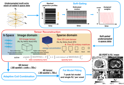 |
Rapid Free-Breathing Volumetric Liver Fat and R2* Quantification using Soft-Gating and Sparsity-Promoting Tensor Reconstruction
Shu-Fu Shih1,2, Tess Armstrong1, Sevgi Gokce Kafali1,2, Xiaodong Zhong3, Kara L. Calkins4, and Holden H. Wu1,2
1Radiological Sciences, University of California, Los Angeles, Los Angeles, CA, United States, 2Bioengineering, University of California, Los Angeles, Los Angeles, CA, United States, 3Siemens Healthcare, Los Angeles, CA, United States, 4Pediatrics, University of California, Los Angeles, Los Angeles, CA, United States
MRI quantification of hepatic proton-density fat fraction (PDFF) and R2* enables non-invasive diagnosis and staging of non-alcoholic fatty liver disease (NAFLD) and liver iron overload, respectively. A recent 3D stack-of-radial technique enables free-breathing quantification, but requires scans of 2-4 minutes and motion may affect R2* accuracy. In this work, we propose an improved free-breathing stack-of-radial technique that combines soft-gating and a sparsity-promoting tensor reconstruction to compensate for motion effects and accelerate the scan to 31 seconds. Data from adult and pediatric NAFLD patients demonstrate good agreement of PDFF and R2* between the proposed method and the conventional breath-held Cartesian scan.
|
0331.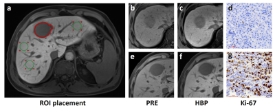 |
Preoperative prediction of HCC with highly aggressive characteristics using quantitative parameters derived from hepatobiliary phase
Zheng Ye1, Yi Wei1, Jie Chen1, and Bin Song1
1West China Hospital, Sichuan University, Chengdu, China
Hepatocellular carcinoma (HCC) with highly aggressive characteristics is usually actively proliferated and easily relapse, thereby requiring adjuvant therapies before surgery, like preoperative TACE, to improve the patients’ prognosis. Ki-67 labeling index (LI) was reported to be highly correlated with aggressive propensity of HCC, and thus could affect the treatment response of the tumor and prognosis directly. Although most of HCC presented hypointensity on hepatobiliary phase (HBP), the absolute signal intensity and relative contrast enhancement ratio are not the same. In this study, we prospectively investigate the usefulness of HBP quantitative parameters for preoperative prediction of aggressiveness in HCC patients.
|
|
0332.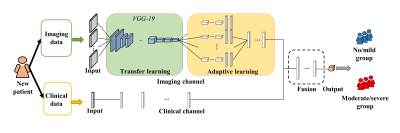 |
A Deep Transfer Learning Model for Liver Stiffness Classification using Clinical and T2-Weighted MRI Data
Hailong Li1,2, Lili He1,2,3, Jonathan Dudley2,4, Thomas Maloney2,4, Elanchezhian Somasundaram4, Samuel L. Brady4,5, Nehal A. Parikh 1,3, and Jonathan R. Dillman2,4,5
1The Perinatal Institute and Section of Neonatology, Perinatal and Pulmonary Biology, Cincinnati Children's Hospital Medical Center, Cincinnati, OH, United States, 2Imaging Research Center, Cincinnati Children's Hospital Medical Center, Cincinnati, OH, United States, 3Department of Pediatrics, University of Cincinnati College of Medicine, Cincinnati, OH, United States, 4Department of Radiology, Cincinnati Children's Hospital Medical Center, Cincinnati, OH, United States, 5Department of Radiology, University of Cincinnati College of Medicine, Cincinnati, OH, United States
Detection and monitoring of chronic liver diseases is typically assessed using a combination of clinical history, physical examination, laboratory testing, biopsy with histopathologic assessment, and imaging. The aim of this study is to develop a deep transfer learning model (DeepLiverNet) to categorically classify the severity of liver stiffening (no/mild vs. moderate/severe) using both anatomic T2-weighted MR images and clinical data. The DeepLiverNet model achieved accuracies of 88.0% and 80.0% on the risk stratification of liver stiffness in internal and external validation datasets, respectively. This demonstrates that a deep learning model may provide a means for stratifying liver stiffness without elastography.
|
|
0333.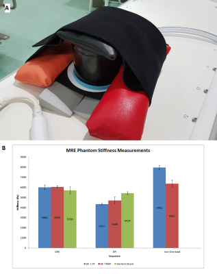 |
Multi-vendor Phantom and Intra-individual Comparison of Liver Stiffness Using Various MR Elastography Sequences at 1.5T & 3T
Justin Yu1, Anshuman Panda1, Kelly Tung-Smith1, Robert Nelson2, Akira Kawashima1, Ming Yang1, Chen Lin3, Aiden McGirr1, Sophia Fasani1, Kristina Flicek1, Sukhdeep Singh1, and Alvin Silva1
1Radiology, Mayo Clinic Arizona, Phoenix, AZ, United States, 2National Institutes of Health, Phoenix, AZ, United States, 3Mayo Clinic Florida, Jacksonville, FL, United States
The variability of stiffness data from GRE and SE EPI MRE sequences is tested on phantoms and in-vivo on 1.5T and 3T MRI scanners from two vendors. Large variability was observed in the phantom measurements for the GRE and EPI sequences on one of the vendor’s scanner, ranging from 20-33% difference. Stiffness measurements were very similar (within 10%) between sequences on the other vendor’s scanner. Similar results were found in several clinical subjects who had GRE and SE EPI sequences performed.
|
|
0334.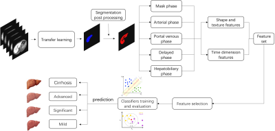 |
Imaging-based Hepatic fibrosis staging of patients with hepatitis B: a radiomics analysis study on Gd-EOB-DTPA-enhanced MRI
Rencheng Zheng1, Tian Qiu2, Nannan Shi2, Yuxin Shi2, Weibo Chen3, Chengyan Wang4, and He Wang4,5
1Fudan University, Shanghai, China, 2ShanghaiPublic Health Clinical Center, Shanghai, China, 3Philips Healthcare, Shanghai, China, 4Human Phenome Institute, Fudan University, Shanghai, China, 5Institute of Science and Technology for Brain-Inspired Intelligence, Fudan University, Shanghai, China
This study proposed an automatic hepatic fibrosis staging model based on transfer learning segmentation and radiomics analysis for hepatitis B patients. The automatic liver ROI extarction Time dimension features of multi DCE phases were included in feature set which played an important role in the classification. The proposed model exhibited a superior performance in significant fibrosis, advanced fibrosis and cirrhosis classification.
|
|
0335.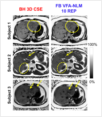 |
Motion-Robust, Free-Breathing, High-SNR Liver Fat Quantification Using a Variable Flip Angle Approach and Motion-Corrected Averaging
Jitka Starekova1, Ruiyang Zhao1,2, Timothy J Colgan1, Kevin M Johnson1,2, Jennifer L Rehm3, Scott B Reeder1,2,4,5,6, and Diego Hernando1,2
1Department of Radiology, University of Wisconsin, Madison, Madison, WI, United States, 2Department of Medical Physics, University of Wisconsin, Madison, Madison, WI, United States, 3Department of Pediatrics, University of Wisconsin, Madison, Madison, WI, United States, 4Department of Biomedical Engineering, University of Wisconsin, Madison, Madison, WI, United States, 5Department of Medicine, University of Wisconsin, Madison, Madison, WI, United States, 6Department of Emergency Medicine, University of Wisconsin, Madison, Madison, WI, United States
Chemical shift-encoded (CSE)-MRI enables accurate and precise quantification of proton density fat-fraction (PDFF) in the liver. Widely used 3D multi-echo spoiled gradient echo (SGRE) CSE-MRI requires reliable breath-holding to avoid motion-related artifacts. This is a major limitation for children, the elderly, and sick patients. Free-breathing 2D sequential CSE-MRI is motion-robust, however, suffers from low signal-to-noise-ratio (SNR). To overcome these limitations, we combined variable flip angle (VFA) 2D acquisitions and nonlocal means (NLM) motion-corrected averaging. In this prospective study, free-breathing multi-repetition VFA-NLM demonstrated high SNR and reduced artifacts compared to the conventional 3D-SGRE, while preserving accuracy of PDFF quantification.
|
|
0336.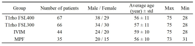 |
A study of sensitivity of quantitative MRI measurements to the presence of iron in the liver
Yurui Qian1, Jian Hou1, Yixiang Wang1, Vincent Wong2, Queenie Chan3, Weibo Chen4, Min Deng1, Franklin Au1, Anthony Chan5, Winnie Chu1, and Weitian Chen1
1Department of Imaging and Interventional Radiology, Chinese University of Hong Kong, Hong Kong, Hong Kong, 2Department of Medicine & Therapeutics, Chinese University of Hong Kong, Hong Kong, Hong Kong, 3Philips Healthcare, Hong Kong, Hong Kong, 4Philips Healthcare, Shanghai, China, 5Department of Anatomical and Cellular Pathology, Chinese University of Hong Kong, Hong Kong, Hong Kong
MRI is widely used as a non-invasive method to diagnose and monitor liver diseases. For certain quantitative MRI techniques, liver iron content may affect the measurement. In this work, we investigated the influence of liver iron content on several quantitative MRI methods, including macromolecular proton fraction, T1rho and intravoxel incoherent motion.
|
|
0337.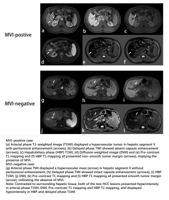 |
Radiomic Analysis Based on Diverse Volumetric Interests Predicts Microvascular Invasion in Solitary Hepatocellular Carcinoma
Huanhuan Chong1, Li Yang1, Yangli Yu1, Dijia Wu2, Chun Yang1, and Mengsu Zeng1
1Department of Radiology, Shanghai Institute of Medical Imaging, Zhongshan Hospital of Fudan University, Shanghai, China, 2Shanghai United Imaging Intelligence Co., Ltd, Shanghai, China
This study aims to construct a preoperative MVI prediction model in solitary HCC derived from gadoxetic acid-enhanced magnetic resonance imaging, and to further investigate its latent association with clinical indexes, imaging features and radiomics signatures based on diverse sequences and volumetric interests (VOIs) of tumor. The conclusion indicated that serum α-fetoprotein, total bilirubin, higher value of tumor margin smoothness (prefer to non-smooth margin), non-intact capsule enhancement and peritumoral enhancement are independent and significant predictors for MVI, and the final nomogram incorporating clinical, imaging and the optimal radiomics model based on VOI_entire_5mm_10mm_liver achieves satisfactory prediction for MVI in solitary HCC.
|
|
0338.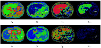 |
Evaluation of liver function by using Hepatocyte fraction of Gd-EOB-DTPA-Enhanced MRI based on MELD score
Mao-Tong LIU1, Xue-Qin ZHANG1, Jian LU1, and Wei-Bo CHEN2
1Third Affiliated Hospital of Nantong University & Nantong Third People's Hospital, Nan Tong, China, 2Philips Healthcare Shanghai, China, Shang Hai, China
The purpose of this study was to identify whether hepatocyte fraction that based on Gadoxetic Acid–enhanced MRI is useful for the assessment of liver function. Firstly, T1 mapping imaging was performed before and 20 minutes after Gd-EOB-DTPA administration, The following parameters are then obtained from the images: pre- and postcontrast T1 values of the liver (T1pre and T1post), increase in the T1 relaxation rate (Δ R1), rate of the decrease of the T1 relaxation time (Δ T1), hepatocyte fraction (HeF), and uptake coefficient (K). Our study showed that hepatocyte fraction can be used to evaluate the liver function of patients with hepatitis B cirrhosis, K value has high diagnostic efficiency.
|
 Watch the Video