
Oral - Power Pitch Session
Machine Learning, Imaging Optimization, and Cancer
Session Topic: Machine Learning, Imaging Optimization, and Cancer
Session Sub-Topic: Image Optimization & Innovation
Oral - Power Pitch
Body
| Wednesday Parallel 3 Live Q&A | Wednesday, 12 August 2020, 14:30 - 15:15 UTC | Moderators: Rebecca Rachow-Pener |
Session Number: PP-12
 |
0816.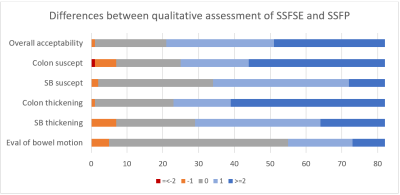 |
Cine SSFSE for reduced susceptibility artifact and increased diagnostic accuracy in MR enterography
Peter Wei1, Anugayathri Jawahar1, Daniel V. Litwiller2, and Andreas M. Loening1
1Department of Radiology, Stanford University, Stanford, CA, United States, 2Global MR applications and Workflow, GE Healthcare, New York, NY, United States
A steady-state free precession (SSFP) sequence is used in many MR enterography (MRE) protocols for acquiring cine images to assess bowel motility, inflammation, and strictures. However, SSFP suffers from susceptibility and banding artifacts that become more significant at high field strengths. In this IRB approved retrospective study, we compared a cine SSFP sequence to a cine T2-weighted single-shot fast spin echo (SSFSE) sequence in 41 patients. We found SSFSE demonstrated significantly superior subjective assessments of image quality, improved diagnostic performance compared to cine SSFP, and successfully mitigated SSFP artifacts that can otherwise limit the exam.
|
|
0817.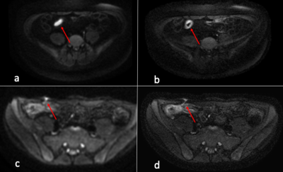 |
The Application of High-resolution Multi-shot DWI with MUSE reconstruction in the Diagnosis of Active Inflammation in Crohn's Disease
Guangtao Chen1, Hing-Chiu Chang1, and Keith Wan-Hang Chiu1
1The Department of Diagnostic Radiology, The University of Hong Kong, Hong Kong, China
Diffusion-weighted imaging(DWI) has been shown to be useful in evaluation of active bowel inflammation. Single-shot diffusion-weighted echo-planar imaging (ssDW-EPI) was commonly used in previous studies. However, ssDW-EPI suffers from geometric distortion and low spatial resolution. Moreover, the data acquisition of ssDW-EPI in bowel region become more difficult due to serve off-resonance effect. A recently developed multiplexed sensitivity-encoding (MUSE) framework can produce multi-shot DW-EPI (msDW-EPI) data with improved spatial resolution and reduced distortions. In this study, we demonstrated that the higher resolution and better overall image quality of msDW-EPI using MUSE framework potentially increased the accuracy in diagnosing active bowel inflammation.
|
0818.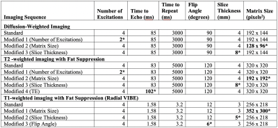 |
Robustness of Texture Features on 3 Tesla Liver MRI.
Vinay Prabhu1, Nicolas Gillingham1, Mary T. Bruno1, James Babb1, Henry Rusinek1, and Hersh Chandarana1
1Radiology, NYU Langone Health, New York, NY, United States
We studied the robustness of liver MRI texture features by scanning five healthy volunteers at 3T, first using standard institutional acquisition parameters, and then introducing slight variation in acquisition parameters. Our results demonstrate that a number of texture features were not robust to acquisition parameter changes.
|
|
0819.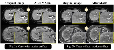 |
Erasing artifacts from arterial phase MRI: Motion Artifact Reduction using a Convolutional network (MARC)
Shinya Kojima1,2, Daiki Tamada 2, Tetsuya Wakayama 3, Shintaro Ichikawa 2, Hiroyuki Morisaka 4, Shigeru Suzuki 1, and Utaroh Motosugi 2
1Department of Radiology, Tokyo Women’s Medical University Medical Center East, Arakawa, Japan, 2Department of Radiology, University of Yamanashi, Yamanashi, Japan, 3MR Collaboration and Development, GE Healthcare, Hino, Japan, 4Department of Radiology, Saitama Medical University International Medical Center, Saitama, Japan
Motion artifact by irregular respiration disturbs accurate diagnosis in dynamic contrast-enhanced MRI of the liver. We developed a motion artifact reduction algorithm using a convolutional network (MARC). The training was performed using U-net with the arterial phase images with and without simulated artifacts. For verifying the ability of MARC algorithm, contrast-to-noise ratio measurement and visual assessment were performed in 120 cases. The image quality of arterial phase images with motion artifacts were significantly improved after applying MARC algorithm, while no particular difference was observed in the images without motion artifacts. MARC provides motion artifacts reduction without variation of image contrast.
|
|
0820.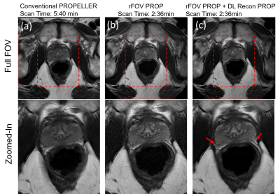 |
High Resolution T2W imaging using Deep Learning Reconstruction and Reduced Field-of-View PROPELLER
Xinzeng Wang1, Daniel Litwiller2, Marc Lebel3, Ali Ersoz4, Lloyd Estkowski4, Jason Stafford5, and Ersin Bayram6
1GE Healthcare, Houston, TX, United States, 2Global MR Applications & Workflow, GE Healthcare, New York, NY, United States, 3Global MR Applications & Workflow, GE Healthcare, Calgary, AB, Canada, 4Global MR Applications & Workflow, GE Healthcare, Waukesha, WI, United States, 5Department of Imaging Physics, MD Anderson Cancer Center, Houston, TX, United States, 6Global MR Applications & Workflow, GE Healthcare, Houston, TX, United States
T2W FSE-PROPELLER is robust to susceptibility artifacts and bulk motion, but requires longer acquisition times compared to conventional FSE methods. Recently, a reduced Field-Of-View PROPELLER sequence using rotating outer volume suppression method has been proposed and optimized to reduce the scan time for small FOV and high-resolution T2W imaging. However, image SNR is comparatively lower compared to the conventional PROPELLER with phase oversampling. In this work, a deep learning based PROPELLER reconstruction method was used to improve the SNR and image quality of the reduced Field-Of-View PROPELLER.
|
|
0821.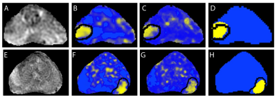 |
End-to-end Deep Learning Strategy To Segment Prostate Cancer From Multi-parametric MR Images
David Hoar1, Peter Lee2, Alessandro Guida3, Steven Patterson3, Chris Bowen3,4, Jennifer Merrimen5, Cheng Wang5, Ricardo Rendon6, Steven Beyea3,4, and Sharon Elizabeth Clarke3,4
1Department of Electrical and Computer Engineering, Dalhousie University, Halifax, NS, Canada, 2Faculty of Computer Science, Dalhousie University, Halifax, NS, Canada, 3Biomedical Translational Imaging Centre, Nova Scotia Health Authority and IWK Health Centre, Halifax, NS, Canada, 4Diagnostic Radiology, Dalhousie University, Halifax, NS, Canada, 5Pathology, Dalhousie University, Halifax, NS, Canada, 6Urology, Dalhousie University, Halifax, NS, Canada
The purpose of this study was to develop a convolutional neural network (CNN) for dense prediction of prostate cancer using mp-MRI datasets. Baseline CNN outperformed logistic regression and random forest models. Transfer learning and unsupervised pre-training did not significantly improve CNN performance; however, test-time augmentation resulted in significantly higher F1 scores over both baseline CNN and CNN plus either of transfer learning or unsupervised pre-training. The best performing model was CNN with transfer learning and test-time augmentation (F1 score of 0.59, AUPRC of 0.61 and AUROC of 0.93).
|
|
0822.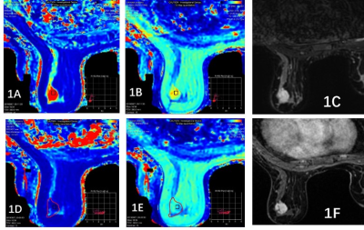 |
T1 and T2 relaxation time in synthetic MRI for differentiating benign and malignant breast lesions
Shi yun SUN1, Zhuo lin Li1, Ying ying Ding1, Yi fan Liu1, Dong xue ZHANG1, Li sha NIE2, Ke XUE1, and Dian Ke DU1
1Radiology, Yunnan Cancer Hospital,The Third Affiliated Hospital of Kunming Medical University, Kunming, China, 2GE Healthcare, MR Research China, Beijing, China, China
It is reported that dynamic contrast imaging and T2 relaxation time can be used to differentiate benign and malignant breast lesions. However, few researches have investigated T1 and T2 relaxation time changes before and after contrast injection. But it's important for the diagnosis of breast diseases. Thus, the study aims to utilize the T1 and T2mapping in synthetic MR to differentiate benign and malignant lesions. Our results demonstrated that T1 and T2 mapping could constitute a new adjunct in the MRI diagnosis of breast diseases.
|
|
0823.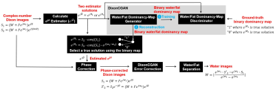 |
Water and Fat Separation with a Dixon Conditional Generative Adversarial Network (DixonCGAN)
Jong Bum Son1, Ken-Pin Hwang1, Marion E. Scoggins2, Basak E. Dogan3, Gaiane M. Rauch2, Mark D. Pagel4, and Jingfei Ma1
1Imaging Physics Department, The University of Texas MD Anderson Cancer Center, Houston, TX, United States, 2Diagnostic Radiology Department, The University of Texas MD Anderson Cancer Center, Houston, TX, United States, 3Department of Diagnostic Radiology, The University of Texas Southwestern Medical Center, Dallas, TX, United States, 4Cancer Systems Imaging Department, The University of Texas MD Anderson Cancer Center, Houston, TX, United States
A Dixon conditional generative adversarial network (DixonCGAN) was developed for Dixon water and fat separation. For the robust water image reconstruction, DixonCGAN performs water and fat separation with three processing steps: (1) phase-correction with DixonCGAN, (2) error-correction for DixonCGAN processing, and (3) the final water and fat separation. A conditional generative adversarial network (CGAN) originally designed to change photo styles could be successfully modified to perform phase-correction with improved global and local image details. Moreover, localized deep-learning processing errors could be effectively recovered with the proposed deep-learning error-correction processes.
|
|
0824.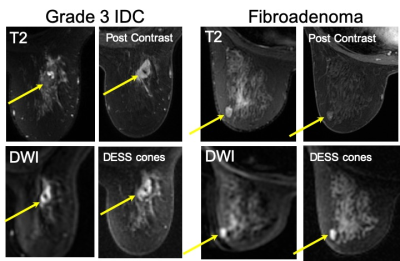 |
Double Echo Steady State (DESS) Cones for Non-Contrast-Enhanced Breast MRI
Catherine Judith Moran1, Christopher M Sandino2, Joseph Cheng1, Marcus T Alley1, Bruce Daniel1, and Brian A. Hargreaves1
1Radiology, Stanford University, Stanford, CA, United States, 2Electrical Engineering, Stanford University, Stanford, CA, United States
Breast MRI without a contrast injection has the potential to increase accessibility and compliance to the method in women who are recommended to undergo annual MRI to screen for breast cancer. Steady-state diffusion weighted methods provide robust image quality in comparison to conventional diffusion weighted methods which can suffer from variable image quality due to distortion, blurring and low-resolution. Double Echo Steady State (DESS) acquisition with a cones k-space trajectory is investigated for non-contrast enhanced breast MRI in 30 women undergoing clinically indicated breast MRIs.
|
|
0825.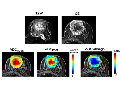 |
Clinical Diffusion Time Dependency of Breast Tumors and Associations with Prognostic Factors
Mami Iima1,2, Masako Kataoka1, Maya Honda1, Ayami Ohno Kishimoto1, Rie Ota1, Akane Ohashi1, Yuta Urushibata3, Thorsten Feiweier4, Masakazu Toi5, and Kaori Togashi1
1Diagnostic Imaging and Nuclear Medicine, Kyoto University Graduate School of Medicine, Kyoto, Japan, 2Clinical Innovative Medicine, Institute for Advancement of Clinical and Translational Science, Kyoto University Hospital, Kyoto, Japan, 3Siemens Healthcare K.K., Tokyo, Japan, 4Siemens Healthcare GMBH, Erlangen, Germany, 5Breast Surgery, Kyoto University Graduate School of Medicine, Kyoto, Japan
We investigated the variation of ADC values obtained at diffusion times that are clinically available for differentiation of human breast tumors. The ADC values in both malignant and benign breast tumors decreased with increased diffusion time, and a larger change in ADC values was found in malignant tumors. The significant association found between ADC change and Ki-67 expression might indicate the potential of diffusion time-dependent ADC values as a tool to differentiate these prognostic biomarkers and assess tumor heterogeneity without the need for contrast agents.
|
|
0826.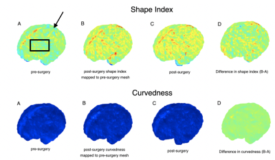 |
Cortical Surface Spectral Matching of the Fetal Brain Pre and Post Fetal Surgery for Open Spina Bifida
Nada Mufti1,2,3, Michael Aertsen4, Michael Ebner2,3, Lucas Fidon2, Tom Vercauteren2,5, Luc De Catte5, Philippe Demaerel4, Jan Deprest1,5, Sebastien Ourselin2, Anna L David1,6, and Andrew Melbourne2,3
1Institute for Women's Health and Department of Medical Physics and Biomedical Engineering, University College London (UCL), London, United Kingdom, 2School of Biomedical Engineering and Imaging Sciences (BMEIS), King's College London, London, United Kingdom, 3Department of Medical Physics and Biomedical Engineering, University College London (UCL), London, United Kingdom, 4Department of Radiology, University Hospitals Katholieke Universiteit (KU) Leuven, Leuven, Belgium, 5Department of Obstetrics and Gynaecology, University Hospitals Katholieke Universiteit (KU) Leuven, Leuven, Belgium, 6University Hospitals KU Leuven, Leuven, Belgium
Comprehensive evaluation of the fetal central nervous system (CNS) is required to select the most suitable candidates, to counsel parents about fetal spina bifida surgery, and to monitor post-op response. In children and adolescents with spina bifida, MRI assessment of gyrification correlates with motor and cognitive function. Our aim is to determine if MRI can quantify fetal brain gyrification and folding before and after fetal spina bifida surgery. If successful, mapping the gyrification changes to different lobes in the brain may prove useful in the prediction of motor and cognitive function after fetal surgery.
|
|
0827.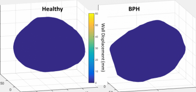 |
Comprehensive Analysis of Bladder Wall Perfusion and Deformation in BPH patients using MRI
Ryan J Pewowaruk1, David R Rutkowski2, Cody J Johnson2, Colin K Kim2, Shane A Wells2, Wade A Bushman3, Diego Hernando2,4, and Alejandro Roldán-Alzate1,2,5
1Biomedical Engineering, University of Wisconsin, Madison, WI, United States, 2Radiology, University of Wisconsin, Madison, WI, United States, 3Urology, University of Wisconsin, Madison, WI, United States, 4Medical Physics, University of Wisconsin, Madison, WI, United States, 5Mechanical Engineering, University of Wisconsin, Madison, WI, United States
Male urogenital disease is a common problem, and non-invasive methods for diagnosis and disease progression tracking are limited. This study was aimed at developing an MRI-based method to characterize urogenital tissue morphology, bladder-prostate interaction, and blood flow perfusion. These methods were tested in three patients with BPH and three healthy volunteers. Strong correlation between void fraction and prostate volume was found. Future work will be aimed at applying these methods to larger cohorts so that clinical utility may be further understood.
|
|
0828.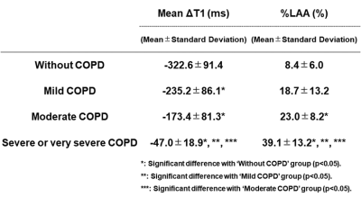 |
3D O2-Enhanced MR Imaging vs. Thin-Section CT: Capability for Pulmonary Functional Loss Assessment and Clinical Stage Classification in Smokers
Yoshiharu Ohno1,2, Masao Yui3, Daisuke Takenaka4, Yoshimori Kassai3, Kazuhiro Murayama1, and Takeshi Yoshikawa2
1Radiology, Fujita Health University School of Medicine, Toyoake, Japan, 2Radiology, Kobe University Graduate School of Medicine, Kobe, Japan, 3Canon Medical Systems Corporation, Otawara, Japan, 4Diagnostic Radiology, Hyogo Cancer Center, Akashi, Japan
No one directly compare this new technique with quantitatively assessed CT for pulmonary functional loss evaluation and The Global Initiative for Chronic Obstructive Lung Disease (GOLD) classification in smokers. We hypothesized that regional ΔT1 change from 3D O2-enhanced MRI has a potential for pulmonary functional loss assessment and clinical stage classification as well as quantitatively assessed thin-section CT in smokers. The purpose of this study was to prospectively and directly compare the quantitative capability for pulmonary functional loss assessment and clinical stage classification between 3D O2-enhanced MRI and thin-section CT in smokers.
|
 Watch the Video