
Oral - Power Pitch Session
Thoracic/Lung MRI
Session Topic: Thoracic/Lung MRI
Session Sub-Topic: Pulmonary Power
Oral - Power Pitch
Body
| Tuesday Parallel 3 Live Q&A | Tuesday, 11 August 2020, 13:45 - 14:30 UTC | Moderators: Peter Thelwall |
Session Number: PP-13
0437.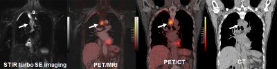 |
Which is the Best Method for NSCLC Recurrence Evaluation among MRI, PET/MRI, PET/CT and Standard Examination in Large Prospective Cohort?
Yoshiharu Ohno1,2, Masao Yui3, Kota Aoyagi3, Yoshimori Kassai3, Daisuke Takenaka4, Kazuhiro Murayama1, and Takeshi Yoshikawa2
1Radiology, Fujita Health University School of Medicine, Toyoake, Japan, 2Radiology, Kobe University Graduate School of Medicine, Kobe, Japan, 3Canon Medical Systems Corporation, Otawara, Japan, 4Diagnostic Radiology, Hyogo Cancer Center, Akashi, Japan
Direct comparison of non-small cell lung cancer (NSCLC) recurrence evaluation among these four methods in large prospective cohort more than 400 patients had not been reported. We hypothesize that whole-body MRI as well as PET/MRI had better potential for postoperative NSCLC recurrence than PET/CT and standard radiological examination in large prospective cohort. The purpose of this study was thus to compare the diagnostic performance of whole-body MRI, PET/MRI, PET/CT and standard radiological examination for postoperative NSCLC recurrence assessment in large prospective cohort more than 400 patients.
|
|
 |
0438.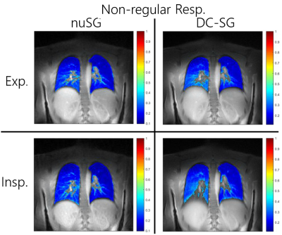 |
Lung Imaging and Proton Fraction Quantification for Highly Irregular Respiratory Patterns Using Nonuniform Self-Gating
Patrick Metze1, Tobias Speidel2, Fabian Straubmüller1, and Volker Rasche1,2
1Department of Internal Medicine II, University Ulm Medical Center, Ulm, Germany, 2Core Facility Small Animal Imaging (CF-SANI), Ulm University, Ulm, Germany Most self-gating methods rely on information that is extracted either from k-space itself or from high temporal resolution sliding-window images. The obtained one-dimensional gating signal is analysed with respect to a dominant and characteristic frequency. These approaches are prone to fail in case of highly non-uniform motion. The presented application of nonuniform Self-Gating based on a two-dimensional correlation matrix is capable of achieving high resolution proton fraction maps in human subjects with non-regular respiratory motion without the need of time consuming respiratory gating during acquisition. |
0439.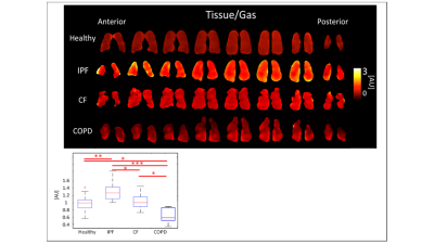 |
3D-SBCSI Xenon-129 lung MRI: Comparison of Healthy, CF, IPF, and COPD Subjects
Vicki Huang1, Steven Guan1, Nick Tustison1, Kun Qing1, Yun Shim2, John Mugler1, Talissa Altes3, Dana Albon2, Deborah Froh2, Borna Mehrad4, James Patrie5, Allan Ropp1, Lucy Gettle2, Mu He1,
and Jaime Mata1
1Radiology & Medical Imaging, University of Virginia, Charlottesville, VA, United States, 2Medicine, University of Virginia, Charlottesville, VA, United States, 3Radiology, University of Missouri, Columbia, MO, United States, 4University of Florida, Gainesville, FL, United States, 5Public Health, University of Virginia, Charlottesville, VA, United States
The results of this study indicated that hyperpolarized Xe-129 MR 3D-SBCSI is sensitive to physiology of lung diseases and can therefore be used to differentiate lung disease and monitor disease progression on regional level and characterizing disease phenotypes and co-morbidities in the future.
|
|
0440.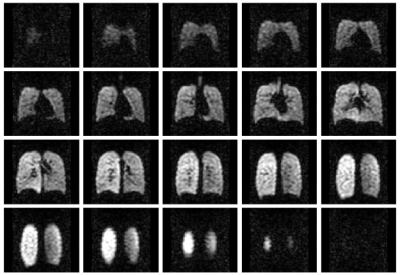 |
19F-MRI of inhaled perfluoropropane for assessment of pulmonary ventilation: a multi-centre reproducibility study in healthy volunteers
Mary Neal1,2, Benjamin Pippard1,2, Adam Maunder3, Rod Lawson4, Holly F. Fisher5, John N. S. Matthews5, Kieren Hollingsworth1,2, A. John Simpson2, Jim M. Wild3, and Pete Thelwall1,2
1Newcastle Magnetic Resonance Centre, Newcastle University, Newcastle upon Tyne, United Kingdom, 2Translational and Clinical Research Institute, Newcastle University, Newcastle upon Tyne, United Kingdom, 3POLARIS, Academic Radiology, University of Sheffield, Sheffield, United Kingdom, 4Sheffield Teaching Hospitals NHS Foundation Trust, Sheffield, United Kingdom, 5Institute of Health and Society, Newcastle University, Newcastle upon Tyne, United Kingdom
19F-MRI of inhaled perfluoropropane can be used to assess regional pulmonary ventilation. We conducted a prospective multi-centre reproducibility study in 40 healthy volunteers. Same-day static breath-hold 19F-MR images with 1 cm isotropic resolution were acquired on four occasions for each volunteer following inhalation of a perfluoropropane/oxygen gas mixture. Percentage ventilated lung volume (%VV) was calculated for all volunteers, reflecting the inhalation protocol, imaging protocol, and image registration and segmentation process applied. Volunteer %VV was determined to within ±1.7% (95% CI). Gas inhalations were well tolerated by all volunteers with no adverse events.
|
|
0441.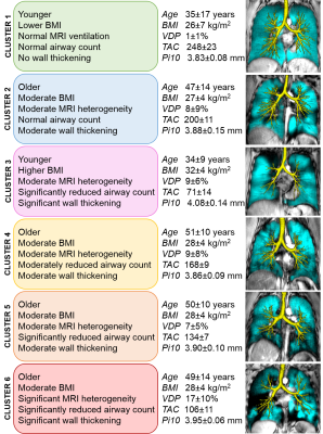 |
Novel MRI-based Clusters of Asthma: Pulmonary Functional MRI and CT
Rachel L Eddy1,2, Christopher Licskai3, David G McCormack3, and Grace Parraga1,2,3
1Robarts Research Institute, London, ON, Canada, 2Department of Medical Biophysics, Western University, London, ON, Canada, 3Division of Respirology, Department of Medicine, Western University, London, ON, Canada
Pulmonary functional MRI measurements have never been evaluated for the generation of imaging-based asthma patient clusters, although computed tomography (CT)-based clusters have been determined. Here we investigated hyperpolarized inhaled gas MRI ventilation in combination with CT airway measurements in 60 patients with asthma and identified 6 pulmonary structure-function imaging-based clusters using MRI ventilation defect percent (VDP) and CT airway measurements. These clusters reflect proximal and distal airway abnormalities in asthma and may be used to stratify patients for treatment decisions.
|
|
0442.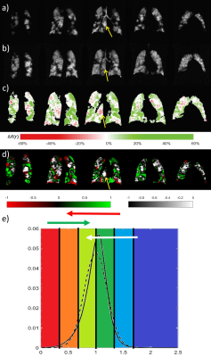 |
Binning method for treatment response mapping with hyperpolarized gas lung MRI: application in subjects with asthma
Guilhem Jean Collier1, Alberto Biancardi1, Paul J Hughes1, Laurie Smith1, Grace T Mussel1, Helen Marshall1, Ho F Chan1, Graham Norquay1, and Jim M Wild1
1Department of Infection, Immunity & Cardiovascular Disease, University of Sheffield, Sheffield, United Kingdom
This work applies a clustering method developed for image analysis of hyperpolarised gas lung ventilation MRI to a cohort of patients referred from a severe asthma clinic for investigation of breathlessness. Patients underwent spirometry tests and imaging at baseline and after inhalation of Salbutamol to assess reversibility. In a subset of patients, images pre and post bronchodilator were registered and a novel treatment response mapping method was applied. Results show significant correlations at baseline between imaging markers and FEV1% and between their percentage changes and the reversibility testing. The treatment response mapping method offers additional robust additional insights.
|
|
|
0443.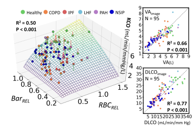 |
A Model for Interpreting Hyperpolarized 129Xe Gas Exchange MRI
Ziyi Wang1, Leith Rankine1, Aparna Swaminathan1, Elianna A Bier1, Matthew P Thorpe2, Robert Tighe1, Yuh Chin Huang1, Sudarshan Rajagopal1, and Bastiaan Driehuys1
1Duke University, Durham, NC, United States, 2Mayo Clinic, Rochester, MN, United States
Hyperpolarized 129Xe gas exchange (GX) MR imaging of pulmonary ventilation, barrier uptake and red blood cell (RBC) transfer has shown sensitivity to a wide range of pathology. However, the physiological interpretation of regional RBC transfer defects is not yet fully established and its connection to conventional measures has yet to be studied in a broad range of pathology. Here we evaluate the extent to which 129Xe RBC transfer reflects local perfusion, by testing its spatial correlation to 99mTc scintigraphy and propose a generalized model connecting 129Xe gas exchange metrics to the membrane and capillary blood volume components of DLCO.
|
0444.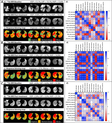 |
Longitudinal lobar analysis to access variable dynamic airflow changes in asthma with hyperpolarized helium-3 MRI
Mu He1, Lindsay A. L. Somerville1, Nicolas J. Tustison2, Jaime F. Mata2, Joanne M. Cassani3, Roselove Nunoo-Asare2, Alan M. Ropp2, Wilson G. Miller2, Yun M. Shim1, Talissa A. Altes3, John P. Mugler2, and Eduard E. de Lange2
1Department of Pulmonary and Critical Care, University of Virginia, Charlottesville, VA, United States, 2Department of Radiology and Medical Imaging, University of Virginia, Charlottesville, VA, United States, 3Department of Radiology, University of Missouri, Columbia, MO, United States
With hyperpolarized 3He MRI regional differences in airflow can be assessed. We sought to evaluate the changes in regional ventilation following bronchodilator therapy in patients with asthma. Baseline and post-treatment 3He/1H scans were co-registered, and normalized to enable serial comparison. Linear binning quantification was applied to the ventilation scan data to obtain quantitative metrics. Baseline and post-treatment scans were compared for regions of ventilation improvement or worsening. Lobar analysis was performed to identify ventilation abnormalities in each lobe. It was found that in asthmatics with bronchodilator response, ventilation improved globally, with most significant improvement in the upper and middle lobes.
|
|
0445.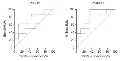 |
Supervised shallow learning of 129Xe MRI texture features to predict response to Anti-IL-5 biologic therapy in severe asthma
Marrissa McIntosh1, Rachel Eddy1, Danielle Knipping2, Tamas Lindenmaier2, David McCormack3, Christopher Licskai3, Cory Yamashita3, and Grace Parraga4
1Department of Medical Biophysics, Robarts Research Institute, Western University, London, ON, Canada, 2Robarts Research Institute, Western University, London, ON, Canada, 3Division of Respirology, Department of Medicine, Western University, London, ON, Canada, 4Department of Medical Biophysics, Division of Respirology, Department of Medicine, Robarts Research Institute, Western University, London, ON, Canada
129Xe MRI ventilation images consist of embedded texture features that help explain abnormal ventilation heterogeneity. We postulated that such texture features may help predict severe asthma patient response to anti-IL-5 therapies. Therefore, we employed supervised shallow learning techniques to identify specific 129Xe MRI features that help predict anti-IL-5 responders. Texture analysis yielded features that were superior to clinical measurements in identifying severe asthma patients that responded to anti-IL-5 therapy after 28 days. These promising results suggest that texture analysis may help predict asthmatics more likely to respond, before treatment is initiated.
|
|
0446.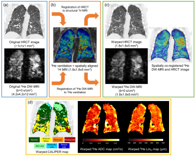 |
Voxel-wise comparison of co-registered quantitative CT and hyperpolarized gas diffusion-weighted MRI measurements in IPF
Ho-Fung Chan1, Alberto Biancardi1, Bilal A Tahir1, Nicholas D Weatherley1, Ronald A Karwoski2, Brian J Bartholmai2, Stephen M Bianchi3, and Jim M Wild1
1Academic Radiology, University of Sheffield, Sheffield, United Kingdom, 2Biomedical Imaging Resource, Mayo Clinic, Rochester, MN, United States, 3Academic Directorate of Respiratory Medicine, Sheffield Teaching Hospitals NHS Foundation Trust, Sheffield, United Kingdom
A framework for spatial co-registration of 3He diffusion-weighted (DW)-MRI and high resolution CT (HRCT) images was developed and implemented in a cohort of fifteen idiopathic pulmonary fibrosis participants. Voxel-wise comparison of 3He DW-MRI derived apparent diffusion coefficient (ADC) and mean alveolar dimension (LmD) with CALIPER image analysis software classifications revealed that the largest DW-MRI metrics are in voxels classified as honeycombing. Furthermore, LmD values in voxels classified as normal by CALIPER were larger than those from age-matched healthy volunteers, suggesting DW-MRI may detect microstructural changes even in areas of the lung determined as macroscopically normal by HRCT.
|
|
0447.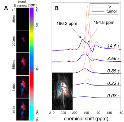 |
Hyperpolarized 129Xe Imaging of Oxygenated and Deoxygenated Blood in a Free-Breathing Mouse
Luis A Loza1, Stephen J Kadlecek1, Mehrdad Pourfathi1, Kai Ruppert1, Tahmina S Achekzai1, Ian F Duncan1, and Rahim R Rizi1
1Radiology, University of Pennsylvania, Philadelphia, PA, United States
129Xe’s high solubility in tissue and blood, coupled with its dramatic change in chemical shift based on local chemical environment, enables quantitative measurements of blood oxygenation. In this work, we demonstrate a technique for distinguishing oxygenated vs. deoxygenated blood in the mouse circulatory system in vivo. Time-resolved dissolved-phase images and spectra were used to identify spectral signatures for 129Xe dissolved in oxygenated and deoxygenated blood, which were then applied to a mouse model of lung cancer to temporally assess regional changes in pulmonary blood oxygenation. The results presented here demonstrate
|
|
0448.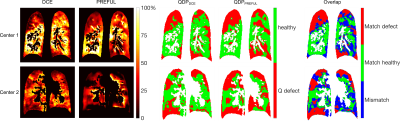 |
Dual Center and Dual Vendor feasibility of perfusion-weighted phase resolved functional lung (PREFUL) MRI in CF patients
Lea Behrendt1,2, Andreas Voskrebenzev1,2, Cristian Crisosto Gonzalez1,2, Marcel Gutberlet1,2, Helen Marshall3, Anna-Maria Dittrich4, Laurie Smith3, Paul Hughes3, Jim Wild3, and Jens Vogel-Claussen1,2
1Institute of Diagnostic and Interventional Radiology, Hannover Medical School, Hannover, Germany, 2Biomedical Research in Endstage and Obstructive Lung Disease Hannover (BREATH), German Center for Lung Research (DZL), Hannover, Germany, 3University of Sheffield, Sheffield, United Kingdom, 4Paediatric Pneumology and Neonatology, Hannover Medical School, Hannover, Germany
Dynamic contrast enhanced (DCE) MRI is an established technique for measurement of lung perfusion, but requires the administration of contrast agents and a breath hold. Thus, methods for contrast agent free assessment of lung perfusion in free breathing, like phase resolved functional lung (PREFUL) MRI, are desirable. Therefore, in this dual center and dual vendor feasibility study, we validated PREFUL MRI against DCE MRI in patients with CF. Perfusion defect percentage (QDP) maps of both methods were calculated, showing an overlap of 61% for the whole lung. Further, a strong correlation between QDPPREFUL and QDPDCE was found (r=0.70, p=0.005).
|
|
0449.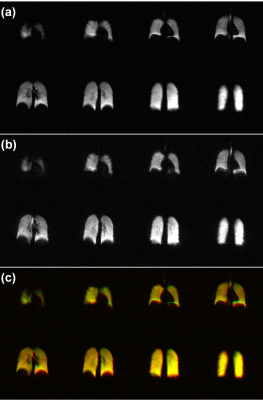 |
Comparison of 3D Stack-of-Spirals and 2D Gradient Echo for Ventilation Mapping using Hyperpolarized 129Xe
Brandon Zanette1, Yonni Friedlander1,2, Samal Munidasa1,2, and Giles Santyr1,2
1Translational Medicine, The Hospital for Sick Children, Toronto, ON, Canada, 2Medical Biophysics, University of Toronto, Toronto, ON, Canada
Hyperpolarized 129Xe MRI is an emergent tool for the quantification of ventilation defects in the lungs. 129Xe is typically imaged with 2D gradient recalled echo (2D-GRE) which may require lengthy breath-holds (up to 16s) to image the lung. This may be problematic in subjects who are not able to comply with these breath-hold constraints. Non-Cartesian spiral imaging samples k-space more efficiently, reducing the acquisition duration. In this work a 3D stack-of-spirals (3D-SoS) imaging sequence was developed and tested in healthy adults alongside conventional 2D-GRE for hyperpolarized 129Xe ventilation mapping, showing equivalent ventilation defect percent quantification in a ~2 s scan.
|
|
0450.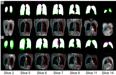 |
Fully-automated 1H MRI Thoracic Cavity Segmentation for Hyperpolarized Gas Imaging using a Convolutional Neural Network
Alexander M Matheson1, Rachel L Eddy1, Jonathan L MacNeil2, Marrissa L McIntosh1, and Grace Parraga1,2
1Medical Biophysics, Robarts Research Institute, Western University, London, ON, Canada, 2School of Biomedical Engineering, Robarts Research Institute, Western University, London, ON, Canada
Thoracic segmentations are crucial for accurate measurements of normalized lung ventilation, perfusion and gas exchange. Current semi-automated methods are time consuming, require experienced readers, and lack the standardization of fully-automated methods, such as convolutional neural networks. We retrospectively pooled data from 449 healthy and respiratory disease participants, resulting in a 55,000 slice augmented data set to train a dense v-net neural network. The network produced segmentations qualitatively matching semi-automated methods, with high Dice scores and an area under the receiver operating characteristic curve of 0.997. Implementation on the NiftyNet platform permits quick model dissemination for multi-site validation.
|
|
0451.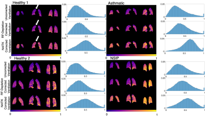 |
Bias Field Correction in Hyperpolarized 129Xe Gas Ventilation MRI
Junlan Lu1, Ziyi Wang2, John C. Nouls3, Kush Gulati2, Elianna Bier2, David Mummy3, and Bastiaan Driehuys3
1Medical Physics Graduate Program, Duke University, Durham, NC, United States, 2Biomedical Engineering, Duke University, Durham, NC, United States, 3Radiology, Duke University, Durham, NC, United States
The presence of RF inhomogeneity in hyperpolarized 129Xe ventilation MRI affects quantitative analysis and interpretation. These inhomogeneities are commonly corrected using the N4ITK algorithm, which retrospectively calculates a smoothly varying bias field with non-uniform intensity normalization. However, this algorithm has not been rigorously validated for functional imaging. Here we show, using flip angle maps derived directly from 3D-radial acquisitions of ventilation, that N4ITK may over-correct bias field and remove inherent physiological gradients. We illustrate this comparison by a combination of simulation, phantom, and in vivo ventilation imaging.
|
 Watch the Video