
Oral - Power Pitch Session
Hot Topics & Cancer
Session Topic: Hot Topics & Cancer
Session Sub-Topic: Musculoskeletal 1
Oral - Power Pitch
Musculoskeletal
| Thursday Parallel 1 Live Q&A | Thursday, 13 August 2020, 15:05 - 15:50 UTC | Moderators: Kimberly Amrami & Valentina Mazzoli |
Session Number: PP-14
 |
1139.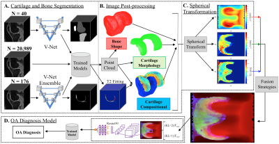 |
Multimodal qMRI Framework for Knee Imaging Biomarker Fusion and Osteoarthritis Prediction
Alejandro Morales Martinez1,2, Francesco Caliva1, Claudia Iriondo1,2, Sarthak Kamat1, Sharmila Majumdar1, and Valentina Pedoia1,2,3
1Department of Radiology and Biomedical Imaging, University of California, San Francisco, San Francisco, CA, United States, 2Graduate Program in Bioengineering, University of California, Berkeley, Berkeley, CA, United States, 3Center for Digital Health Innovation (CDHI), University of California San Francisco, San Francisco, CA, United States
Bone and cartilage segmentation models were trained and validated with a segmented dataset of 40 and 176 3D DESS MRI volumes respectively. The trained models were used to run inference on 20,989 3D DESS MRI volumes from the Osteoarthritis Initiative dataset. Biomarkers such as femoral bone shape, cartilage thickness and cartilage T2 average values were extracted from the segmentations. Point clouds representing each biomarker were transformed into spherical coordinates and merged using different fusion strategies. The spherical maps were used to train an OA diagnosis model with a test specificity, sensitivity and AUC was 84.1%, 78.7%, and 89.7% respectively.
|
1140.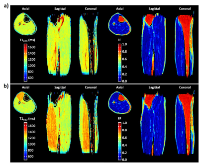 |
3D MR fingerprinting with water and fat separation
Benjamin Marty1,2
1NMR Laboratory, Neuromuscular Investigation Center, Institute of Myology, Paris, France, 2NMR Laboratory, CEA/DRF/IBFJ/MIRCen, Paris, France
In this study, a fast 3D MR fingerprinting sequence with water and fat separation (3D MRF T1-FF) was developed for simultaneous measurement of FF and water T1 in the skeletal muscles of patients with fat infiltrations. The precision and accuracy of the sequence was evaluated on a multi-vial phantom and in vivo proofs of concept were obtained in the legs of a healthy volunteer before and after plantar dorsi-flexions and at rest in a patient suffering from inclusion body myositis.
|
|
 |
1141.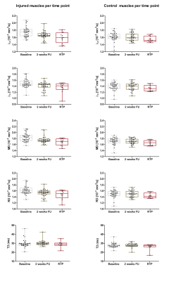 |
Diffusion Tensor Imaging detects recovery after acute muscle injury
Melissa Tamara Hooijmans1, Jithsa R. Monte2, Martijn Froeling3, Jos Oudeman4, Johannes L. Tol5, Mario Maas2, Gustav J. Strijkers1, and Aart J. Nederveen2
1Department of Biomedical Engineering & Physics, Amsterdam University Medical Centers, University of Amsterdam, Amsterdam, Netherlands, 2Department of Radiology & Nuclear Medicine, Amsterdam University Medical Centers, University of Amsterdam, Amsterdam, Netherlands, 3Department of Radiology, University Medical Center Utrecht, Utrecht, Netherlands, 4Department of Orthopaedic Surgey, University Medical Center Utrecht, Utrecht, Netherlands, 5Department of Orthopaedic Surgey, Amsterdam University Medical Centers, University of Amsterdam, Amsterdam, Netherlands
41 athletes with an acute hamstring injury underwent MRI examination of their injured leg and the uninjured contralateral leg at three different time points: (1) within one week after the index injury (baseline), (2) two weeks after baseline, and at (3) Return to Play (RTP). Baseline DTI values (MD, RD and the three eigenvalues) were elevated compared to control hamstring muscles and decreased during the RTP phase. qT2 values were elevated after the index injury and did not change over time. DTI is promising for monitoring recovery of hamstring injuries.
|
1142.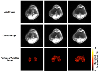 |
Knee Epiphyseal Bone Marrow Perfusion Imaging Using FAIR RESOLVE
Xiufeng Li1, Casey P. Johnson1, and Jutta Ellermann1
1Center for Magnetic Resonance Research, University of Minnesota, Minneapolis, MN, United States
Perfusion imaging can provide critical information to assess vasculature status and tissue viability of knee bone marrow. Arterial spin labeling, as a non-invasive and non-contrast-enhanced perfusion imaging approach, is well-suited for routine assessment of bone marrow perfusion, longitudinal monitoring of disease progression and repeated evaluation of therapy response. Recently, knee epiphyseal bone marrow ASL imaging has been demonstrated at 3T with promising results by using FAIR ss-FSE method. However, the ss-FSE image readout only supports single-slice acquisitions. To overcome this limitation, we implemented and evaluated FAIR RESOLVE for multi-slice knee epiphyseal bone marrow perfusion imaging.
|
|
|
1143.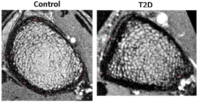 |
Multi-modality in vivo imaging of cortical bone vasculature: Comparison of diabetes patients to healthy controls
Po-hung Wu1, Misung Han1, Roland Krug1, Jing Liu1, Gabby B. Joseph1, Thomas Link1, and Galateia Kazakia1
1Department of Radiology and Biomedical Imaging, University of California - San Francisco, San Francisco, CA, United States
Type 2 diabetes is known to increase fracture risk, possibly through the development of pathological cortical bone porosity. However, the mechanisms of pathological pore growth are not understood. We hypothesize that T2D patients will display altered vascularization within cortical pores due to microvascular disease. In this study, 15 T2D patients and 22 controls were imaged by HR-pQCT and DCE-MRI to analyze vessel and perfusion metrics (eg. vessel density, transition time). The study results suggest that T2D patients have altered vessel distribution and perfusion characteristics, and that microvascular disease may be a factor in diabetic bone disease.
|
1144.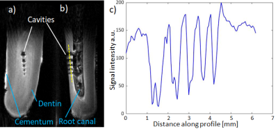 |
Visibility of artificial dental side root canals using MRI
Agazi Samuel Tesfai1, Andreas Vollmer2, Moritz Braig1, Johannes Fischer1, Ute Ludwig1, and Michael Bock1
1Dept. of Radiology, Medical Physics, Medical Center - University of Freiburg, Freiburg, Germany, 2Dept. of Oral and Craniomaxillofacial Surgery, Center for Dental Medicine, Medical Center - University of Freiburg, Freiburg, Germany
Detection of root canals is vital for dental diagnosis, however it is difficult to locate these anatomies within sub-millimeter dimension. To determine the ability of MRI to display such structures, a bovine tooth with different sized artificial cavities was prepared. It was evaluated with a preclinical 7T system and a clinical 3T MR system against cone beam CT. 7T measurements with UTE offer precise distinction of cavities up to 200µm. 3T UTE allows only differentiation of 1000µm cavities due to blurring. Tooth immersed in contrast agent solution allows localization up to 200µm cavity with spin echo sequence and negative contrast.
|
|
1145.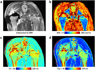 |
Quantitative T2, T1ρ, and Diffusion Mapping of Early-Stage Ischemic Osteonecrosis of the Femoral Head: An In Vivo Piglet Model Study at 3T MRI
Casey P. Johnson1,2, Ferenc Toth1, Alexandra R. Armstrong1, Harry K. W. Kim3,4, and Jutta M. Ellermann2,5
1Veterinary Clinical Sciences Department, University of Minnesota, Saint Paul, MN, United States, 2Center for Magnetic Resonance Research, University of Minnesota, Minneapolis, MN, United States, 3Texas Scottish Rite Hospital for Children, Dallas, TX, United States, 4Department of Orthopaedic Surgery, UT Southwestern Medical Center, Dallas, TX, United States, 5Department of Radiology, University of Minnesota, Minneapolis, MN, United States
This study tested whether T2 and T1ρ relaxation times are sensitive in detecting early-stage osteonecrosis of the femoral head in piglet model under in vivo conditions and at clinical 3T MRI. This study builds on recent ex vivo 9.4T studies assessing T2 and T1ρ in the piglet model. We also evaluated apparent diffusion coefficient for comparison. We found that T2, T1ρ, and ADC were all significantly increased in the ischemic vs. contralateral control femoral heads in n=6 piglets one week after onset of ischemia. These methods may be clinically useful to detect and characterize early-stage osteonecrosis to inform treatment decisions.
|
|
|
1146.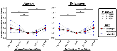 |
Estimating contraction of individual muscles during isometric wrist torque using multi-muscle MR elastography (MM-MRE)
Daniel Smith1, Andrea Zonnino1, Fabrizio Sergi1, and Curtis Johnson1
1University of Delaware, Newark, DE, United States
In this study, we propose to use a technique called MM-MRE to identify states of contraction of individual forearm muscles based on the measurement of muscle shear wave speed. Using a custom protocol and passive driver device, we scanned four subjects through 45 MRE scans each in three wrist positions during five torque application states. We found significant correlations between increased wave speed and higher applied torques, in both agonist and antagonist motions. The results indicate that MM-MRE is an effective measurement tool for analyzing the contractile state of individual forearm muscle during isometric contractions of the wrist joint.
|
|
1147.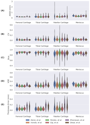 |
A Report on the International Workshop on Osteoarthritis Imaging Segmentation Challenge: A Multi-Institute Evaluation on a Standard Dataset
Arjun D. Desai1,2, Francesco Caliva3, Claudia Iriondo3, Naji Khosravan4, Aliasghar Mortazi4, Sachin Jambawalikar5, Drew Torigian6, Jutta Ellerman7, Mehmet Akçakaya8, Ulas Bagci4, Radhika Tibrewala3, Io Flament3, Matt O'Brien3,
Sharmila Majumdar3, Mathias Perslev9, Akshay Pai9, Christian Igel9, Erik B. Dam9, Sibaji Gaj10, Mingrui Yang10, Kunio Nakamura10, Xiaojuan Li10, Cem M. Deniz11, Vladimir Juras12, Ravinder Regatte11, Garry E. Gold2, Brian
A. Hargreaves2, Valentina Pedoia3, and Akshay S. Chaudhari2
1Electrical Engineering, Stanford University, Stanford, CA, United States, 2Radiology, Stanford University, Stanford, CA, United States, 3Radiology, University of California San Francisco, San Francisco, CA, United States, 4University of Central Florida, Orlando, FL, United States, 5Radiology, Columbia University Medical Center, New York, NY, United States, 6Radiology, University of Pennsylvania, Philadelphia, PA, United States, 7Radiology, University of Minnesota, Minneapolis, MN, United States, 8Electrical and Computer Engineering, University of Minnesota, Minneapolis, MN, United States, 9Computer Science, University of Copenhagen, Copenhagen, Sweden, 10Biomedical Engineering, Cleveland Clinic, Cleveland, OH, United States, 11Radiology, New York University Langone Health, New York, NY, United States, 12Biomedical Imaging and Image-Guided Therapy, Medical University of Vienna, Vienna, Austria
Cartilage thickness can be predictive of joint health. However, manual cartilage segmentation is tedious and prone to inter-reader variations. Automated segmentation using deep-learning is promising; yet, heterogeneity in network design and lack of dataset standardization has made it challenging to evaluate the efficacy of different methods. To address this issue, we organized a standardized, multi-institutional challenge for knee cartilage and meniscus segmentation. Results show that CNNs achieve similar performance independent of network architecture and training design and, given the high segmentation accuracy achieved by all models, only a weak correlation between segmentation accuracy metrics and cartilage thickness was observed.
|
1148.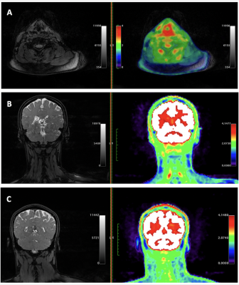 |
Abnormal [18F]FDG PET/MRI Findings in Paraspinal Structures of Patients with Suspected Cerebrospinal Fluid Leak
Peter Cipriano1, Daehyun Yoon1, Ryan Penticuff1, Yingding Xu2, Ian Carroll3, and Sandip Biswal1
1Stanford University, Stanford, CA, United States, 2Stanford University, Palo Alto, CA, United States, 3Stanford University, Redwood City, CA, United States
Six patients with suspected cerebrospinal fluid leak but in whom no site of leakage had been identified and six controls underwent simultaneous, whole-body [18F]FDG PET/MRI imaging. Increased [18F]FDG uptake was found in paraspinal structures in all six patients and was significantly greater than in the corresponding areas of controls. Temporary but significant relief in symptoms resulted from blood patches placed at locations coinciding with PET/MRI abnormalities.
|
|
|
1149. |
Classification of Spinal Metastases Coming from Different Primary Cancer Origin by Using Quantitative Radiomics Analysis with Multi-Class SVM
Yongye Chen1, Yang Zhang2, Enlong Zhang1, Xiaoying Xing1, Qizheng Wang1, Huishu Yuan1, Min-Ying Su2, and Ning Lang1
1Department of Radiology, Peking University Third Hospital, Beijing, China, 2Department of Radiological Sciences, University of California, Irvine, CA, United States
For patients suspected to have spinal metastasis, a confirmed pathological diagnosis is needed to proceed with appropriate treatment. This study applied quantitative radiomics to differentiate 5 groups of patients with metastatic cancers in the spine, including 28 lung, 11 breast, 7 kidney, 11 prostate and 18 thyroid. The analysis was done on post-contrast images. A total of 107 features, including 32 first order and 75 texture, were extracted for each case by using PyRadiomics. The group differentiation was done by using multi-class support vector machine (SVM). The overall accuracy was 80%, with the highest accuracy of 27/28=96% for lung mets.
|
|
1150.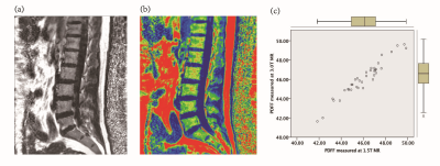 |
Evaluation of the risk of osteoporosis by using IDEAL-IQ in diabetic patients
Yu Song1, Qingwei Song1, Aibo Wang2, Ailian Liu1, Yanwei Miao1, Nan Zhang1, Haonan Zhang1, and Lizhi Xie3
1Department of Radiology, the First Affiliated Hospital of Dalian Medical University, Dalian, China, 2Department of Radiology, Peking University Third Hospital, Beijing, China, 3GE Healthcare, MR Research, Beijing, China
Diabetes is a metabolic disease that leads to a high risk of fracture related to osteoporosis. Noninvasive and reliable assessment of osteoporosis is essential for clinical practice. The aim of this study was to explore the agreement of the Proton Density Fat Fraction (PDFF) values of lumbar vertebra measured by Magnetic Resonance Imaging (MRI) IDEAL-IQ sequences at different field strengths and to investigate the value of IDEAL-IQ in the assessment of osteoporosis risk in diabetic.
|
|
1151.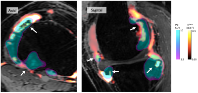 |
Imaging of Bone-Synovium Interactions Using Dynamic Contrast Enhanced MRI and 18F-Sodium Fluoride PET
James MacKay1,2, Lauren Watkins3, Garry Gold3, and Feliks Kogan4
1Radiology, University of East Anglia, Norwich, United Kingdom, 2Radiology, University of Cambridge, Cambridge, United Kingdom, 3Radiology, Stanford University, Stanford, CA, United States, 4Stanford University, Stanford, CA, United States Synovial inflammation is hypothesised to play a role in the development and progression of osteophytes in osteoarthritis (OA).
Here we use hybrid 3T PET-MRI to perform simultaneous bilateral knee MR imaging of 11 participants (22 knees) with knee OA. We use 18F-NaF PET to quantify osteophyte metabolic activity and dynamic contrast enhanced MR imaging to quantify synovitis.
We demonstrate that synovitis adjacent to osteophytes is more intense (as quantified by the DCE parameter Ktrans) than the whole-joint average, and that there is a significant association between increased osteophyte metabolic activity (as quantified by PET SUVmax) and intensity of adjacent synovitis. |
 Watch the Video