
Oral - Power Pitch Session
Hot Topics & Cancer
Session Topic: Hot Topics & Cancer
Session Sub-Topic: Musculoskeletal 2
Oral - Power Pitch
Musculoskeletal
| Thursday Parallel 1 Live Q&A | Thursday, 13 August 2020, 15:05 - 15:50 UTC | Moderators: Jutta Ellermann & Hiroshi Yoshioka |
Session Number: PP-15
 |
1152.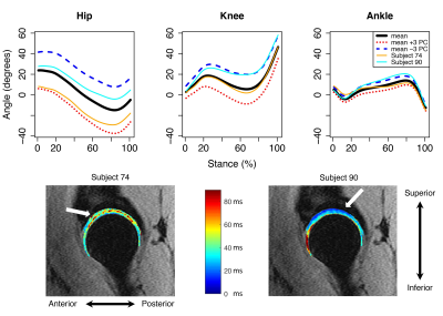 |
Linking multi-joint biomechanics with cartilage composition from qMRI
Koren Roach1, Valentina Pedoia1, Jinhee J Lee1, Tijana Popovic1, Sharmila Majumdar1, and Richard B Souza1
1UCSF, San Francisco, CA, United States
Hip osteoarthritis is likely caused by changes in gait biomechanics and cartilage biochemistry and composition. In this study, we employed multivariate functional principal component analysis to identify gait waveform characteristics that were related to T1ρ and T2 relaxation times in the femoral and acetabular cartilage. Our results indicated that transverse and sagittal plane waveform characteristics are significantly related to T1ρ and T2 relaxation times in the femoral and acetabular cartilage and may be key planes of motion on which to focus preventative therapies for hip osteoarthritis.
|
|
1153.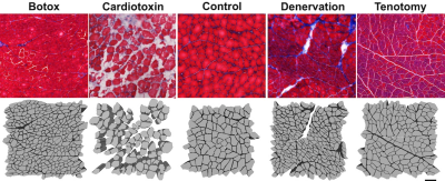 |
Using the random permeable barrier model to predict fiber size in histology informed simulated skeletal muscle models
David B Berry1, Erin K Englund2, Vitaly Galinsky3, Lawrence R Frank3, and Samuel R Ward2,3,4
1Nanoengineering, University of California, San Diego, La Jolla, CA, United States, 2Orthopaedic Surgery, University of California, San Diego, La Jolla, CA, United States, 3Radiology, University of California, San Diego, La Jolla, CA, United States, 4Bioengineering, University of California, San Diego, La Jolla, CA, United States
There is growing interest in using the Random Permeable Barrier Model (RPBM) to measure muscle microstructure. The goal of this study was to evaluate the accuracy of RPBM in predicting muscle fiber size in histology informed models of healthy and injured skeletal muscle from simulated DTI data. RPBM was found to systematically underestimate fiber size, but accurately predicted surface area to volume ratio (S/V) of the simulated muscle fibers. While the clinical interpretation of S/V ratio is unclear, this indicates that accurate measurement of S/V may serve as a proxy to changes in muscle fiber size and therefore function.
|
|
1154.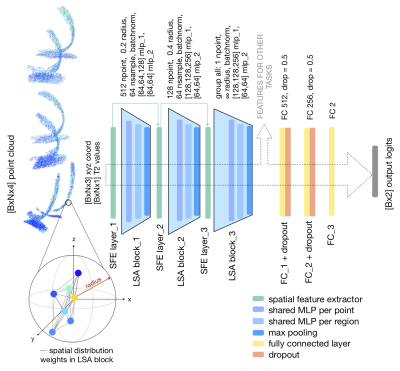 |
To the point: deep learning on dense T2 point clouds for improved feature extraction
Claudia Iriondo1, Alaleh Razmjoo2, Francesco Caliva2, Sharmila Majumdar2, and Valentina Pedoia2
1Radiology and Biomedical Imaging, University of California, San Francisco, San Francisco, CA, United States, 2University of California, San Francisco, San Francisco, CA, United States
To-the-point (TTP) is a novel approach for analyzing compositional MR imaging data. By representing tibial and femoral cartilage T2 values as a dense point cloud, our approach can leverage the data's inherent sparsity while maintaining local geometric properties, leading to improved feature extraction and faster image processing times. Experiments on the whole OAI T2 dataset show strong performance in an OA diagnosis task 82.44% sens, 82.59% spec, with extracted features even identifying patients who would become diagnosed with OA 1 to 2 years in the future.
|
1155.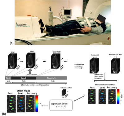 |
3D Internal dynamic strains in the intervertebral disc (IVD) of the lumbar spine with GRASP-MRI under mechanical loading
Rajiv G Menon1, Marcelo V. W. Zibetti1, and Ravinder R Regatte1
1Center for Biomedical Imaging, Department of Radiology, New York University Langone Health, New York, NY, United States
The goal of this study was to develop a non-invasive MRI technique to measure 3D dynamic internal strains in the intervertebral discs (IVDs) of lumbar spine during loading and recovery phases. For this purpose, a combination of static mechanical loading of the IVD using MR-compatible ergometer and continuous MRI-acquisition with a 3D-GRASP acquisition was used. Data was acquired on five healthy volunteers, and dynamic strains under loading and recovery conditions were calculated in 5-IVD segments from L1/L2 to L5/S1. By measuring temporal evolution of strain during rest, loading and recovery phases, dynamic strain information in the IVD may be investigated.
|
|
1156.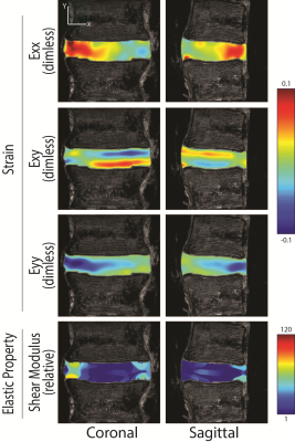 |
Intervertebral Disc Elastography: Physiological Strain, Stiffness, and Relaxometry in Axial Compression and Bending
Deva D. Chan1,2, Paull C. Gossett2, Robert L. Wilson3, Woong Kim2, Yue Mei4,5,6, Kent Butz2, Nancy Emery7, Eric A. Nauman2, Stéphane Avril6, and Corey P. Neu2,3
1Biomedical Engineering, Rensselaer Polytechnic Institute, Troy, NY, United States, 2Biomedical Engineering, Purdue University, West Lafayette, IN, United States, 3Mechanical Engineering, University of Colorado Boulder, Boulder, CO, United States, 4Engineering Mechanics, Dalian University of Technology, Dalian, China, 5International Research Center for Computational Mechanics, Dalian University of Technology, Dalian, China, 6Center for Biomedical and Healthcare Engineering, MINES Saint-Étienne, Saint-Étienne, France, 7Ecology and Evolutionary Biology, University of Colorado Boulder, Boulder, CO, United States
IVD degeneration is the most recognized cause of low back pain, characterized by the decline of tissue structure and mechanics. MRI relaxometry is one quantitative measure of IVD degeneration, yet MRI metrics of mechanics have not been fully explored. We quantified patterns of IVD strain and mechanics during physiological compression and bending. Strains patterns depended on the loading mode, and shear modulus in the nucleus pulposus was typically an order of magnitude lower than the annulus fibrosis, except in bending, where the apparent stiffness depended on the loading direction. Strain and material properties provide new possible biomarkers for IVD degeneration.
|
|
1157.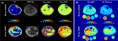 |
Quantitative 1H and 23Na NMR imaging in the skeletal muscles of patients with fascioscapulohumeral muscular dystrophy
Benjamin Marty1,2, Teresa Gerhalter1,2,3, Lena V. Gast3, Katharina Porzelt4, Matthias Türk4, Matthias Hammon3, Michael Uder3, Rolf Schröder5, Pierre G. Carlier1,2, and Armin M. Nagel3,6,7
1NMR Laboratory, Neuromuscular Investigation Center, Institute of Myology, Paris, France, 2NMR Laboratory, CEA/DRF/IBFJ/MIRCen, Paris, France, 3Institute of Radiology, University Hospital, FAU, Erlangen, Germany, 4Institute of Neurology, FAU, Erlangen, Germany, 5Department of Neuropathology, University Hospital Erlangen, FAU, Erlangen, Germany, 6Division of Medical Physics in Radiology, DKFZ, Heidelberg, Germany, 7Institute of Medical Physics, FAU, Erlangen, Germany
Facioscapulohumeral muscular dystrophy (FSHD) is a neuromuscular disorder characterized by structural changes affecting skeletal muscle tissues, resulting in muscle wasting and dysfunction. Here, we determined the value of quantitative 1H and 23Na muscle MRI approaches for providing variables related to disease severity (fat fraction) and disease activity (water T2, water T1, total sodium content and inversion-recovery 23Na) in patients with FSHD. We found that MRI variables related to water mobility and ion homeostasis were increased at an early stage of the degeneration process in several muscles of FSHD patients and represent potential candidates for assessing treatment response in clinical trials.
|
|
1158.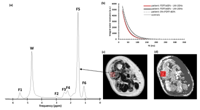 |
About the origin of decreased 1H NMRS-based water T2 in highly fatty infiltrated skeletal muscles of subjects with neuromuscular disorders
Harmen Reyngoudt1,2, Ericky Caldas de Almeida Araujo1,2, Pierre-Yves Baudin3, Benjamin Marty1,2, and Pierre G. Carlier1,2
1NMR Laboratory, Neuromuscular Investigation Center, Institute of Myology, Paris, France, 2NMR Laboratory, CEA/DRF/IBFJ/MIRCen, Paris, France, 3Consultants for Research in Imaging and Spectroscopy, Tournai, Belgium
1H NMRS-based water T2 (T2w) has shown to be decreasing when muscle fat fraction levels are elevated (>60%). Here, two myopathy patient groups with similar fat fraction levels (>60%) emerged, being a group with T2w>30 ms and a group with T2w<30 ms, which seemed to be correlated to the respective water resonance linewidths. Interpretation of these reduced T2w values at high fat fractions needs to be handled cautiously. The larger linewidths observed in the spectra characterized by shorter T2w may be due to the local B0 gradients induced by susceptibility differences between muscle and fat.
|
|
1159.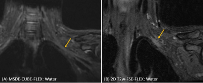 |
Radiologic evaluation of MSDE-CUBE-FLEX for imaging brachial plexus
Daehyun Yoon1, Neha Antil1, Sandip Biswal1, and Amelie Lutz1
1Radiology, Stanford university, Stanford, CA, United States
The conventional MRI examination of brachial plexus using 2D fast spin-echo sequences suffers from 1) long scan time due to a large field of view, 2) insufficient fat suppression due to strong off-resonance, 3) confusion between nerves and blood vessels. We recently presented a novel MSDE-CUBE-FLEX sequence addressing these issues by combining 1) outer volume suppression, 2) fast triple-echo Dixon technique, 3) magnitude-preparation to suppress blood signal. In this work, we compared the MSDE-CUBE-FLEX and 2D FSE sequences for imaging brachial plexus, which shows clear improvements in brachial plexus visualization with the MSDE-CUBE-FLEX sequence.
|
|
1160.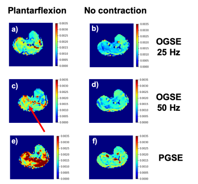 |
Imaging of skeletal muscle contraction using Oscillating Gradient Spin Echo (OGSE)
Valentina Mazzoli1, Kevin Moulin1, Feliks Kogan1, Brian Hargreaves1, and Garry E. Gold1
1Department of Radiology, Stanford University, Stanford, CA, United States
The apparent diffusion coefficient measured using DTI in skeletal muscles depends on the time allowed for diffusing water molecules to probe the local environment and on the size of muscle cells. Here we explore the use of Oscillating Gradient Spin Echo diffusion to obtain information on skeletal muscle microstructure over smaller distances than conventionally probed using PGSE. Our results show the ability to image skeletal muscle during active contraction (foot dorsiflexion and plantarflexion) using OGSE, and diffusion values are dependent on the oscillation frequency and on the contraction status of the muscle
|
|
1161.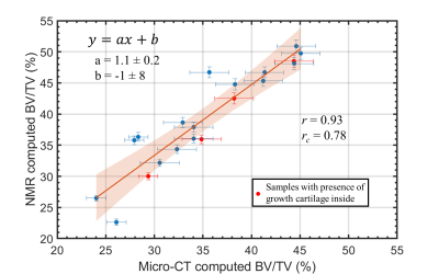 |
A new Trabecular BV/TV Estimation Method using Single-Sided NMR Devices through the Separation between Intra- and Inter-trabecular 1H Signals
Marco Barbieri1, Paola Fantazzini1, Anna Festa2, Fabio Baruffaldi2, Claudia Testa1, and Leonardo Brizi1
1Physics and Astronomy, University of Bologna, Bologna, Italy, 2IRCCS Istituto Ortopedico Rizzoli, Bologna, Italy
Methods to improve the early detection of diseases associated with an increased bone fragility are objects of investigation. Single-sided NMR is an appealing approach for medical applications. In this work we propose a new methodology to assess the bone volume fraction (BV/TV) of trabecular bone (TB), without the need of using a reference sample, exploiting the separation between intra- and inter-trabecular 1H signals from quasi-continuous T2 distributions. BV/TV of TB samples estimated using NMR were found in strong agreement with micro-CT estimations. This is promising for the application of single-sided NMR scanners to in-vivo assessing of bone micro-structure.
|
|
1162.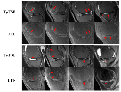 |
Osteochondral Junction (OCJ) Imaging Using a Fast T1-weighted 3D Ultrashort Echo Time Cones Sequence at 3T
Zhenyu Cai1,2, Zhao Wei2, Mingxin Chen2, Saeed Jerban2, Hyungseok Jang2, Eric Chang2,3, Jiang Du2, and Yajun Ma2
1Radiology, Fuwai Hospital Chinese Academy of Medical Sciences, Shenzhen, shenzhen, China, 2Radiology, University of California San Diego, San Diego, CA, United States, 3VA San Diego Healthcare System, San Diego, CA, United States
The osteochondral junction (OCJ) is the region where calcified cartilage meets subchondral bone (SCB), and is likely to be highly related to osteoarthritis (OA). However, it is difficult to image OCJ tissues due to their relatively short transverse relaxation times, which cause little or no signal to appear with conventional imaging sequences. In this study, we developed a 3D T1-weighed fast ultrashort echo time cones sequence with fat saturation (FS-UTE-Cones) to generate a high OCJ contrast image of the human knee on a clinical 3T MRI scanner.
|
|
1163.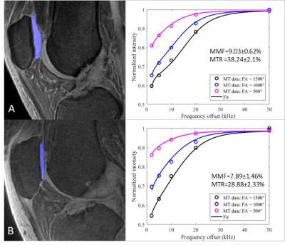 |
Quantitative 3D ultrashort echo time Cones magnetization transfer (3D UTE-Cones-MT) magnetic resonance imaging of knee cartilage degeneration
Yanping Xue1,2, Yajun Ma1, Zhao Wei1, Francis Tang1, Mei Wu1, Saeed Jerban1, Eric Y Chang1,3, and Jiang Du1
1University of California, San Diego, San Diego, CA, United States, 2Radiology, Beijing Chao-Yang Hospital, Beijing, China, 3VA San Diego Healthcare System, San Diego, CA, United States
Quantitative MRI biomarkers, such as T2, T2*, and T1rho have been used to detect cartilage degeneration. However, these biomarkers are sensitive to the magic angle effect. Magnetization transfer (MT) modeling provides magic angle insensitive parameters such as macromolecular proton fraction (MMF). This study focuses on the clinical evaluation of cartilage degeneration using 3D ultrashort echo time cones MT (3D UTE-Cones-MT) modeling in osteoarthritis (OA) patients. Both MMF and MT ratio (MTR) show significant negative correlations with WORMS grading of knee cartilage. This study highlights the potential of 3D UTE-Cones-MT techniques for detection of early cartilage degeneration in OA.
|
|
1164.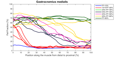 |
Muscular fat infiltration in FSHD starts with a “fat burst” near the distal tendon and advances towards the proximal tendon
Linda Heskamp1, Augustin C. Ogier2,3, David Bendahan3, and Arend Heerschap1
1Radiology and Nuclear Medicine, Radboud university medical center, Nijmegen, Netherlands, 2Aix Marseille Univ, Université de Toulon, CNRS, LIS, Marseille, France, 3Aix Marseille Univ, CNRS, CRMBM, Marseille, France
In patients with facioscapulohumeral muscular dystrophy (FSHD) it is known what the genetic origin of the disease is, but unknown how it is initiated and propagates along muscles. In a cross-sectional and longitudinal study we analyzed fat infiltration in lower leg muscles, tendon-to-tendon, with a 3D Dixon method. This revealed that fat infiltration starts with a distal “fat burst” in the first years of its initiation after which fat replacement further proceeds in a slower pace towards the proximal tendons. This indicates that the disease is triggered by an event typical for the distal parts of lower extremity muscles.
|
|
1165. |
Efficient Measurement of Composite Metric R2-R1ρ in Knee Cartilage
Misung Han1, Radhika Tibrewala1, Emma Bahroos1, Valentina Pedoia1,2, and Sharmila Majumdar1,2
1Radiology and Biomedical Imaging, University of California, San Francisco, San Francisco, CA, United States, 2Center for Digital Health Innovation, University of California, San Francisco, San Francisco, CA, United States
Cartilage degeneration, characterized by collagen structure disruption, proteoglycan depletion, and increased water content, has been shown to alter T2 and T1ρ relaxation times. A composite metric, R2-R1ρ, which further reflects an anisotropic component of R2, has recently demonstrated high sensitivity to cartilage degeneration; however, quantification of R2 and R1ρ respectively requires long scan times. In this work, we validated the potential of assessing R2-R1ρ using one pair of signals with T1ρ preparation and T2 preparation
from a combined T1ρ/T2 quantification sequence for in vivo knee MRI at 3T.
|
 Watch the Video