
Oral - Power Pitch Session
Educational Q&A: Preclinical
Session Topic: Educational Q&A: Preclinical
Session Sub-Topic: Preclinical Imaging Advances
Oral - Power Pitch
Preclinical/Animal Studies
| Wednesday Parallel 1 Live Q&A | Wednesday, 12 August 2020, 15:15 - 16:00 UTC | Moderators: Simon Robinson |
Session Number: PP-16
0897.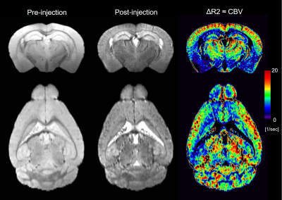 |
Toward high spatial resolution functional MRI: In vivo 50-micron cerebral blood volume mapping of the mouse brain
Akira Sumiyoshi1, Keigo Hikishima2, and Ichio Aoki1
1National Institutes for Quantum and Radiological Science and Technology (QST), Chiba, Japan, 2Okinawa Institute of Science and Technology Graduate University (OIST), Okinawa, Japan
We report here in vivo 50-micron cerebral blood volume (CBV) mapping of the mouse brain using intraperitoneal injection protocol of gadolinium-based contrast agent. Based on k-means clustering we identified different vascular clusters that separately range from macro- to micro-vasculature. The CBV map demonstrated layer-dependent macro- and micro-vascular densities in the cortex where different cortical regions exhibited different vascular patterns. The CBV map also identified different vascular densities and patterns in the hippocampus. These results suggest that CBV map would be a useful and alternative tool that assesses brain function and metabolism at extremely high spatial resolution.
|
|
0898.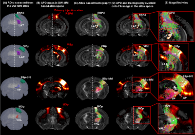 |
A quantitative approach to validate the mouse thalamo-cortical structural network reconstructed using diffusion MRI tractography
Tanzil Mahmud Arefin1, Choong Heon Lee1, Zifei Liang1, and Jiangyang Zhang1
1Radiology, NYU School of Medicine, New York, NY, United States
In this work, we used our previously reported high-resolution dMRI-based mouse brain atlas8 to trace node-to-node thalamo-cortical structural connectivity in the mouse brain. Taking advantage of the rich viral tracer data from the Allen Mouse Brain Connectivity Atlas (AMBCA)4, the tractography results were examined using the tracer data as ground truth. Our findings pinpoint the potentiality of mapping reliable structural connectivity in gray matter structures using tractography and at the same time, highlight the necessity of further investigation on determining the imaging and tractography parameters for accurate estimation of such connectivity.
|
|
0899.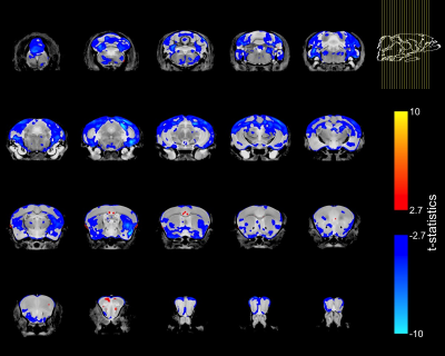 |
Brain structure in the homozygous FUSDelta14 mouse recapitulates amyotrophic lateral sclerosis phenotypes
Aurea B. Martins Bach1, Lily Qiu1, Jacob Ellegood2, Nick Wang2, Brian J. Nieman2, John G. Sled2, Remya R. Nair3, Elizabeth M. C. Fisher3,4, Thomas J. Cunningham3, Jason Lerch1, and Karla L. Miller1
1Wellcome Centre for Integrative Neuroimaging, FMRIB, Nuffield Department of Clinical Neurosciences, University of Oxford, Oxford, United Kingdom, 2Mouse Imaging Centre, The Hospital for Sick Children, Toronto, ON, Canada, 3Mammalian Genetics Unit, MRC Harwell Institute, Oxfordshire, United Kingdom, 4Department of Neuromuscular Diseases, Institute of Neurology, University College London, London, United Kingdom
This study assesses changes in brain anatomy with MRI in the homozygous humanized FUSDelta14 mouse model of amyotrophic lateral sclerosis (ALS). Post-mortem brain T2w-images were acquired at 7T, with 40μm isotropic resolution. After registration, the deformation fields were compared between mutant and wild-type mice. Homozygous FUSDelta14 mice exhibited atrophy in multiple grey and white matter structures. These results are in agreement with observations such as cortical thinning and alterations in white matter microstructure in ALS patients. Homozygous humanized FUSDelta14 mice show an early brain phenotype and are therefore a promising model for the study of ALS pathogenic mechanisms.
|
|
0900.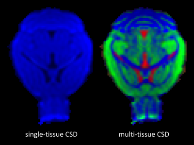 |
Multi-tissue constrained spherical deconvolution in a murine brain
Steven Jillings1, Jan Morez2, Nicholas Vidas-Guscic3, Johan Van Audekerke3, Floris Wuyts1, Marleen Verhoye3, Jan Sijbers2, and Ben Jeurissen2
1Lab for Equilibrium Investigations and Aerospace, Dept. of Physics, University of Antwerp, Antwerp, Belgium, 2imec-Vision Lab, Dept. of Physics, University of Antwerp, Antwerp, Belgium, 3Bio Imaging Lab, Dept. of Biomedical Sciences, University of Antwerp, Antwerp, Belgium
Multi-tissue constrained spherical deconvolution (MT-CSD) leverages the unique b-value dependency of each brain tissue type to estimate the full white matter (WM) fiber orientation density function (fODF) as well as the apparent densities of gray matter (GM) and cerebrospinal fluid (CSF), directly from multi-shell diffusion MRI (dMRI) data. Currently, its adoption is focussed almost entirely on imaging of the human brain. In this work, we demonstrate that the sequence, the pipeline and the advantages that are now well established for human brains, can be transferred to murine brains, bringing the technique into the preclinical realm.
|
|
 |
0901.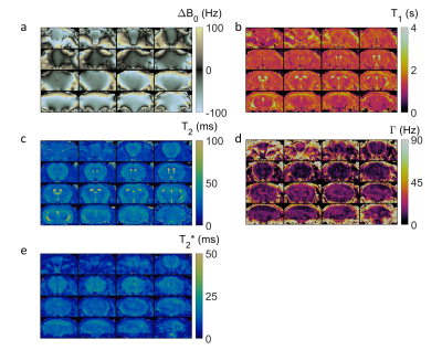 |
3D Magnetic Resonance Fingerprinting with Quadratic Phase in Mouse Brain on Preclinical 7T System
Charlie Wang1, Yuning Gu2, Rasim Boyacioglu2, Charlie Androjna3, Mark Alan Griswold2, and Xin Yu2
1Metrohealth Hospital, Cleveland, OH, United States, 2Case Western Reserve University, Cleveland, OH, United States, 3Cleveland Clinic, Cleveland, OH, United States
Magnetic Resonance Fingerprinting with Quadratic Phase (qRF-MRF) was previously validated in 2D clinical imaging for simultaneous off-resonance, T1, T2, and T2* mapping. Translation of qRF-MRF to high-field preclinical systems for small animal imaging is challenging due to the higher field inhomogeneity and the higher spatial resolution required. Here, a 3D qRF-MRF method was explored to address these challenges. High-resolution simultaneous mapping of off-resonance, T1, T2, and T2* on in vivo mouse brain at 7T was demonstrated. Computational limitations for large dictionary parameter space and reconstruction times were addressed using randomized SVD time compression and quadratic fitting methods.
|
0902.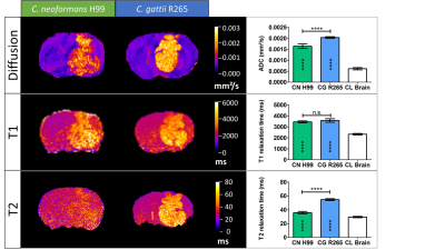 |
In vivo MRI can assess differences in cell density and size of different Cryptococcus species in brain lesions.
Liesbeth Vanherp1, Kristof Govaerts1, Amy Hillen2, Jennifer Poelmans3, Katrien Lagrou4, Greetje Vande Velde1, and Uwe Himmelreich1
1Biomedical MRI, University of Leuven, Leuven, Belgium, 2Department of Cell and Molecular Biology, Karolinska Institutet, Stockholm, Sweden, 3Janssen Pharmaceutical Companies of Johnson & Johnson, Beerse, Belgium, 4Laboratory of Clinical Bacteriology and Mycology, University of Leuven, Leuven, Belgium
Multi-parametric MRI was correlated with in vivo Fibred Confocal Fluorescence Microscopy and histology in two preclinical models of cryptococcosis. Increased ADCs and T2 relaxation times were linked to differences in capsule size and associated fungal density in brain lesions caused by the two pathogenic fungi Cryptococcus neoformans and C. gattii. This provides not only a non-invasive means to assess one of the most important virulence factors of Cryptococci in preclinical research but may also affect patient management by providing a method for differential diagnosis.
|
|
0903.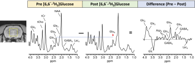 |
Investigating cerebral energetics and neurotransmission using in vivo 1H MRS and [6,6′-2H2]glucose in a preclinical model
Puneet Bagga1, Laurie J Rich1, Neil E Wilson1, Mark Elliott1, Mitch D Schnall1, Mohammad Haris2,3, John A Detre4, and Ravinder Reddy1
1Department of Radiology, University of Pennsylvania, Philadelphia, PA, United States, 2Sidra Medicine, Doha, Qatar, 3LARC, Qatar University, Doha, Qatar, 4Department of Neurology, University of Pennsylvania, Philadelphia, PA, United States
Energy metabolism and neurotransmission are two crucial processes affecting almost all aspects of cerebral function and 1H MRS allows the non-invasive detection and quantification of neurochemicals. In this study, we performed 1H MRS in conjuction with the administration of [6,6′-2H2]glucose to measure turnover kinetics of glutamate, glutamine and GABA in rat brain. As 2H is invisible on 1H MRS, the turnover of metabolites leads to a corresponding drop in their 1H MR signal visualized by subtraction of the Post-[6,6′-2H2]glucose administration from the Pre-administration 1H MR spectra. The fractional enrichment
data can be fitted to evaluate the rates of cerebral energetics.
|
|
0904.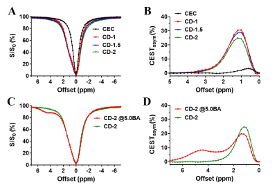 |
CEST imaging of self-healing hydrogels for drug delivery to the brain
Xiongqi Han1, Jianpan Huang1, and Kannie Wai Yan Chan1,2
1Department of Biomedical Engineering, City University of Hong Kong, Hong Kong, Hong Kong, 2Russell H. Morgan Department of Radiology and Radiological Science, Johns Hopkins Medicine, Baltimore, MD, United States
Self-healing hydrogels can adapt to the dynamic and mechanically demanding environment in the brain when compare to conventional brittle hydrogels. Herein, we developed a series of self-healing chitosan-dextran based hydrogels (CDgels) which were mechanically soft and its composition could be detected via CEST-MRI. Once crosslinked, CEST-MRI contrast at 1.2 ppm decreased when the crosslinking density increased. Interestingly, this phenomenon was observed when we further incorporated barbituric acid (BA) into CDgels to form part of the Schiff-base interaction. The resultant BA-loaded CDgels showed both CEST contrast at 3T, demonstrating a robust approach for imaging-guided hydrogel-based therapy in brain.
|
|
0905.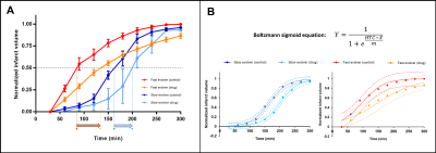 |
Oxygen carrier therapy slows infarct growth in large vessel occlusion dog model based on perfusion- and diffusion-weighted MRI analysis
Mohammed Salman Shazeeb1,2, Robert King1,2, Josephine Kolstad1, Christopher Raskett1, Natacha Le Moan3, Jonathan A. Winger3, Lauren Kelly3, Ana Krtolica3, Nils Henninger1, and Matthew Gounis1
1University of Massachusetts Medical School, Worcester, MA, United States, 2Worcester Polytechnic Institute, Worcester, MA, United States, 3Omniox Inc., San Carlos, CA, United States
The dog large vessel occlusion (LVO) model mimics the clinical trend observed in patients where the brain infarct follows either a slow or fast progression. The dog LVO model can be used in the design of new therapeutics to improve clinical outcome in patients. This study examined the effect of an oxygen carrier in its ability to slow infarct growth in the dog LVO model. In fast evolvers, the oxygen carrier therapy prolonged infarct progression and reduced the final normalized infarct volume. Delaying infarct progression can potentially extend the time-window for thrombectomy enabling more patients to receive this critical treatment.
|
|
0906.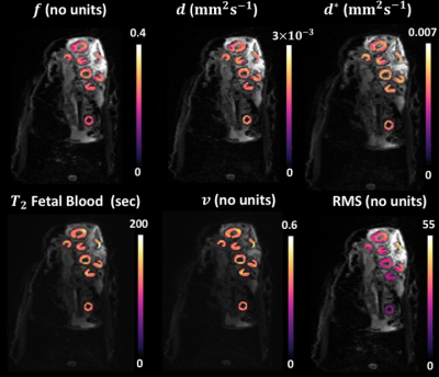 |
A Bayesian Approach for Diffusion-Weighted Imaging to study placenta development and function in pregnancy in a large animal model
Dimitra Flouri1,2, Jack RT Darby3, Stacey L Holman3, Sunthara R Perumal4, Anna L David5,6, Janna L Morrison3, and Andrew Melbourne1,2
1School of Biomedical Engineering & Imaging Sciences, King's College London, London, United Kingdom, 2Department of Medical Physics & Biomedical Engineering, University College London, London, United Kingdom, 3Early Origins of Adult Health Research Group, University of South Australia, Adelaide, Australia, 4Preclinical Imaging and Research Laboratories, South Australian Health and Medical Research Institute, Adelaide, Australia, 5Institute for Women's Health, University College London, London, United Kingdom, 6NIHR University College London Hospitals Biomedical Research Center, London, United Kingdom
Abnormalities of placental development and function result in fetal growth restriction. There is growing interest in understanding placenta structure and function throughout pregnancy to gain better understanding of placenta dysfunction. Advances in technology enables derivation of quantitative indices that reflect tissue microcapillary perfusion and tissue diffusivity from MRI. Despite recent progress, in-vivo diffusion-weighted MRI remains challenging due to long scan times, respiratory motion and low signal-to-noise ratio. Sheep provide a relevant large-scale model for invasive validation studies for MRI measurements. We aimed to improve parameter mapping using Bayesian inference. Bayesian analysis yields improved parameter maps relative to conventional least-squares fitting.
|
|
|
0907.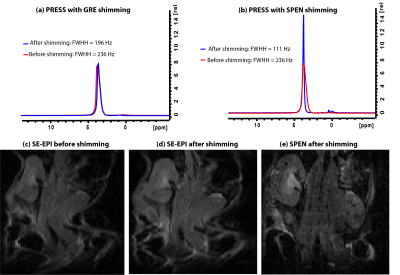 |
A robust shimming method for in vivo abdominal mice studies based on ultrafast pulse sequences
Qingjia Bao1, Ricardo Martinho 1, and Lucio Frydman1
1Weizmann Institute of Science, Rehovot, Israel
A challenge in functional, diffusivity and spectroscopic MRI studies of mouse body regions is B0 inhomogeneity, especially at ultrahigh fields. To map and correct these DB0s, phase difference images are usually acquired using gradient echo sequences. These, however, are hard to obtain with good quality in abdominal studies due to motion artifacts. In this study we report a fully automated 3D map-based shimming method based on an ultrafast sequence that can overcome these artifacts, delivering optimal B0 homogeneity over the targeted ROIs. This technique is exemplified with mice studies at 15.2T, where its usefulness and reproducibility is demonstrated.
|
0908.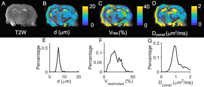 |
In vivo Quantification of Restriction Sizes in Gray Matter of Rat Brain Using Temporal Diffusion Spectroscopy
xiaoyu jiang1, junzhong xu1, sean p. devan1, and john c. gore1
1Vanderbilt University Institute of Imaging Science, nashville, TN, United States
The diffusion time (tdiff) dependence of diffusion MRI signals provides a means to characterize tissue microstructure at cellular levels. Several studies have reported the diffusion time dependence of diffusion signals from rodent brains in health and disease, as well as human brains. Here, we apply IMPULSED (Imaging Microstructural Parameters Using Limited Spectrally Edited Diffusion), a temporal diffusion spectroscopy-based method that we have described previously, to map the mean restriction size for water diffusion in gray matter of the rat brain in vivo by fitting the tdiff dependence of diffusion signals to a simple biophysical model.
|
 Watch the Video