
Oral - Power Pitch Session
Cancer Imaging: Machine Learning & Advanced Imaging
| Monday Parallel 5 Live Q&A | Monday, 10 August 2020, 14:30 - 15:15 UTC | Moderators: Nima Gilani |
0279.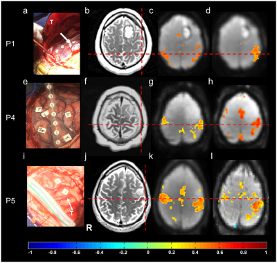 |
Presurgical Mapping in Brain Tumors with High-Speed Resting-State fMRI: Comparison with Task-fMRI and Intra-Operative Mapping
Stefan Posse1, Kishore Vakamudi1, Bruno Sa De La Rocque Guimaraes1, Rex Jung2, and Muhammad Omar Chohan2
1Neurology, University of New Mexico, Albuquerque, NM, United States, 2Neurosurgery, University of New Mexico, Albuquerque, NM, United States
We investigated presurgical resting-state fMRI (rsfMRI) in 9 patients with brain tumors using high-speed multiband-EPI (TR:400ms) with real-time data quality monitoring for seed-based localization of sensorimotor and language networks. The Euclidean distance between intra-operative electrocortical-stimulation (ECS) and rsfMRI connectivity and task-activation in motor cortex, Broca’s and Wernicke’s areas was 5-13mm, except for discordant rsfMRI localization of Wernicke’s area in one patient due to possible altered neurovascular coupling. A secondary objective was to accelerate encoding using echo-volumar-imaging. This study demonstrates the potential of high-speed rsfMRI for presurgical mapping and clinically-acceptable concordance with task-based fMRI and ECS localization.
|
|
 |
0280.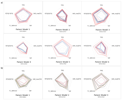 |
Patient Specific Planning of Thermal Magnetic Resonance: An EMF Simulation Study in Realistic Glioblastoma Multiforme Models
Eva Oberacker1, Cecilia Diesch1, Jacek Nadobny2, Andre Kuehne3, Thomas Wilhelm Eigentler1, Pirus Ghadjar2, Peter Wust2, and Thoralf Niendorf1,3,4
1Berlin Ultrahigh Field Facility (B.U.F.F.), Max Delbrück Center for Molecular Medicine in the Helmholtz Association, Berlin, Germany, 2Clinic for Radiation Oncology, Charité Universitätsmedizin, Berlin, Germany, 3MRI.TOOLS GmbH, Berlin, Germany, 4Experimental and Clinical Research Center (ECRC), joint cooperation between the Charité Medical Faculty and the Max-Delbrück-Center for Molecular Medicine in the Helmholtz Association, Berlin, Germany
Ultrahigh-field MR employs higher radio frequencies (RF) than conventional MR and has unique potential to provide focal temperature manipulation and high resolution imaging in an integrated device (ThermalMR). The advantage of integrated therapy monitoring and guidance benefits thermal interventions in brain tumor treatments. To approach this goal this work examines the inter-patient variability in a heterogeneous group of glioma multiforme patients using EMF simulations. Our findings provide useful indicators as potential patient inclusion criteria for thermal treatment of brain tumors and form the technological basis for treatment planning and RF applicator developments en route to clinical applications of Thermal MR.
|
0281. |
Characterization of CT-2A neurosphere-derived high-grade glioma in mice.
Uwe Himmelreich1, Matteo Riva2, Sarah Belderbos1, Roxanne Wouters2, Akila Weerasekera1, Thais Baert2, Willy Gsell1, and An Coosemans2
1Biomedical MRI, University of Leuven, Leuven, Belgium, 2Department of Oncology, University of Leuven, Leuven, Belgium Several promising treatments against high-grade gliomas (HGGs) failed to provide significant benefit when translated from the preclinical setting to patients. Improving animal models is fundamental to overcome this translational gap. We have developed and comprehensively characterized in-vivo model based on the orthotopic implantation of CT-2A cells cultured in neurospheres (NS). Anatomical, metabolic (MRS) and perfusion MRI indicated that CT-2A NS-derived tumors showed a more HGG-like behavior, which was supported by survival data, increased glioma stem cell population and enhanced neoangiogenesis. Because of these specific features, the CT-2A NS-derived model represents a high-translational platform for the search of new HGG treatments. |
|
0282.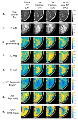 |
Magnetization Transfer and Chemical Exchange Saturation Transfer in Glioblastoma at 1.5T: Comparison of Early and Late Tumor Progression
Rachel W Chan1, Sten Myrehaug2, Greg J Stanisz1,3,4, James Stewart2, Mark Ruschin2, Arjun Sahgal2,3, and Angus Z Lau1,3
1Physical Sciences, Sunnybrook Research Institute, Toronto, ON, Canada, 2Department of Radiation Oncology, Sunnybrook Health Sciences Centre, University of Toronto, Toronto, ON, Canada, 3Medical Biophysics, University of Toronto, Toronto, ON, Canada, 4Department of Neurosurgery and Pediatric Neurosurgery, Medical University, Lublin, Poland
This study aims to quantify the MT and CEST parameters in glioblastoma at 1.5T over four time points of chemoradiation. Previous approaches to quantify the MT/CEST signal in terms of response to treatment were at higher field strengths. Additionally, we analyzed different subregions within the clinical target volume and gross tumor volume, generated by thresholding based on the T1-weighted, FLAIR and DWI scans, for comparing the MT/CEST parameters between the early and late tumor progression. Results indicated that MT/CEST at 1.5T can be used for monitoring therapy and that the results depend on the specific tumor region analyzed.
|
|
0283.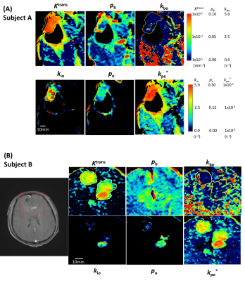 |
A Comprehensive, Adaptive Approach for Shutter-Speed DCE-MRI Analyses of Heterogeneous Brain Tumor Data
Ruiliang Bai1, Bao Wang2, Yinhang Jia1, Zhaoqing Li3, Yi-Cheng Hsu4, Charles S. Springer Jr.5, and Yingchao Liu6
1Interdisciplinary Institute of Neuroscience and Technology, School of Medicine, Zhejiang University, Hangzhou, China, 2Department of Radiology, Shandong University Qilu Hospital, Jinan, China, 3College of Biomedical Engineering and Instrument Science, Zhejiang University, Hangzhou, China, 4MR Collaboration, Siemens Healthcare, Shanghai, China, 5Advanced Imaging Research Center, Oregon Health & Science University, Portland, OR, United States, 6Department of Neurosurgery, Provincial Hospital Affiliated to Shandong University, Jinan, China
The [Shutter-Speed-Model-Dynamic-Contrast-Enhanced] SSM-DCE-MRI pharmacokinetic analysis has a metabolic dimension. However, SSM must be applied thoughtfully, especially in Glioblastoma multiforme [GBM], because of strong tissue vascular heterogeneity across the brain field-of-view Here, we present a method to select the appropriate SSM-DCE-MRI version to analyze such tissue, on a pixel-by-pixel basis. The supra-intensive parameters, vascular water efflux (kbo), cellular water efflux (kio), and vascular CA efflux (kpe) rate constants could be reliably determined. Pilot data on one recurrent and one pre-diagnosis GBM patient are presented to demonstrate method performance.
|
|
0284.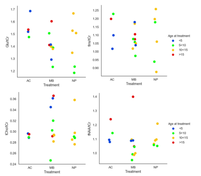 |
Hippocampal MR Spectroscopy show chronic metabolic effects following cranial radiotherapy in childhood cancer survivors
Maria Ljungberg1,2, Erik Fernström3, Oscar Jalnefjord1,2, Mikael Montelius1, Thomas Björk-Eriksson3, Marie Kalm4, and Marianne Jarfelt5
1Radiation Physics, Institute of Clinical Sciences, Sahlgrenska Academy, Gothenburg University, Göteborg, Sweden, 2Medical Physics and Biomedical Engineering, Sahlgrenska University Hospital, Göteborg, Sweden, 3Oncology, Institute of Clinical Sciences Sahlgrenska Academy Gothenburg University, Göteborg, Sweden, 4Health and Rehabilitation, Institute of neuroscience and physiology, Sahlgrenska Academy Gothenburg University, Göteborg, Sweden, 5Pediatrics, Institute of Clinical Sciences, Sahlgrenska Academy Gothenburg University, Göteborg, Sweden
Modern cranial radiotherapy (CRT) achieves high targeted doses of radiation to brain tumours, which has resulted in an increased population of long-term survivors of childhood cancer. Unfortunately, the CRT cause radiation damage to the healthy brain which results in cognitive deficits. In a group of adult childhood cancer survivors lower ratios of tNAA/tCho, Glu/tCho and Glu/tCr were obtained in the hippocampus for the patient group that received highest radiation doses to target volume as compared to the group that did not receive any CRT, indicating a still ongoing process of e.g inflammation, re-myelination and gliosis due to CRT
|
|
0285.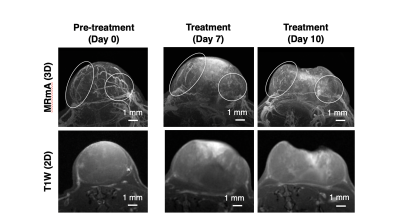 |
Longitudinal 3D intratumoral evaluation of an anti-angiogenic tumor treatment using a Gd-nano liposomal contrast agent and MR micro angiography
Nobuhiro Nitta1, Yoichi Takakusagi1, Daisuke Kokuryo2, Sayaka Shibata1, Akihiro Tomita3, Tatsuya Higashi1, Ichio Aoki1, and Masafumi Harada4
1Department of Molecular Imaging and Theranostics, National Institutes for Quantum and Radiological Science and Technology, Chiba, Japan, 2Graduate School of System Informatics, Kobe University, Hyogo, Japan, 3Department of Hematology, Fujita Health University School of Medicine, Aichi, Japan, 4Graduate School of Medicine, Tokushima University, Tokushima, Japan
The enhanced permeability and retention (EPR) effect depends on nanoparticle properties and tumor/vessel conditions. We aimed to develop a tumor vasculature evaluation method and high-resolution nanoparticle-delivery imaging technique using magnetic resonance micro-imaging technology and a gadolinium (Gd)-dendron assembled liposomal contrast agent. We achieved 50-μm isotropic MR angiography with clear visualization of tumor micro-vessel structure. The Gd-liposome-enhanced MR micro-imaging revealed differences in the vascular structures between two different types of grafted mice models. The MR micro-imaging methods facilitate the evaluation of intratumoral vascularization patterns, the quantitative assessment of vascular-properties that alter tumor malignancy, particle retentivity, and the effects of treatment.
|
|
0286.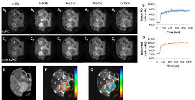 |
Radial sampling and KWIC-reconstruction reduce motion artifacts and improve spatial resolution for DCE-MRI of mouse pancreatic cancer
Jianbo Cao1, Stephen Pickup1, Hee Kwon Song1, and Rong Zhou1,2
1Department of Radiology, University of Pennsylvania, Philadelphia, PA, United States, 2Abramson Cancer Center, University of Pennsylvania, Philadelphia, PA, United States
Genetically engineered mouse (GEM) model of pancreatic ductal adenocarcinoma (PDA) recapitulates a dense stroma exhibited in human disease and is thus relevant for testing stroma-directed drugs. However, the lesion location makes it highly susceptible to motion. We present a multi-slice 2D saturation-recovery technique using golden-angle radial k-space sampling combined with KWIC reconstruction for DCE-MRI of PDA. This method minimizes respiration motion artefacts in PDA tumors and increases the effective temporal resolution of the AIF. Ktrans and kep maps derived from PK model fitting of DCE series exhibit adequate spatial resolution to reveal permeability and perfusion heterogeneity in the PDA tumor.
|
|
0287.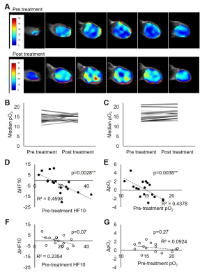 |
Imaging assessment of pancreatic ductal adenocarcinoma xenograft treated with hypoxia activated prodrug Evofosfamide
Shun Kishimoto1, Nallathamby Devasahayam1, Yu Saida1, Yasunori Otowa1, Kazutoshi Yamamoto1, Jeffrey R Brender1, and Murali C Krishna1
Video Permission Withheld
1NCI, Bethesda, MD, United States
TH-302 is designed to release cytotoxic bromo-isophosphoramide (Br-IPM) moiety in hypoxic microenvironment. Therefore, this drug preferentially attacks the hypoxic region in cancer where other standard anti-cancer treatment such as chemotherapy and radiation therapy are ineffective. Here, we monitored the change in tumor hypoxia and perfusion in response to TH-302 treatment by EPR oximetry and DCE MRI using two pancreatic ductal adenocarcinoma xenograft models. The result showed improved oxygenation only in treatment sensitive MIA Paca-2 tumors without modulating tumor blood perfusion, suggesting that intratumor pO2 is a useful biomarker to evaluate treatment response to TH-302.
|
|
0288.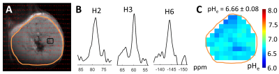 |
Extracellular pH Changes Induced by Immuno-Thermal Ablation in a Murine Colorectal Cancer Model
Daniel Coman1, Ryan J Slovak2,3, Fahmeed Hyder1,4,5, and Hyun S Kim1,2,5,6
1Radiology & Biomedical Imaging, Division of Bioimaging Sciences, Yale University School of Medicine, New Haven, CT, United States, 2Radiology & Biomedical Imaging, Section of Interventional Radiology, Yale University School of Medicine, New Haven, CT, United States, 3University of Connecticut School of Medicine, Farmington, CT, United States, 4Department of Biomedical Engineering, Yale University School of Medicine, New Haven, CT, United States, 5Yale Cancer Center, Yale University School of Medicine, New Haven, CT, United States, 6Department of Internal Medicine, Section of Medical Oncology, Yale University School of Medicine, New Haven, CT, United States
Acidification of tumor microenvironment is associated with aggressive tumor growth and facilitate resistance to anti-cancer therapies. Extracellular pH (pHe) mapping with BIRDS is used to differentiate ablated and non-ablated tumors in the setting of systemic immunotherapy of murine colorectal cancer. Combination of Cryoablation with Dual Immune Checkpoint Blockade (DICB) resulted in a significant pHe increase compared to control tumors. This work demonstrates the feasibility of measuring pHe with BIRDS in a murine colorectal cancer model. pHe imaging could serve as a non-invasive imaging biomarker for tumor microenvironment assessment and monitoring of metabolic changes after immuno-thermal ablation therapy.
|
|
0289.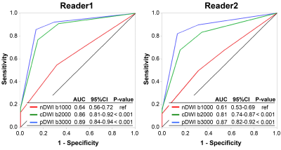 |
Utility of computed diffusion-weighted imaging for evaluating primary prostate cancer in whole-body MRI
Yuki Arita1,2, Yuma Waseda3, Soichiro Yoshida2,3, Taro Takahara2,4, Chikako Ishii2, Thomas C Kwee5, Ryota Ishii6, Shigeo Okuda1, Yasuhisa Fujii3, and Masahiro Jinzaki1
1Department of Radiology, Keio University School of Medicine, Tokyo, Japan, 2Department of Radiology, Advanced Imaging Center Yaesu Clinic, Tokyo, Japan, 3Department of Urology, Tokyo Medical and Dental University Graduate School, Tokyo, Japan, 4Department of Biomedical engineering, Tokai University School of Engineering, Kanagawa, Japan, 5Department of Radiology, Nuclear Medicine and Molecular Imaging, University Medical Center Groningen, Groningen, Netherlands, 6Department of Biostatistics Unit, Clinical and Translational Research Center, Keio University Hospital, Tokyo, Japan
The purpose of this study is to determine the value of applying computed DWI to a whole-body MRI/DWI protocol to acquire computed high b-value (2000 s/mm2) diffusion-weighted images for local prostate cancer evaluation. Based on the results, computed diffusion-weighted images obtained from whole-body MRI provide a similar diagnostic performance compared to pelvic bi-parametric MRI for the detection of primary prostate cancer. Computed DWI is a straightforward postprocessing technique without the need for additional image acquisition time. It can be recommended for use in routine clinical practice in whole-body MRI protocols for concurrent evaluation of primary and metastatic prostate cancer.
|
|
0290.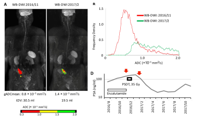 |
Progressive site-directed therapy for oligo-progressive castration-resistant prostate cancer
Soichiro Yoshida1, Taro Takahara2, Yuki Arita3,4, Chikako Ishii4, Kazuma Toda5, Kazuma Toda5, Tsuyoshi Sakamoto6, Toshiki Kijima1, Minato Yokoyama1, Junichiro Ishioka1, Yoh Matsuoka1, Kazutaka Saito1, Ryoichi Yoshimura5,
and Yasuhisa Fujii1
1Urology, Tokyo Medical and Dental University, Tokyo, Japan, 2Biomedical Engineering, Tokai University School of Engineering, Kanagawa, Japan, 3Radiology, Keio University School of Medicine, Tokyo, Japan, 4Radiology, Advanced Imaging Center, Yaesu Clinic, Tokyo, Japan, 5Radiation Therapeutics and Oncology, Tokyo Medical and Dental University, Tokyo, Japan, 6PixSpace, Ltd., Fukuoka, Japan
Locoregional therapy for oligometastatic prostate cancer has generated great interest. However, the benefit for castration-resistant prostate cancer (CRPC) has not been fully demonstrated. According to the current study, whole body-MRI incorporating DWI identified a substantial number of oligo-progressive CRPC patients (OP-CRPC) with a number of progressive lesions 3 or less. Progressive site-directed therapy (PSDT) to OP-CRPC provided a high treatment effect in terms of prostate-specific antigen (PSA) response, especially for patients with longer PSA-doubling time. Furthermore, this study identified the site-dependencies of the PSDT treatment effect; patients whose progressive site was localized in the pelvis were good candidates for PSDT.
|
|
 |
0291.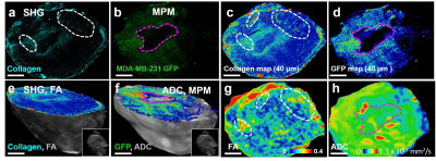 |
A Multiscale, Multimodality Pipeline for “Image-based” Cancer Systems Biology
Akanksha Bhargava1, Manisha Aggarwal1, and Arvind Pathak1
1Russell H. Morgan Department of Radiology and Radiological Science, Johns Hopkins University, Baltimore, MD, United States
Preclinical imaging methods such as magnetic resonance microscopy (µMRI), micro-computed tomography (µCT) and optical techniques such as multiphoton microscopy (MPM) have been instrumental in advancing our understanding of the role of vasculature in cancer1. However, integrating vascular microenvironmental data across modalities and spatial scales for novel “image-based” cancer systems biology applications2 remains a challenge. Therefore, we developed a multimodality imaging pipeline that achieves multiscale data integration, image co-registration and results in the generation of “cancer atlases” for systems and computational biology applications.
|
|
0292.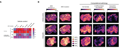 |
Evaluating the sensitivity of ADC to childhood neuroblastoma pathology in vivo using Gaussian mixture modeling and computational pathology
Konstantinos Zormpas-Petridis1, Matthew D. Blackledge1, Louis Chesler2, Yinyin Yuan3, Simon P. Robinson1, and Yann Jamin1
1Radiotherapy and Imaging, Institute of Cancer Research, London, Sutton, United Kingdom, 2Clinical studies, Institute of Cancer Research, London, Sutton, United Kingdom, 3Molecular pathology, Institute of Cancer Research, London, Sutton, United Kingdom
We use Gaussian mixture modeling, computational pathology and MRI-histopathology registered datasets to evaluate i) the sensitivity of diffusion-weighted imaging to the rich histopathology of childhood neuroblastoma and ii) the sensitivity of the derived apparent diffusion coefficient, ADC, as a biomarker of response to a potent MYCN-targeted small molecule inhibitor in the Th-MYCN mouse, a faithful model of high-risk MYCN-driven disease.
|
0293.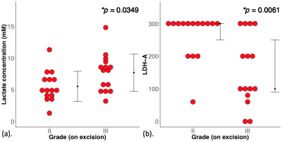 |
Lactate concentration from double quantum filtered (DQF) MRS in human breast tumour is associated with tumour grading and patient prognosis
Sai Man Cheung1, Ehab Husain2, Yazan Masannat3, Klaus Wahle4, Steven Heys3, and Jiabao He1
1Aberdeen Biomedical Imaging Centre, University of Aberdeen, Aberdeen, United Kingdom, 2Pathology Department, Aberdeen Royal Infirmary, Aberdeen, United Kingdom, 3Breast Unit, Aberdeen Royal Infirmary, Aberdeen, United Kingdom, 4Institute of Pharmacy and Biological Sciences, University of Strathclyde, Glasgow, United Kingdom Upregulation of aerobic glycolysis and an elevated lactate accumulation have been linked to tumour aggressiveness. However, current evidence drawn from cell culture and small animal models remains controversial. Since lactate and lipid share the same spectral frequency, conventional MRS is inadequate in quantifying lactate under overwhelming lipid signal. Double quantum filtered (DQF) MRS allows excellent suppression of lipid signal from adipose breast tissues. We examined prognostic role of lactate concentration through a cross sectional study in grade II and III whole tumours freshly excised from patients with breast cancer using DQF MRS for the quantification of lactate concentration. |
 Watch the Video