
Oral - Power Pitch Session
MRS and Molecular Imaging, Development and Applications
Session Topic: MRS and Molecular Imaging, Development and Applications
Session Sub-Topic: Molecular Imaging Technical Developments & Novel Applications
Oral - Power Pitch
Molecular Imaging
| Tuesday Parallel 5 Live Q&A | Tuesday, 11 August 2020, 14:30 - 15:15 UTC | Moderators: René Botnar |
Session Number: PP-19
0623.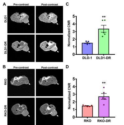 |
MRMI of Extradomain-B Fibronectin for Assessing Drug Resistance in Colon Cancer
Zheng-Rong Lu1, Amita Vaidya1, Nadia Ayat1, Helen Wang1, and Megan Buford1
1Biomedical Engineering, Case Western Reserve University, Cleveland, OH, United States
Non-invasive active surveillance and risk-stratification of drug-resistant colon cancer is imperative, in order to facilitate disease management and tailor therapeutic interventions. This research demonstrates that acquired drug resistance in colon cancer is associated with enhanced expression of extracellular matrix oncoprotein extradomain-B fibronectin (EDB-FN). MR molecular imaging of EDB-FN at a subclinical dose of macrocyclic ZD2-targeted contrast agent ZD2-N3-Gd(HP-DO3A) facilitates effective assessment of drug-resistant colon cancer in two independent models, highlighting the potential of EDB-FN as a diagnostic molecular marker for invasive colon cancer.
|
|
 |
0624.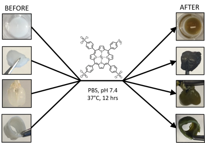 |
An MRI Method for Labeling and Imaging Decellularized Extracellular Matrix Scaffolds for Tissue Engineering
Daniel Andrzej Szulc1,2, Mohammadali Ahmadipour1,3, Fabio Gava Aoki3,4, Thomas K. Waddell1,3,5, Golnaz of Karoubi5,6,7, and Hai-Ling Margaret Cheng1,2,7,8,9
1Institute of Biomaterials and Biomedical Engineering, University of Toronto, Toronto, ON, Canada, 2Translational Biology & Engineering Program, Ted Rogers Centre for Heart Research, Toronto, ON, Canada, 3Latner Thoracic Surgery Laboratories, Toronto General Hospital Research Institute, University Health Network, Toronto, ON, Canada, 4Biomedical Engineering Laboratory, University of Sao Paulo, Sao Paulo, Brazil, 5Department of Mechanical and Industrial Engineering, University of Toronto, Toronto, ON, Canada, 6Latner Thoracic Surgery Laboratories, University of Toronto, Toronto, ON, Canada, 7Ontario Institute for Regenerative Medicine, Toronto, ON, Canada, 8Heart & Stroke/Richard Lewar Centre of Excellence for Cardiovascular Research, Toronto, ON, Canada, 9Edward S. Rogers Sr. Department of Electrical & Computer Engineering, University of Toronto, Toronto, ON, Canada
Extracellular matrix (ECM) forms the underlying complex structure of bodily tissues, for this reason, ECM has been greatly explored as an ideal scaffold for tissue engineering. To better understand and optimize scaffold-based therapies we require sensitive and non-invasive imaging techniques. In this study, a novel and facile method for labelling and imaging decellularized ECM scaffolds is presented. A series of tissue-specific (bladder, lung, and tracheal smooth muscle and cartilage) dECM scaffolds are labelled with a small molecule manganese porphyrin, MnPNH2. The labelled scaffolds are biocompatible and exhibit significant and sustained signal in vitro and in vivo.
|
|
0625.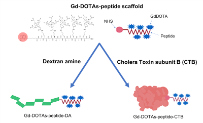 |
Multivalent Gadolinium-decorated Peptides for Versatile Bioconjugation of Molecular MRI Probes
Nikorn Pothayee1, Deepak Sail2, Stephen Dodd1, Rolf Swenson2, and Alan Koretsky1
1Laboratory of Functional and Molecular Imaging, National Institutes of Health, Bethesda, MD, United States, 2Imaging Probe Development Center, National Institutes of Health, Rockville, MD, United States
One of the most important goals of brain imaging is to define the anatomical connections within the brain. In addition to revealing normal circuitry, studies of neural connections and their transports can show rewiring and outgrowth during degeneration following brain injury and diseases. Ultrasensitive agents that can reveal neuroconnectivity and axonal transport dynamics in vivo will be very useful and allow for the interrogation of changes in brain connections and circuitry. In this work, we report two novel MR-visible neural tracers that can be used to visualize neuroconnectivity in vivo.
|
0626.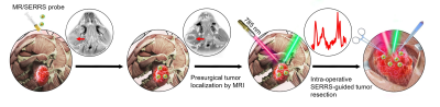 |
Image-guided tumor resection of head and neck carcinoma (HNSCC) in rabbit models with targeting MRI-Raman nanoprobe
Pengpeng Sun1, Yunfei Zhang2, Kaicheng Li1, Cong Wang3, Feng Zeng3, Jinyu Zhu1, Yongming Dai2, Xiaofeng Tao1, and Yinwei Wu1
1Shanghai Ninth People’s Hospital, Shanghai, China, 2United Imaging Healthcare, Shanghai, China, 3Fudan University, Shanghai, China
Accurately defining infiltrative tumor margin is extremely crucial for complete resection and avoiding mistaken removal of normal tissue. Currently, pre-operative MRI imaging is the most widely-used strategy for defining tumor margin. However, there are plenty of insurmountable disadvantages in terms of accuracy, low resolution, mismatch and so on. This research aims to develop one targeting MRI-Raman nanoprobe able to pre-operatively and intra-operatively evaluate infiltrative margin of head and neck carcinoma (HNSCC) and real-timely guide tumor resection. The results showed that the nanoprobe greatly benefits the complete resection of HNSCC. As a result, the prognosis of tumor-bearing rabbits were considerably improved.
|
|
|
0627.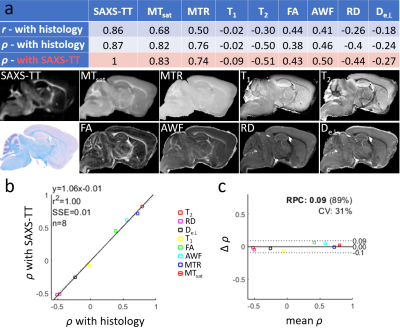 |
Myelin-specific imaging using synchrotron X-ray scattering and comparison to MRI myelin-sensitive methods and histology
Marios Georgiadis1,2,3, Els Fieremans2, Aileen Schroeter3, Manuel Guizar-Sicairos4, Zirui Gao4, Aleezah Balolia5, Piotr Walczak6,7, Lin Yang8, Gergely David9, Jiangyang Zhang2, Dmitry S. Novikov10, Markus Rudin3,11, and Michael Zeineh1
1Radiology, Stanford University, Stanford, CA, United States, 2NYU School of Medicine, New York, NY, United States, 3Institute for Biomedical Engineering, ETH Zurich, Zurich, Switzerland, 4Swiss Light Source, Paul Scherrer Institute, Villigen, Switzerland, 5Psychology, University of Colorado Denver, Denver, CO, United States, 6Radiology, Johns Hopkins Medicine, Baltimore, MD, United States, 7Diagnostic Radiology & Nuclear Medicine, University of Maryland School of Medicine, Baltimore, MD, United States, 8National Synchrotron Light Source II, Brookhaven National Laboratory, Upton, NY, United States, 9Balgrist University Hospital, University of Zurich, Zurich, Switzerland, 10Center for Biomedical Imaging, NYU School of Medicine, New York, NY, United States, 11Institute of Pharmacology and Toxicology, University of Zurich, Zurich, Switzerland
Axonal myelination is an important indicator of brain development and is implicated in many neurologic diseases. However, MRI methods to probe myelin are sensitive but not specific. Small-angle X-ray scattering (SAXS) produces signal specific to myelin’s nanostructural periodicity. Here we apply the recently developed SAXS tensor tomography (SAXS-TT) to non-invasively retrieve myelin levels in mouse brains, and compare them to myelin-sensitive MRI methods. We demonstrate SAXS-TT myelin specificity i) using myelin histology, ii) on a dysmyelination model and iii) by selectively probing central and peripheral nervous system myelin. We propose SAXS-TT as quantitative tomographic method for validating MRI myelin-sensitive sequences.
|
0628.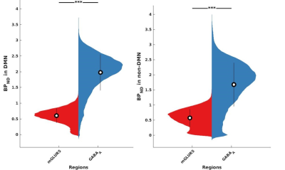 |
mGLUR5 and GABAA Receptor’s Association with fMRI BOLD Signals in the Default Mode Network as Assessed via Simultaneously recorded PET/MR data
Ravichandran Rajkumar1,2,3, Claudia Régio Brambilla1,2,3, Christine Wyss1,4, Shukti Ramkiran1,2, Linda Orth1,2, Joshua Lewis Bierbrier1,5, Elena Rota Kops1, Jürgen Scheins1, Bernd Neumaier6, Johannes Ermert6, Hans Herzog1, Karl Joseph Langen1,3,7, Christoph Lerche1,
N. Jon Shah1,3,8,9, and Irene Neuner1,2,3
1Institute of Neuroscience and Medicine 4 (INM-4), Forschungszentrum Jülich, Jülich, Germany, 2Department of Psychiatry, Psychotherapy and Psychosomatics, RWTH Aachen University, Aachen, Germany, 3JARA – BRAIN – Translational Medicine, Aachen, Germany, 4Department of Psychiatry, Psychotherapy and Psychosomatics, University of Zurich, Zürich, Switzerland, 5Department of Electrical and Computer Engineering, McMaster University, Hamilton, ON, Canada, 6Institute of Neuroscience and Medicine 5 (INM-5), Forschungszentrum Jülich, Jülich, Germany, 7Department of Nuclear Medicine, RWTH Aachen University, Aachen, Germany, 8Institute of Neuroscience and Medicine 11 (INM-11), Forschungszentrum Jülich, Jülich, Germany, 9Department of Neurology, RWTH Aachen University, Aachen, Germany
fMRI-BOLD signals reflect the synaptic activity and glucose energy metabolism in the brain. This study investigated the association between excitatory (mGLUR5), inhibitory (GABAA) neuroreceptors, and glucose metabolism using PET imaging with resting-state fMRI for the first time. The significantly higher mGLUR5 and GABAA neuroreceptor availability and glucose metabolism within the DMN and its correlations show a possible association between increased energy requirements and neuronal activity in the DMN. Further correlations with fMRI measurements show that higher energy demand is utilised for higher functional connectivity, and consecutively higher connectivity within the DMN is more strongly associated with inhibitory receptors.
|
|
0629.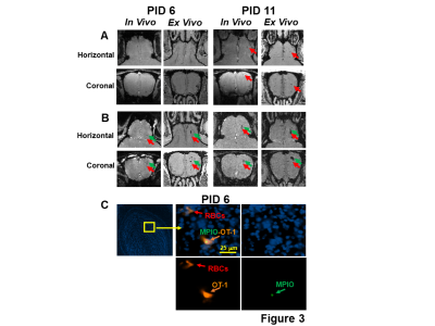 |
MRI Investigation of Adoptive T Cell Transfer and Microbleeds during Vesicular Stomatitis Virus Infection of the Brain
Li Liu1, Stephen Dodd1, Ryan Hunt1, Nikorn Pothayee1, Nadia Bouraoud1, Dragan Maric1, E Ashley Moseman1, Dorian B McGavern1, and Alan P Koretsky1
1National Institute of Neurological Disorders and Stroke, National Institutes of Health, Bethesda, MD, United States
A method to label non-phagocytic CD8 T cells with a micron-sized iron oxide particle (MPIO) has been developed which enables MRI single cell detection. Adoptive transfer of T cells showed therapeutic effects in mice causing less bleeding after intranasal virus infection. MPIO-labeled CD8 T cells entered the brain and the combination of labeled T cells and blood made sites of microbleeds easily detectable by MRI. The ability to track individual immune cells by MRI should open new possibilities for early detection of inflammation as well as to monitor the rapidly expanding field of immune cell therapies.
|
|
0630.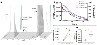 |
Non-invasive Detection of M1 Activation in Macrophages using Hyperpolarized 13C MRS of Pyruvate and DHA at 1.47 Tesla
Kai Qiao1,2, Lydia Le Page1,2, Celine Taglang1,2, and Myriam M Chaumeil1,2
1Physical Therapy and Rehabilitation Science, University of California, San Francisco, San Francisco, CA, United States, 2Radiology and Biomedical Imaging, University of California, San Francisco, San Francisco, CA, United States
The immune system plays an essential role in various diseases, and macrophage activation patterns can vary greatly - impacting intervention. We propose that 13C Magnetic Resonance Spectroscopy (MRS) of hyperpolarized [1-13C] Pyruvate and [1-13C] Dehydroascorbic acid (DHA) can differentiate between non-activated and M1 classically activated macrophages at the clinically-relevant field strength of 1.47T. In M1-activated macrophages we report increased HP Lactate from Pyruvate, and increased HP Ascorbic Acid from DHA compared to Control cells. This study is a first in differentiating between activated and non-activated macrophages with HP probes at this field strength and could become a powerful translational tool.
|
|
|
0631.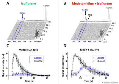 |
Cerebral metabolism of hyperpolarized [2H7, U-13C6]D-glucose in the healthy mouse under different anesthetic conditions
Emmanuelle Flatt1, Bernard Lanz1, Andrea Capozzi1, Magnus Karlsson2, Mathilde H.Lerche2, Rolf Grütter1,3, and Mor Mishkovsky1
1LIFMET, Ecole Polytechnique Fédérale de Lausanne, Lausanne, Switzerland, 2Danmarks Tekniske Universitet, Lyngby, Denmark, 3CIBM, Ecole Polytechnique Fédérale de Lausanne, Lausanne, Switzerland
Glucose is the primary fuel for the brain and its metabolism is linked with cerebral function. Isoflurane anesthesia is commonly employed in preclinical MRS but influences functional connectivity. The combination of isoflurane and medetomidine is regularly used in rodent fMRI and show similar functional connectivity as in awake animals. Here we compared the cerebral metabolism of hyperpolarized [2H7,U-13C6]-D-glucose under these two anesthetic conditions. When using the combination, the [1-13C]lactate signal and lactate-to-glucose ratio were more than doubled compared to isoflurane solely, showing that the change of anesthesia had a high impact on cerebral glucose uptake and glycolytic flux.
|
0632.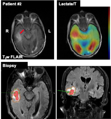 |
Hyperpolarized 13C Pyruvate Imaging of Glioblastoma Patients
Jun Chen1, Toral Patel2, Crystal E Harrison1, Galen D Reed3, Craig R Malloy1, Bruce Mickey2, and Jae Mo Park1,4,5
1Advanced Imaging Research Center, University of Texas Southwestern Medical Center, Dallas, TX, United States, 2Neurosurgery, University of Texas Southwestern Medical Center, Dallas, TX, United States, 3GE Healthcare, GE Healthcare, Dallas, TX, United States, 4Radiology, University of Texas Southwestern Medical Center, Dallas, TX, United States, 5Electrical Engineering, University of Texas Dallas, Richardson, TX, United States
Noninvasive tumor characterization is extremely beneficial for brain tumor patients for establishing surgical procedure and treatment plans. In this study, we imaged newly diagnosed glioblastoma patients using hyperpolarized [1-13C]pyruvate few days prior to surgical procedures and compared the imaging and biopsy results to evaluate the diagnostic values of hyperpolarized pyruvate imaging. Brain regions with increased 13C-lactate production are confirmed as glioblastoma from stereotactic tissue-biopsy. This pilot study with treatment-naïve or newly diagnosed brain tumor patients suggest that preoperative metabolic imaging with hyperpolarized [1-13C]pyruvate may have strong diagnostic value with potential to be an alternative method for tissue biopsy.
|
|
0633.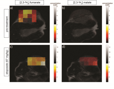 |
Assessing tumor cell death in vivo using 2H-labeled fumarate and deuterium magnetic resonance spectroscopic imaging
Friederike Hesse1, Vencel Somai1,2, Flaviu Bulat1, Felix Kreis1, and Kevin Brindle1,3
1Cancer Research UK Cambridge Institute, Cambridge, United Kingdom, 2Department of Radiology, University of Cambridge, Cambridge, United Kingdom, 3Department of Biochemistry, University of Cambridge, Cambridge, United Kingdom
Monitoring tumor responses to treatment using metabolic imaging can give an early indication of outcome. We show here that Deuterium Metabolic Imaging (DMI) with 2H-labelled fumarate can be used to detect early evidence of cell death following drug treatment in a pre-clinical murine lymphoma model. 2H spectra were acquired from tumors with a time resolution of 5 min, following a bolus injection of 2H-labelled fumarate. Within 48 h of etoposide treatment the rate of tumor malate production from the labelled fumarate increased significantly. Increased levels of labelled malate were also evident in spectroscopic images of the tumors.
|
|
0634.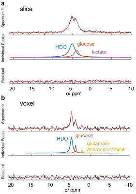 |
Deuterium MR spectroscopy to probe Krebs cycle metabolism of the in-vivo heart
Felix Kreis1, Grzegorz Kwiatkowski1, and Sebastian Kozerke1
1Institute for Biomedical Engineering, University and ETH Zurich, Zurich, Switzerland
We investigated whether deuterium MR spectroscopy using 2H-labeled glucose can be used to assess Krebs cycle metabolism in the in-vivo heart. Localized 2H spectra were acquired from a slice containing the whole heart and from a voxel containing only the left ventricle of the heart using a temporal resolution of ~12 min following a bolus injection of [6,6′-2H2]glucose. Single voxel spectra show the production of labelled Glutamate/Glutamine (Glx) ˜36 min after the administration of the glucose. The Glx concentration reflects Krebs cycle activity which holds potential to probe various metabolic states of the heart.
|
 Watch the Video