
Oral - Power Pitch Session
Quantitative Tissue Properties
Session Topic: Quantitative Tissue Properties
Session Sub-Topic: Contrast Mechanisms: Beyond the Usual Suspects
Oral - Power Pitch
Contrast Mechanisms
| Monday Parallel 1 Live Q&A | Monday, 10 August 2020, 14:30 - 15:15 UTC | Moderators: Janine Lupo and Stefano Mandija |
Session Number: PP-20
 |
0171.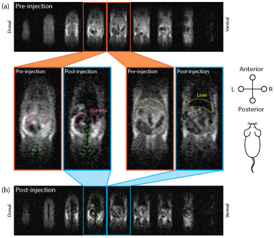 |
High-sensitivity in vivo Contrast Agent Imaging at Ultra-low Magnetic Fields with SPIONs
David Waddington1,2,3, Thomas Boele2,4, Richard Maschmeyer1, Zdenka Kuncic1,5, and Matthew Rosen2,6,7
1Institute of Medical Physics, School of Physics, The University of Sydney, Sydney, Australia, 2A. A. Martinos Center for Biomedical Imaging, Charlestown, MA, United States, 3ACRF Image X Institute, Faculty of Medicine and Health, The University of Sydney, Sydney, Australia, 4ARC Centre of Excellence for Engineered Quantum Systems, School of Physics, The University of Sydney, Sydney, Australia, 5Sydney Nano Institute, The University of Sydney, Sydney, Australia, 6Department of Physics, Harvard University, Cambridge, MA, United States, 7Harvard Medical School, Boston, MA, United States
MRI scanners operating at ultra-low fields (ULF) promise to reduce the cost and expand the clinical accessibility of MRI. Here, we use a 6.5 mT MRI scanner and an efficient balanced steady-state free precession MRI protocol to image superparamagnetic iron oxide nanoparticles (SPIONS) in vivo by leveraging the extremely high magnetization of SPIONs at ULF. Further, we show how positive contrast imaging of SPIONs can be performed at ULF with susceptibility-based techniques. These advances overcome a key limitation of ULF MRI by enabling high-contrast in vivo imaging of clinically safe contrast agents with short acquisition times.
|
 |
0172.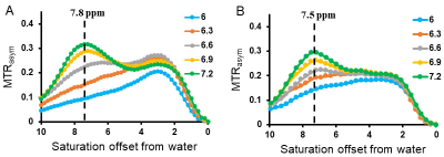 |
Developing imidazoles for performing functional MRI of kidneys
Shaowei Bo1, KowsalyaDevi Pavuluri1, Yunkou Wu2, Farzad Sedaghat1, Martin G. Pomper3, Max Kates4, and Michael T. McMahon5
1The Russell H. Morgan Department of Radiology and Radiological Science, Division of MR Research, The Johns Hopkins University School of Medicine, Baltimore, MD, United States, 2Russell H. Morgan Department of Radiology and Radiological Science, The Johns Hopkins University School of Medicine, Baltimore, MD, United States, 3Russell H. Morgan Department of Radiology and Radiological Science, The Johns Hopkins University School of Medicine, Department of Psychiatry, The Johns Hopkins University School of Medicine, Baltimore, MD, United States, 4Department of Urology, The James Buchanan Brady Urological Institute, The Johns Hopkins University School of Medicine, Baltimore, MD, United States, 5The Russell H. Morgan Department of Radiology and Radiological Science, Division of MR Research, The Johns Hopkins University School of Medicine, F.M. Kirby Research Center for Functional Brain Imaging, Kennedy Krieger Institute, Baltimore, MD, United States
Urinary tract obstructions (UTOs) are blockages that inhibit the flow of urine through its normal path, which can lead to kidney injury and infection. Chemical exchange saturation transfer (CEST) MRI is a fast, noninvasive molecular MRI technique which has shown promise for clinical applications. In this study, we designed and tested a series of imidazoles as CEST MRI contrast agents and tested these for performing functional kidney imaging on a UTO mouse model. The results demonstrate that CEST MRI can facilitate early detection of loss in kidney function.
|
|
0173.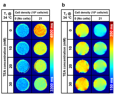 |
A Change in Membrane Potential Induces Measurable Changes in Relaxation Times
Kyeongseon Min1, Tan Toi Phan2,3, Sungkwon Chung4, Jongho Lee1, Seung-Kyun Lee2,3, and Jang-Yeon Park2,3
1Laboratory for Imaging Science and Technology, Department of Electrical and Computer Engineering, Seoul National University, Seoul, Korea, Republic of, 2Department of Biomedical Engineering, Sungkyunkwan University, Suwon, Korea, Republic of, 3Center for Neuroscience Imaging Research, Institute for Basic Science, Suwon, Korea, Republic of, 4Department of Physiology, Samsung Biomedical Research Institute, Sungkyunkwan University School of Medicine, Suwon, Korea, Republic of
In this study, the effects of membrane potential on T1 and T2 were examined using Jurkat T lymphocytes. We applied tetraethylammonium ion (TEA) to depolarize Jurkat cell membrane potential. Significant changes in T1 and T2, which were measured to be -10.39 ms/mM and 0.920 ms/mM, respectively, were observed. One potential explanation for the changes of T1 and T2 is the depolarization of membrane potential, while the underlying mechanism needs to be explored. Further studies are expected to utilize the membrane potential as a new contrast mechanism for MRI.
|
|
0174.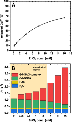 |
Quantifying the Transchelation of Gd3+ Ions from Linear and Macrocyclic GBCA to Glycosaminoglycans using MR Relaxometry
Patrick Schuenke1, Patrick Werner1,2, Matthias Taupitz3, and Leif Schröder1
1Leibniz-Forschungsinstitut fuer Molekulare Pharmakologie (FMP), Berlin, Germany, 2BIOQIC, Charité Berlin, Berlin, Germany, 3Department of Radiology, Charité Berlin, Berlin, Germany
In this study, we quantified the dissociation of GBCAs at different ZnCl2 concentrations and the subsequent chelation of Gd3+ to glycosaminoglycans (GAGs) like heparin. We showed that the relaxivity of the resulting Gd-GAG complexes is about seven times higher compared to that of GBCAs. Under physiological conditions, we further showed that ~20% of the Gd3+-ions transchelated from linear GBCAs to heparin and that these are accountable for more than 50% of the observed relaxation rate. Therefore, Gd-GAG complexes should be considered as the Gd-containing macromolecular substances with high relaxivity that are needed to explain the observed long-term enhancements in vivo.
|
|
0175.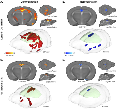 |
Multi-T1D weighting ihMT imaging in the Cuprizone mouse model
Andreea Hertanu1, Lucas Soustelle1, Arnaud Le Troter1, Julie Buron1,2, Victor Carvalho1,3, Myriam Cayre2, Pascale Durbec2, Gopal Varma4, David C. Alsop4, Olivier M. Girard1, and Guillaume Duhamel1
1Aix-Marseille Univ, CNRS, CRMBM, Marseille, France, 2Aix-Marseille Univ, CNRS, IBDM, Marseille, France, 3Aix-Marseille Univ, CNRS, ICR, Marseille, France, 4Division of MR Research, Radiology, Beth Israel Deaconess Medical Center, Harvard Medical School, Boston, MA, United States
Inhomogeneous magnetization transfer (ihMT) is a myelin sensitive MRI technique that provides access to multiple contrast regimes by tuning the amount of dipolar relaxation time (T1D) weighting of the sequence. This opens new perspectives to characterize the sensitivity and specificity of ihMT for myelin in a pathological context. In this study, multiple T1D-weighting ihMT imaging was investigated in the cuprizone mouse model. IhMT signals compared to myelin imaging with fluorescence microscopy demonstrate that ihMT techniques that are weighted towards long T1D values are more specifically related to myelin content during the demyelinating/remyelinating phases of the cuprizone model.
|
0176.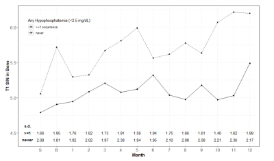 |
Gadolinium-induced Marrow Signal Changes with Metabolic Correlation: Are Cell Lines Directly Affected?
John J DeBevits1, Devin Bageac1, Leo Wolansky1, Paul Dicamillo2, Rong Wu1, Chaoran Hu1, and David Karimeddini1
1UConn Health, Farmington, CT, United States, 2University of Iowa, Iowa City, IA, United States
In the BECOME trial, subjects experienced progressively increased T1W signal changes in deep grey matter nuclei. This retrospective analysis concluded these signal changes also can be seen in the diploic space bone marrow. While increasing trends in hypophosphatemia and leukopenia were also seen in the original study, this analysis has shown that these metabolic abnormalities are not associated with increased marrow signal and that low phosphate and low WBC were not associated with one another. This rejects our hypothesis that gadolinium deposition might interact with a common osteoclast-WBC progenitor cell to result in the metabolic abnormalities.
|
|
0177.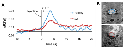 |
Dynamic Susceptibility Contrast with Undersampled Golden-Angle Radial Imaging in the Rodent Spinal Cord
Briana Meyer1 and Matthew Budde2
1Biophysics, Medical College of Wisconsin, Milwaukee, WI, United States, 2Neurosurgery, Medical College of Wisconsin, Milwaukee, WI, United States
Dynamic susceptibility contrast (DSC) to monitor spinal cord perfusion and hemodynamics has the potential to inform the clinical care of spinal cord injury and other disorders. Acquisition of high spatial and temporal resolution images of the rodent spinal cord for DSC perfusion measurements was achieved using a golden-angle radial gradient-echo acquisition combined with iGRASP iterative undersampled reconstruction.
|
|
|
0178.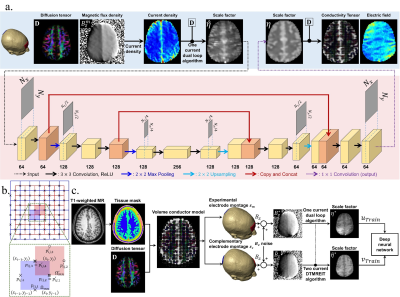 |
In-vivo Electromagnetic Field Mapping for Transcranial Electrical Stimulation (tES) using Deep Learning
Saurav Zaman Khan Sajib1, Munish Chauhan1, and Rosalind J Sadleir1
1School of Biological and Health Systems Engineering, Arizona State University, Tempe, AZ, United States
Magnetic flux densities induced by tES currents can be measured from MR phase and used to reconstruct current density, electric field and conductivity tensor distributions, via diffusion tensor magnetic resonance electrical impedance tomography (DT-MREIT). Determination of tES electric field distributions from DT-MREIT conductivities is challenging, because DT-MREIT requires data from two independent current administrations, increasing acquisition time. We demonstrate a deep-learning model for DT-MREIT reconstruction, showing that conductivity tensors and electric fields can be measured in human subjects in-vivo using a single current administration. This strategy can be used to directly monitor tES electric fields and verify treatment precision.
|
|
0179.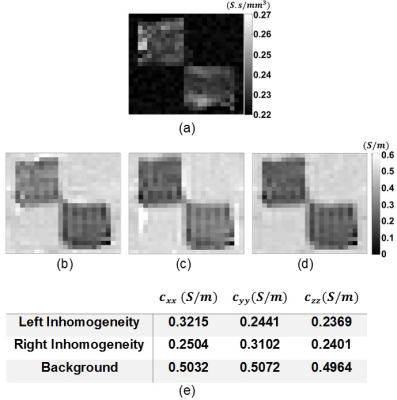 |
Experimental Realization of Single Current Diffusion Tensor Magnetic Resonance Electrical Impedance Tomography
Mehdi Sadighi1, Mert Şişman1, Berk Can Açıkgöz1, and B. Murat Eyüboğlu1
1Electrical and Electronics Engineering Dept., Middle East Technical University (METU), Ankara, Turkey
To obtain low-frequency anisotropic conductivity distribution of biological tissues recently Diffusion Tensor Magnetic Resonance Electrical Impedance Tomography (DT-MREIT), which is combination of the DTI and MREIT techniques, is proposed. There are two in vivo applications of DT-MREIT in the literature where two linearly independent current injection patterns are used. Decreasing the number of current injection patterns to one improves the feasibility of DT-MREIT in clinical applications. In this study, DT-MREIT using a single current injection pattern is experimentally realized. The obtained results approve the validity of the proposed single current DT-MREIT method.
|
|
0180.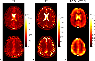 |
Configuration-based Electrical Properties Tomography
Santhosh Iyyakkunnel1,2, Jessica Schäper1,2, and Oliver Bieri1,2
1Department of Radiology, University Hospital Basel, Basel, Switzerland, 2Department of Biomedical Engineering, University of Basel, Basel, Switzerland
Only recently, phase imaging with balanced steady-state free precession (bSSFP) has been suggested for electrical properties tomography (EPT). Here we suggest exploring the SSFP configuration space retrieved from multiple phase-cycled bSSFP scans used for relaxometry also for electrical conductivity mapping. Consequently, the conductivity can be estimated in conjunction with standard quantitative tissue properties requiring no additional scan time.
|
|
0181.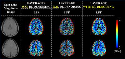 |
Improving Phase-based Conductivity Reconstructions by Means of Deep Learning-based Denoising of B1+ Phase Data
Kyu-Jin Jung1, Stefano Mandija2,3, Jun-Hyeong Kim1, Kanghyun Ryu1, Soozy Jung1, Mina Park4, Mohammed A. Al-masni1, Cornelis A.T. van den Berg2, and Dong-Hyun Kim1
1Department of Electrical and Electronic Engineering, Yonsei University, Seoul, Korea, Republic of, 2Department of Radiotherapy, Division of Imaging & Oncology, University Medical Center Utrecht, Utrecht, Netherlands, 3Computational Imaging Group for MR diagnostics and therapy, Center for Image Sciences, University Medical Center Utrecht, Utrecht, Netherlands, 4Department of Radiology, Gangnam Severance Hospital, Seoul, Korea, Republic of
Electrical Properties Tomography reconstruction technique is highly sensitive to noise, as it requires Laplacian calculations of phase data. To alleviate the noise amplification, large derivative kernels combined with image filters are used. However, this leads to severe errors at tissue boundaries. In this study, we employ a deep learning-based denoising network allowing for noise robust conductivity reconstructions obtained using smaller derivative kernels sizes. This comes with the intrinsic advantage of reduced boundary errors. The feasibility study was performed using cylindrical numerical simulations. Then, the proposed technique was tested using spin echo in-vivo data, and clinical patient data.
|
|
0182.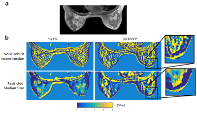 |
Protocol Optimization for in vivo Electrical Propertes Tomography of the Human Breast at 3T
Wyger Brink1, Loes Huijnen1, Reijer Leijsen1, Remco Overdevest1, Andrew Webb1, and Lucia Bossoni1
1C.J. Gorter Center for High Field MRI, dept. Radiology, Leiden University Medical Center, Leiden, Netherlands
This work demonstrates a clinically feasible protocol and reconstruction pipeline which is relatively straightforward to implement, and achieves reliable conductivity reconstructions of the human breast. We aim to establish a reliable MR protocol with a scan time of 6 minutes to further develop the clinical potential of this technique.
|
|
0183.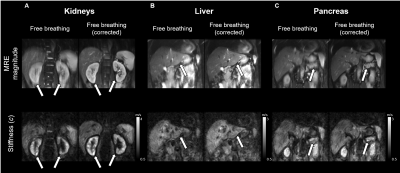 |
Reduction strategies for breathing motion artifacts in abdominal MR elastography
Mehrgan Shahryari1, Helge Herthum1, Gergely bertalan1, Tom Meyer1, Heiko Tzschätzsch1, Carsten Warmuth1, Jürgen Braun2, and Ingolf Sack1
Video Permission Withheld
1Department of Radiology, Charité - Universtitätsmedizin Berlin, Berlin, Germany, 2Institute of Medical Informatics, Charité - Universtitätsmedizin Berlin, Berlin, Germany
MR elastography can provide high-resolution stiffness maps of abdominal organs. However, MRE – in particular when applied with multiple drive frequencies – requires measure times which significantly exceed single breath holds. Therefore, reduction strategies for motion artifacts are required including breath-holds, navigators and image registration, which all were consistently applied and analyzed in this in-vivo study. Our results show that displacement of organs is smallest during breath-hold MRE while, remarkably, mean stiffness values are not significantly affected by breathing. Overall image quality is comparable between breath-hold and free-breathing MRE when the latter is corrected by 2D-image registration during post processing.
|
|
0184.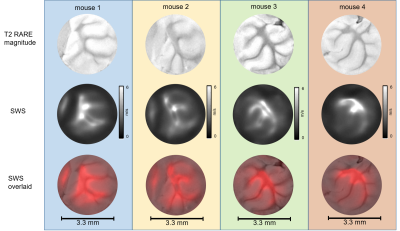 |
Microscopic multifrequency MR elastography with 40 micrometer spatial resolution: Application to murine neural tissue specimens
Gergely Bertalan1, Bettina Müller2, Leif Schröder3, Heiko Tzschätzsch1, Mehrgan Shahryari1, Helge Herthum1, Jing Guo1, Jürgen Braun4, and Ingolf Sack1
1Department of Radiology, Charite Universitätsmedizin Berlin, Berlin, Germany, 2Tierhaltung CCM, Charite Universitätsmedizin Berlin, Berlin, Germany, 3Molecular Imaging, Leibniz Forschungsinstitut für Molekulare Pharmakologie, Berlin, Germany, 4Department of medical Informatics, Charite Universitätsmedizin Berlin, Berlin, Germany
The purpose of this study was the development of multifrequency MR elastography (MRE) of tissue samples with 40 micrometer pixel edge size for analyzing the mechanical properties of murine neural tissue. The new technique revealed in specimens of cerebellum and cortical brain areas that white matter is significantly stiffer than gray matter. Microscopic multifrequency MRE provides insight into micro mechanical structures of ex-vivo soft tissues and might be used in the future to investigate fresh biopsy samples.
|
0185.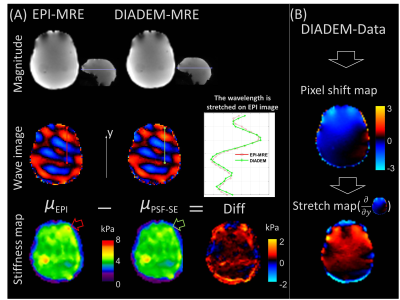 |
High-Resolution Distortion-Free Whole-Brain MR Elastography using Multiband DIADEM (DIADEM-MRE)
Yi Sui1, MyungHo In1, Ziying Yin1, Matthew A. Bernstein1, Richard L. Ehman1, and John III Huston1
1Radiology, Mayo Clinic, Rochester, MN, United States |
 Watch the Video