
Oral - Power Pitch Session
Diffusion Acquisition & Reconstruction
Session Topic: Diffusion Acquisition and Reconstruction
Session Sub-Topic: Diffusion: Acquisition, Reconstruction & Processing
Oral - Power Pitch
Diffusion
| Wednesday Parallel 4 Live Q&A | Wednesday, 12 August 2020, 15:15 - 16:00 UTC | Moderators: Daan Christiaens & Muge Karaman |
Session Number: PP-26
0963.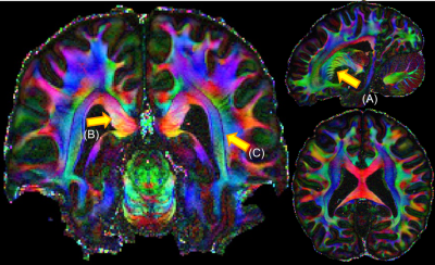 |
Acquisition of a reference Connectom diffusion MRI dataset: In vivo whole-brain diffusion MRI at 760 µm isotropic averaged over 18 hours
Fuyixue Wang1,2, Zijing Dong1,3, Qiyuan Tian1, Congyu Liao1, Qiuyun Fan1, W. Scott Hoge4, Chanon Ngamsombat1, Boris Keil5, Jonathan R. Polimeni1,2, Lawrence L. Wald1,2, Susie Y. Huang1,2, and Kawin Setsompop1,2
1A. A. Martinos Center for Biomedical Imaging, Massachusetts General Hospital, Charlestown, MA, United States, 2Harvard-MIT Health Sciences and Technology, MIT, Cambridge, MA, United States, 33Department of Electrical Engineering and Computer Science, MIT, Cambridge, MA, United States, 4Department of Radiology, Brigham and Women's Hospital, Boston, MA, United States, 5Department of Life Science Engineering, Institute of Medical Physics and Radiation Protection, Giessen, Germany
We present a whole brain in vivo diffusion MRI dataset acquired at 760um isotropic resolution and sampled at 1260 q-space points across 9 two-hour sessions. The creation of this benchmark in vivo diffusion MRI dataset is possible through use of the Connectom scanner, custom-built 64-channel phased-array coil, gSlider acquisition, dual-polarity GRAPPA reconstruction, reverse phase encoding for distortion mitigation, and personalized motion-robust stabilizer. The data will enhance our understanding of gray and white matter structure with fine detail revealed at sub-mm resolution and serve as a reference dataset for new modeling and processing algorithms.
|
|
0964.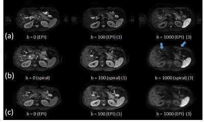 |
Towards Single-shot Diffusion-weighted Spiral Abdominal Imaging on a Clinical Scanner
Peter Börnert1,2, Holger Eggers1, Kay Nehrke1, Peter Mazurkewitz1, Jürgen Rahmer1, Johan van den Brink 3, and Silke Hey3
1Philips Research Hamburg, Hamburg, Germany, 2Radiology, LUMC, Leiden, Netherlands, 3Philips Healtcare Best, Best, Netherlands
Single-shot diffusion-weighted imaging is predominantly performed with echo planar imaging today. Spiral imaging allows shorter echo times and thus promises higher SNR, but is more sensitive to various system imperfections. Previous work showed the feasibility of single-shot diffusion-weighted spiral imaging in the brain. This work explores the applicability to abdominal imaging. It shows that good image quality is achievable in volunteers, using the system demand trajectory for reconstruction, parallel imaging for acceleration, and static main field inhomogeneity mapping for corresponding deblurring.
|
|
0965. |
Diffusion Imaging with Very High Resolution and Very Short Echo Time
Bertram Jakob Wilm1, Manuela Roesler1, Franciszek Hennel1, Markus Weiger1, and Klaas Paul Pruessmann1
Video Permission Withheld
1ETH and University of Zürich, Zürich, Switzerland
To achieve high-resolution diffusion imaging with short echo times, single-shot spiral DWI using a recently developed gradient insert (strength=200 mT/m, slew=600 T/m/s) was implemented. The high gradient strength in combination with the spiral readout allowed for an echo time as short as 19 ms at a b-factor of 1000 s/mm2. The high slew rate enabled shortening of the spiral readout duration which reduces sensitivity against static off-resonance and T2* blurring artifacts, and allowed imaging with an in-plane image resolution of only 0.69 mm. First in-vivo results are presented.
|
|
0966.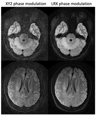 |
An enhanced turboPROP+ technique for diffusion weighted imaging
Zhiqiang Li1, Melvyn B Ooi1,2, and John P Karis1
1Neuroradiology, Barrow Neurological Institute, Phoenix, AZ, United States, 2Philips Healthcare, Gainesville, FL, United States
Diffusion weighted MRI is a useful technique for the diagnosis of neurological disorders. DW EPI is time efficient but suffer from geometric distortions. DW PROPELLER and its variants, including turboPROP and turboPROP+, have been proposed to generate distortion free images. This project improves the turboPROP+ technique by incorporating LRX RF phase modulation approach to improve SNR and signal stability, and by revising the phase correction algorithm to minimize residual artifacts. Volunteer results demonstrate reduced artifacts compared to the original phase correction algorithm, and increased SNR/image quality compared to original XY2 phase modulation.
|
|
0967.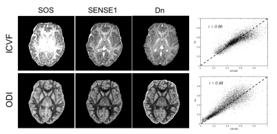 |
Denoising of DWI signal using deep learning
Hu Cheng1, Jian Wang1,2, Shreyas Sanjeev Fadnavis3, Eleftherios Garyfallidis3, and Sharlene Newman1
1Psychological and Brain Sciences, Indiana University, Bloomington, IN, United States, 2School of Information Science and Engineering, Shandong Normal University, Jinan, China, 3Indiana University, Bloomington, IN, United States
We developed a simple deep learning method for DWI data denoising and tested it on correcting sum of square (SoS) noise. By acquiring two sets of diffusion images reconstructed with SoS and SENSE1 coil combination schemes on one subject as training data, the learned model can effectively denoise any SoS data acquired with the same DWI protocol. The denoised data produces similar results in diffusion tensor analysis and NODDI analysis as the SENSE1 data. This method also shed light on denoising techniques for diffusion imaging if a low-noise DWI dataset is available.
|
|
0968.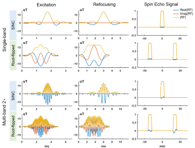 |
SNR-Enhanced High-Resolution Diffusion Imaging Using 3D Simultaneous Multi-Slab (SMSlab) with Root-flipped RF Pulse Design
Simin Liu1, Erpeng Dai1,2, and Hua Guo1
1Center for Biomedical Imaging Research, Department of Biomedical Engineering, School of Medicine, Tsinghua University, Beijing, China, 2Department of Radiology, Stanford University, Stanford, CA, United States
3D simultaneous multi-slab (SMSlab) is a technique to increase SNR efficiency in high-resolution diffusion imaging. However, it still suffers from the intrinsic low SNR of diffusion MRI, especially when using multi-band RF pulses, which increases the pulse duration and thus lengthens the echo time. In this study, root-flipped RF pulses were used in SMSlab to acquired 1 mm isotropic 3D diffusion images. With a multi-band factor of 2, the root-flipped pulses brought about 12 ms reduction of TE (from 91 to 79 ms), and 16% SNR gain, compared to traditional SINC pulses.
|
|
 |
0969.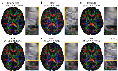 |
DeepDTI: Six-direction diffusion tensor MRI using deep learning
Qiyuan Tian1,2, Berkin Bilgic1,2, Qiuyun Fan1,2, Congyu Liao1,2, Chanon Ngamsombat1, Yuxin Hu3, Thomas Witzel1, Kawin Setsompop1,2, Jonathan R. Polimeni1,2, and Susie Y. Huang1,2
1Martinos Center for Biomedical Imaging, Massachusetts General Hospital, Charlestown, MA, United States, 2Department of Radiology, Harvard Medical School, Boston, MA, United States, 3Department of Electrical Engineering, Stanford University, Stanford, CA, United States
Diffusion tensor imaging (DTI) is widely used clinically but typically requires acquiring diffusion-weighted images (DWIs) along many diffusion-encoding directions for robust model fitting, resulting in lengthy acquisitions. Here, we propose a joint denoising and q-space angular super-resolution method called “DeepDTI” achieved using data-driven supervised deep learning that minimizes the data requirement for DTI to the theoretical minimum of one b=0 image and six DWIs. Metrics derived from DeepDTI’s results are equivalent to those obtained from three b=0 and 19 to 26 DWI volumes for different scalar and orientational DTI metrics, and superior to those derived from state-of-the-art denoising methods.
|
0970.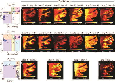 |
Multidimensional correlation MRI of the brain
Kristofor Pas1,2, Michal Komlosh2,3, Daniel Perl4, Peter Basser2, and Dan Benjamini2,3
1University of Texas at Arlington, Arlington, TX, United States, 2National Institutes of Health, Bethesda, MD, United States, 3Uniformed Service University of the Health Sciences, Bethesda, MD, United States, 4Uniformed Services University of the Health Sciences, Bethesda, MD, United States
Multidimensional correlation MRI is an emerging imaging modality that is capable of disentangling highly heterogeneous systems, according to chemical and physical interactions of water within them. Using this approach, the conventional three dimensional MR scalar images are replaced with spatially resolved multidimensional spectra. The ensuing abundance in microstructural and chemical information is a blessing that incorporates a real challenge: how does one distill and refine it into images? Here we introduce a method that robustly identifies the multidimensional spectral components in the image domain, defines the spectral regions of interest, and uses them to reconstruct images of sub-voxel components.
|
|
|
0971.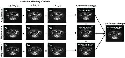 |
Accelerating clinical diffusion-weighted MRI using deep learning: Potential utility in metastatic prostate cancer and malignant mesothelioma
Konstantinos Zormpas-Petridis1, Nina Tunariu1, Andra Curcean1, Christina Messiou1, David Collins1, Yann Jamin1, Dow-Mu Koh1, and Matthew D. Blackledge1
1Radiotherapy and Imaging, Institute of Cancer Research, London, Sutton, United Kingdom
Diffusion-weighted MR-imaging (DWI) is an attractive non-invasive tool for staging and response evaluation of myeloma and metastatic bone disease. However, scans can last up to 30 minutes in whole body studies, which can hinder the adoption of DWI in clinical practice, especially in patients who are unwell. Here, we use a deep learning approach to establish that sub-sampled, but rapidly acquired images, could be used to reconstruct ‘clinical-grade’ DWI images, potentially reducing acquisition times (from ~30 to ~5 minutes). Such time savings could reduce scanning costs and spare patient time/discomfort.
|
0972.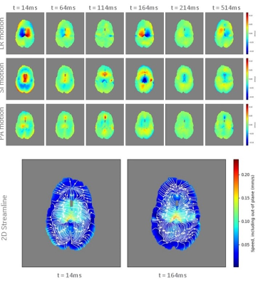 |
Simultaneous Imaging of Diffusion and Coherent Motion in Slow-Flow Compartments in the Brain
Isabelle Heukensfeldt Jansen1, Luca Marinelli1, Ek Tsoon Tan2, Robert Y Shih3,4, J Kevin DeMarco3,4, J Kent Werner3,4, Vincent B Ho3,4, and Thomas Foo1
1GE Global Research Center, Niskayuna, NY, United States, 2Hospital for Special Surgery, New York, NY, United States, 3Uniformed Services University of the Health Sciences, Bethesda, MD, United States, 4Walter Reed National Military Medical Center, Bethesda, MD, United States
We demonstrate a method for simultaneous imaging of diffusion and slow motion in vivo. We use both the magnitude and phase information from image data to reconstruct coherent and incoherent motion (flow and diffusion). We modified a PGSE diffusion imaging sequence so that b-value and encoded velocity can be set independently. We imaged healthy volunteers with a 2-shell sequence with bmax=2000 sec/mm2 and venc=0.24 mm/s at multiple phases during the cardiac cycle using peripheral gating. Results show a distinct periodic motion around the ventricles with RMS speed 0.065 mm/s, moving laterally during systole and medially during diastole
|
|
0973.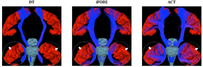 |
Mitigating Gyral Bias via Active Cortex Tractography
Ye Wu1, Yoonmi Hong1, and Pew-Thian Yap1
1Department of Radiology and BRIC, University of North Carolina, Chapel Hill, Chapel Hill, NC, United States
We propose a tractography method, called active cortex tractography (ACT), to overcome gyral bias by enabling fiber streamlines to curve naturally into the cortex. We show that the cortex can play an active role in cortical tractography by affording anatomical knowledge to overcome orientation ambiguities as the streamlines enter the superficial white matter in gyral blades and approach the cortex.
|
|
0974.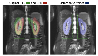 |
Simultaneous distortion and motion correction in abdominal DW-MRI using dual echo EPI and slice-to-volume registration
Jaume Coll-Font1,2, Onur Afacan1,2, Scott Hoge2,3, Bahram Marami4, Ali Gholipour1,2, Jeanne Chow1,2, Simon Warfield1,2, and Sila Kurugol1,2
1Radiology, Boston Children's Hospital, Boston, MA, United States, 2Harvard Medical School, Boston, MA, United States, 3Radiology, Brigham and Women's Hospital, Boston, MA, United States, 4Icahn School of Medicine at Mount Sinai, New York, NY, United States
Diffusion-weighted MRI (DW-MRI) has been increasingly used in abdominal applications. However, unavoidable respiratory motion, as well as B0 field inhomogeneities reduce the accuracy of the quantitative parameters and hinders clinical applicability. In this work, we present a dual echo EPI DW-MRI and slice-to-volume registration method to jointly correct for geometric distortion and motion of the kidneys. The results show that our method effectively reduced geometric distortions, improved alignment of the DW-MR volumes and increased the precision of the estimated quantitative parameters.
|
|
0975.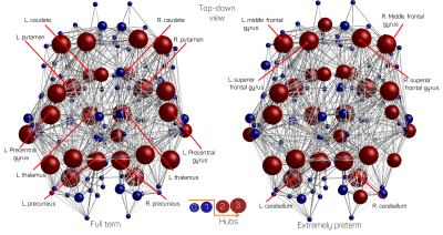 |
Analysis of hub-regions from the structural connectomes of preterm-born and control adolescents
Hassna Irzan1,2, Michael Hütel2, Sebastien Ourselin2, Neil Marlow3, and Andrew Melbourne1,2
1Medical Physics and Biomedical Engineering, University College London, London, United Kingdom, 2Biomedical Engineering and Imaging Sciences, King's College London, London, United Kingdom, 3Institute for Women's Health, London, United Kingdom
Preterm birth has been linked to white matter abnormalities in infants, however the functional implications of these abnormalities are poorly understood. Thus, the long-term effect of such alterations needs further investigation. By combining graph theory and statistical analysis methods, we identify and investigate the hub structure of the preterm brain. The results suggest that while the hub structure is preserved, the connectivity strength and capacity of information flow is reduced and that is linked to reduced brain volume as well as preterm birth.
|
|
0976. |
Structural Analysis of Whole Mouse Brain by Magnetic Resonance Histology
Nian Wang1, Leonard E. White2, Gary Cofer1, Yi Qi1, and G. Allan Johnson1
1Department of Radiology, Duke University, Durham, NC, United States, 2Department of Neurology, Duke University, Durham, NC, United States
Diffusion MRI (dMRI) encompasses a broad range of scales, physical mechanisms and models and applications from clinical to the basic sciences. The recent development of compressed sensing allowed us to extend the spatial and contrast resolution to define more subtle brain architecture beyond the meso scale, solidly in the microscopic domain. We report here dMRI at spatial resolution down to 25 μm i.e. voxels that are more 500,000 times smaller than that of the routine clinical scans.
|
|
0977. |
Semi-automated tractography analysis using a Allen mouse brain atlas : comparing DTI acquisition between NEX and SNR
SangJIn Im1 and Hyeon-Man Baek2
1Gachon Advanced Institute for Health Sciences & Technology, Gachon university, Incheon, Korea, Republic of, 2Gachon university, Incheon, Korea, Republic of
Although tractography research was focused primarily on the human brain, tractography was integrated into animal models to benefit from various preclinical experiments. Accurate segmentation is required for proper connectome of animal models. The Allen mouse brain atlas can provide accurate coordinates and segmentation information to the mouse brain, but it is difficult to use because it is not MRI data. In this study, we use the ABA to accurately segment the mouse brain and examine tractography. In addition, various NEX are used to determine the changes in tractography caused by an increase in the SNR of the DTI.
|
 Watch the Video