
Oral - Power Pitch Session
Diffusion Microstructure Modeling & Validation
Session Topic: Diffusion Microstructure Modeling and Validation
Session Sub-Topic: Diffusion & Microstructure: Modelling & Validation
Oral - Power Pitch
Diffusion
| Wednesday Parallel 4 Live Q&A | Wednesday, 12 August 2020, 13:45 - 14:30 UTC | Moderators: Daan Christiaens |
Session Number: PP-27
0731.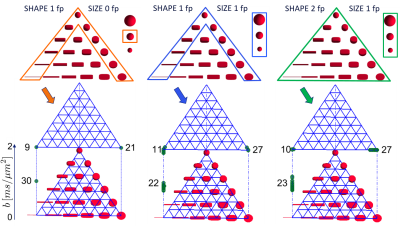 |
Optimal experimental design in multidimensional diffusion MRI for parameter estimation of biophysical tissue models
Santiago Coelho1,2, Jose M Pozo1, Sune N Jespersen2,3,4, Alejandro F Frangi1, Dmitry S Novikov2, and Els Fieremans2
1Centre for Computational Imaging & Simulation Technologies in Biomedicine (CISTIB), School of Computing and School of Medicine, University of Leeds, Leeds, United Kingdom, 2Radiology, School of Medicine, New York University, New York City, NY, United States, 3Center of Functionally Integrative Neuroscience (CFIN) and MINDLab, Department of Clinical Medicine, Aarhus University, Aarhus, Denmark, 4Department of Physics and Astronomy, Aarhus University, Aarhus, Denmark
It was recently shown that multidimensional diffusion MRI enables well-posed estimation of the Standard Model (SM) for diffusion in white matter. However, various multidimensional acquisitions can achieve this, and there are currently no criteria for efficient data acquisition for SM. We propose an optimal experiment design framework based on Cramér-Rao bounds to maximise accuracy and precision of SM parameter estimation. We explore the high-dimensional continuous acquisition space and identify the optimal combination of b-tensors that minimises estimation error. Simulations and in vivo experiments demonstrate that our optimised acquisition has a reduced estimation error on all SM microstructural parameters.
|
|
0732. |
Towards unconstrained compartment modeling in white matter using diffusion-relaxation MRI with tensor-valued diffusion encoding
Björn Lampinen1, Filip Szczepankiewicz2,3, Johan Mårtensson4, Danielle van Westen2, Oskar Hansson5, Carl-Fredrik Westin3, and Markus Nilsson2
1Clinical Sciences Lund, Medical Radiation Physics, Lund University, Lund, Sweden, 2Clinical Sciences Lund, Diagnostic Radiology, Lund University, Lund, Sweden, 3Brigham and Women's Hospital, Harvard Medical School, Boston, MA, United States, 4Clinical Sciences Lund, Department of Logopedics, Phoniatrics and Audiology, Lund University, Lund, Sweden, 5Clinical Sciences Malmö, Clinical Memory Research Unit, Lund University, Lund, Sweden
Microstructure imaging aims to estimate specific quantities such as the axonal density through modeling of diffusion MRI (dMRI) data. However, the low information content of conventional dMRI necessitates assumptions limiting the estimates’ accuracy. Here, we show how to replace model assumptions with independent information from tensor-valued diffusion encoding and diffusion-relaxation experiments. We present sampling protocols optimized using Cramér-Rao lower bounds allowing precise whole-brain estimation of compartment-specific fractions, diffusivities and T2 values in 15 minutes and show results from subjects of different ages. The approach greatly expands the set of parameters measurable with dMRI and provides parameter relations informing model constraints.
|
|
0733.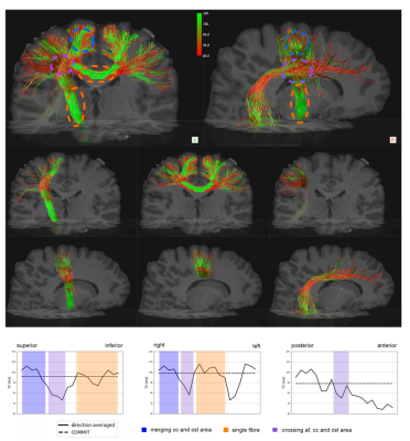 |
Resolving bundle-specific intra-axonal T2 within a voxel using a microstructure-informed approach
Muhamed Barakovic1,2,3, Chantal MW Tax1, Umesh S Rudrapatna1, Jonathan Rafael-Patino2, Cristina Granziera3, Jean-Philippe Thiran2,4, Alessandro Daducci5, Erick J Canales-Rodriguez2,4,6, and Derek K Jones1,7
1Cardiff University Brain Research Imaging Centre, Cardiff University, Cardiff, United Kingdom, 2Signal Processing Laboratory 5 (LTS5), Ecole Polytechnique Fédérale de Lausanne, Lausanne, Switzerland, 3Translational Imaging in Neurology (ThINk) Basel, Department of Biomedical Engineering, University Hospital Basel and University of Basel, Basel, Switzerland, 4Radiology Department, Centre Hospitalier Universitaire Vaudois and University of Lausanne, Lausanne, Switzerland, 5Department of Computer Science, University of Verona, Verona, Italy, 6FIDMAG Germanes Hospitalaries Research Foundation, Barcelona, Spain, 7Mary MacKillop Institute for Health Research, Faculty of Health Sciences, Australian Catholic University, Melbourne, Australia
At the typical spatial resolution of MRI, approximately 60-90% of voxels in the human brain contain multiple fibre populations. Quantifying microstructural properties of distinct fibre bundles within a voxel is challenging. While progress has been made for diffusion and T1-relaxation properties, resolving intra-voxel T2 heterogeneity remains an open question. Here we proposed a novel framework, COMMIT-T2, that uses tractography-based spatial regularization. Unlike previously-proposed voxel-based methods, COMMIT-T2 can recover bundle-specific T2 values within a voxel. Adding this new dimension to the microstructural characterisation of white matter pathways improves the power of tractometry to detect subtle differences in tissue properties.
|
|
0734. |
Orientation-dependent biases in powder averaging caused by inhomogeneous distributions of magnetic susceptibility in white matter
Sidsel Winther1,2, Henrik Lundell2, Mariam Andersson1,2, and Tim Dyrby1,2
1DTU Compute, Technical University of Denmark, Kgs. Lyngby, Denmark, 2Danish Research Center for Magnetic Resonance, Functional and Diagnostic Imaging and Research, Copenhagen University Hospital Hvidovre, Hvidovre, Denmark
Numerical experiments show orientation dependency of the powder averaged diffusion signal (commonly assumed orientation-invariant) when susceptibility-induced inhomogeneity in white matter is taken into account. This implies an axon-orientation-dependent bias of the diffusion signal from white matter regions containing non-uniformly dispersed myelinated axons, which would lead to an over-estimation of the anisotropy. This implies a potential bias between the interpretation of the signal from eg. the internal capsule (oriented inferior-superior) and corpus callosum (oriented left-right).
|
|
0735.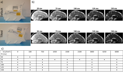 |
Incorporating T2-orientational dependence into diffusion-T2 correlation experiments using a tiltable coil
Chantal M.W. Tax1, Elena Kleban1, Muhamed Barakovic1,2,3, Maxime Chamberland1, and Derek K. Jones1
1CUBRIC, Cardiff University, Cardiff, United Kingdom, 2Signal Processing Laboratory 5, Ecole Polytechnique Federale de Lausanne, Lausanne, Switzerland, 3Translational Imaging in Neurology Basel, Department of Biomedical Engineering, University Hospital Basel, Basel, Switzerland
The anisotropy of white matter is reflected in various white matter contrasts. Transverse relaxation rates can be probed as a function of fibre-orientation with respect to the main magnetic field, while diffusion properties are probed as a function of fibre-orientation with respect to the gradient field. While the latter is easy to obtain in the same head position, the former involves reorientation of the subject’s head inside the scanner. In this work we deployed a tiltable RF-coil to study R2 anisotropy of the brain white matter in diffusion-T2 correlation experiments.
|
|
0736. |
Tortuosity assumption not the cause of NODDI’s incompatibility with tensor-valued diffusion encoding
Michele Guerreri1, Filip Szczepankiewicz2,3, Björn Lampinen4, Marco Palombo1, Markus Nilsson2, and Hui Zhang1
1Computer Science & Centre for Medical Image Computing, University College London, London, United Kingdom, 2Clinical Sciences Lund, Department of Radiology, Lund University, Lund, Sweden, 3Radiology, Brigham and Women’s Hospital, Harvard Medical School, Boston, MA, United States, 4Clinical Sciences Lund, Department of Medical Radiation Physics, Lund University, Lund, Sweden
This work shows that the tortuosity assumption in NODDI can not be identified as the source of incompatibility when the model is extended to data acquired with tensor-valued diffusion encoding. NODDI, originally developed for multi-shell linear tensor encoded (LTE) data, was shown to be inadequate when extended to LTE and spherical tensor encoded (STE) data jointly. The adoption of tortuosity assumption by NODDI has been suggested as a plausible explanation. We conduct a systematic model-comparison study to show that this explanation is inaccurate. We identify a different assumption of the model, the equal-axial-diffusivity, as a source of incompatibility.
|
|
0737.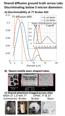 |
Diffusion ground truth quantification of axon scale phantom: Limits of diffusion MRI on 7T, 3T and Connectome 1.0
Sudhir Pathak1, Walter Schneider1, Anthony Zuccolotto2, Susie Huang3, Qiuyun Fan4, Thomas Witzel5, Lawrence Wald4, Els Fieremans6, Michal E. Komlosh7, Dan Benjamini7, Alexandru V Avram7, and Peter J. Basser7
1University of Pittsburgh, Pittsburgh, PA, United States, 2Psychology Software Tools, Inc, Pittsburgh, PA, United States, 3Athinoula A. Martinos Center for Biomedical Imaging, Massachusetts General Hospital, Boston, MA, United States, 4Department of Radiology, Massachusetts General Hospital, Boston, MA, United States, 5Massachusetts General Hospital, Boston, MA, United States, 6Department of Radiology, New York University, New York City, NY, United States, 7National Institutes of Health, Bethesda, DC, United States
We have constructed a novel Taxon (textile water filled tubes) anisotropic diffusion phantom to provide “ground truth” verification of the current limits of diffusion imaging.This phantom is designed to contain 0.8 micron ID tubes with, a packing density of 106 per mm2 , matched to human axon histology, and allows parametric control of diameters, density, and angle dispersion. On a 7T small-bore scanner, we report the ability to distinguish fine Taxon diameter changes between 2-5 micron diameters and approximate 5 micron ID tubes on the 3T Connectome. This is approaching the anatomical scale of axons found in human brain.
|
|
 |
0738. |
Comparison of microstructural models of Sodium and Diffusion Basis Spectrum Images
Simona Schiavi1,2, Lazar Fleysher3, Peng Sun4, Nicole Graziano1, Arielle Falcone1, Yongxian Qian5, Fernando E. Boada5, Sheng-Kwei Song4,6,7, and Matilde Inglese1,2,3
1Department of Neurology, Icahn School of Medicine at Mount Sinai, New York, NY, United States, 2DINOGMI, University of Genoa/IRCCS AOU San Martino-IST, Genoa, Italy, 3Department of Radiology, Icahn School of Medicine at Mount Sinai, New York, NY, United States, 4Biomedical MR Laboratory, Department of Radiology, Washington University School of Medicine, St. Louis, MO, United States, 5Department of Radiology, NYU Langone Medical Center, New York, NY, United States, 6Hope Center for Neurological Disorders, Washington University School of Medicine, St. Louis, MO, United States, 7Biomedical Engineering, Washington University, St. Louis, MO, United States
Both diffusion weighted MRI and Sodium MRI are imaging techniques sensitive to tissue microstructure. The two methods provide complementary information and use tissue models to interpret observed signals to obtain tissue-specific parameters. We investigated the relationship between several tissue-parameters of the two models in the white matter and in the Corpus Callosum. Our results suggests that, the measured cell volume fraction of sodium agrees with diffusion basis spectrum images features. This opens to the possibility of using sodium MRI to investigate pathological tissues and recover complementary information to those we can retrieve with diffusion MRI.
|
0739.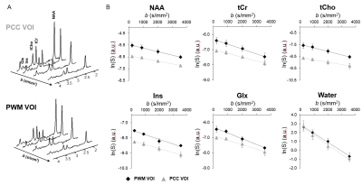 |
Single-shot isotropic diffusion-weighted NMR spectroscopy in the human brain at 7T using tetrahedral encoding
Chloé Najac1, Henrik Lundell2, Hermien E. Kan1, Andrew G. Webb1, and Itamar Ronen1
1C.J. Gorter Center for High Field MRI, Department of Radiology, Leiden University Medical Center, Leiden, Netherlands, 2Danish Research Centre for Magnetic Resonance, Copenhagen, Denmark
We propose a single-shot isotropic diffusion-weighted magnetic resonance spectroscopy (DW-MRS) sLASER-based sequence which enables single-shot measurement of metabolite apparent diffusion coefficient (ADC) at relatively short diffusion times and reasonable echo times in the human brain at 7T. Five brain metabolites and water ADC values were measured in two brain regions that differs significantly in white (WM) and grey matter (GM) content. Significantly higher ADCmetabolites and lower ADCwater were observed in WM compared to GM, illustrating microstructural tissue-specific differences.
|
|
0740. |
Whole-brain mapping of cortical architectonic features with high-resolution MAP-MRI
Alexandru V. Avram1,2, Kadharbatcha Saleem1,2, Frank Q Ye3, Cecil Yen4, Michal E Komlosh1,2, and Peter J Basser1
1NICHD, National Institutes of Health, Bethesda, MD, United States, 2The Henry Jackson Foundation, Bethesda, MD, United States, 3NIMH, National Institutes of Health, Bethesda, MD, United States, 4NINDS, National Institutes of Health, Bethesda, MD, United States
We apply high-resolution mean apparent propagator (MAP)-MRI to quantify cortical architectonic features in a fixed rhesus macaque brain. Cortical depth profiles of MAP-derived parameters, such as the propagator anisotropy (PA), correlate well with histological stains in corresponding brain regions, and may be used to automatically detect boundaries between cortical areas with distinct cyto- and myeloarchitectonic organization. Mapping cortical architectonic features non-invasively could provide a new radiological tool for diagnosis of developmental and neurodegenerative disorders and improve our understanding of how the human brain is organized and connected.
|
|
0741. |
Towards Clinical Translation of Microscopic Diffusion Spectrum Imaging
Enrico Kaden1, Noemi G Gyori1,2, Iulius Dragonu3, Chris A Clark2, and Daniel C Alexander1
1Centre for Medical Image Computing, University College London, London, United Kingdom, 2Great Ormond Street Institute of Child Health, University College London, London, United Kingdom, 3Siemens Healthcare Ltd, Frimley, United Kingdom
Conventional wisdom suggests that it is necessary to average the diffusion signal over the gradient directions to map microstructural features in the presence of orientational heterogeneity. Contrary to this belief, we show that powder-averaging the signal is redundant and leverage this insight to perform, within the same scan time, diffusion experiments with many rather than few b-values and with many rather than few gradient waveforms for b-tensor encoding and beyond, facilitating the translation of advanced techniques such as microscopic diffusion spectrum imaging to clinical practice.
|
|
|
0742.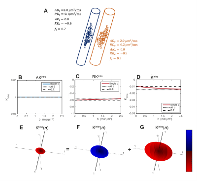 |
Measuring the full diffusional intra- and inter-compartmental kurtosis tensors using double diffusion encoding
Rafael Neto Henriques1, Jonas Lynge Olesen 2,3, Sune Nørhøj Jespersen2,3, and Noam Shemesh1
1Champalimaud Research, Champalimaud Centre for the Unknown, Lisbon, Portugal, 2Center of Functionally Integrative Neuroscience (CFIN) and MINDLab, Clinical Institute, Aarhus University, Aarhus, Denmark, 3Department of Physics and Astronomy, Aarhus University, Aarhus, Denmark
Diffusional kurtosis imaging (DKI) quantifies the non-Gaussian degree of diffusion using the kurtosis tensor. However, kurtosis can depend on conflicting sources of non-Gaussian diffusion such as Gaussian diffusion variance (inter-compartmental kurtosis) or the presence of restricted and hindered effects inside compartments (intra-compartmental kurtosis). Here, we develop and apply a novel double diffusion encoding method that is capable of providing the full directional dependence of both inter- and intra-compartmental kurtosis which can be summarized into two distinct kurtosis tensors and thereby improving kurtosis specificity and potentially providing information for diffusion model validation.
|
0743.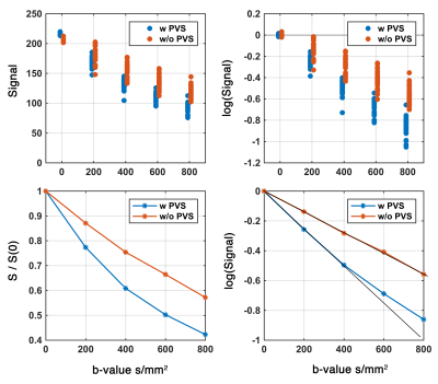 |
Perivascular space fluid contribute to diffusion signal attenuation at low b-value, revisiting extra-cellular space diffusion
Farshid Sepehrband1
1Stevens Neuroimaging and Informatics Institute, Keck School of Medicine, USC, Los Angeles, CA, United States
Perivascular space (PVS) is a pial-lined, fluid-filled structure that surrounds vessels in the cerebral cortex 1,2, and occupies a large portion of the cerebral tissue. The contribution of PVS fluid diffusion signal is less known and could be challenging distinguish from perfusion-related signal changes (or IVIM); because both are expected to affect diffusion at low b-values. Here we showed that PVS fluid significantly contributes to diffusion signal at low b-values dominating tissue or perfusion-related signal changes.
|
|
0744.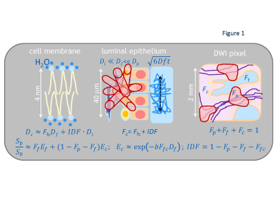 |
Impeded diffusion fraction model for multi-exponential DWI: demonstration in kurtosis phantom and prostate cancer
Dariya Malyarenko1, Scott D Swanson1, and Thomas L Chenevert1
1University of Michigan, Ann Arbor, MI, United States
Majority of current clinical MRI protocols continue to use DWI qualitatively, as an indicator of impeded diffusion evident from sustained signal at high b-values. Quantitative microenvironment description relying on multi-exponential diffusion models is precluded by required prolonged multi-b acquisition and high resolution/SNR not routinely achievable in clinical setting. This study presents a model based on multi-compartment formalism to quantify impeded diffusion fraction (IDF, of water coordinated by macromolecules) from conventional clinical DWI acquisition. The physical origin for IDF is verified using two-compartment diffusion kurtosis phantom, and application example is demonstrated for prostate cancer.
|
 Watch the Video