
Oral - Power Pitch Session
fMRI Physiology
| Thursday Parallel 4 Live Q&A | Thursday, 13 August 2020, 14:20 - 15:05 UTC | Moderators: Paula Croal |
 |
1105.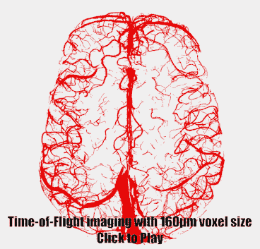 |
Geometrically accurate imaging of the pial arterial vasculature of the human brain in vivo with high-resolution time-of-flight angiography at 7T
Saskia Bollmann1,2, Michael I. Bernier1,2, Simon Daniel Robinson3,4,5, and Jonathan R. Polimeni1,2,6
1Athinoula A. Martinos Center for Biomedical Imaging, Massachusetts General Hospital, Charlestown, MA, United States, 2Department of Radiology, Harvard Medical School, Charlestown, MA, United States, 3Centre for Advanced Imaging, The University of Queensland, Brisbane, Australia, 4High Field MR Centre, Department of Biomedical Imaging and Image-guided Therapy, Medical University of Vienna, Vienna, Austria, 5Department of Neurology, Medical University of Graz, Graz, Austria, 6Division of Health Sciences and Technology, Massachusetts Institute of Technology, Cambridge, MA, United States
Non-invasive imaging of the pial arterial vasculature using the inflow-based contrast provided by moving blood water spins requires sufficiently small voxel sizes (160 μm) to maintain high contrast in small pial arteries (200 μm diameter). Additional acquisition of quantitative susceptibility values allows the differentiation of veins and arteries, turning magnetic resonance angiography into true arteriography. Importantly, flow compensation in all phase encoding directions is necessary to assure geometric accuracy, even for small vessels.
|
1106.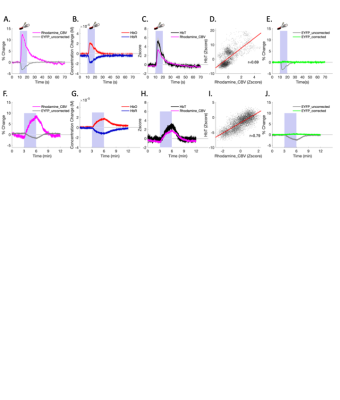 |
Correcting hemodynamic crosstalk effects in fluorescent fiber-photometry signals for quantitative neurovascular coupling studies
Weiting Zhang1,2,3, Tzu-Hao Chao1,2,3, Yue Yang1,4, Tzu-Wen Wang1,2,3, Esteban Oyarzabal1,2,3, SungHo Lee1,2,3, Brittany Katz1,2,3, Guohong Cui5, and Yan-Yu Ian Shih1,2,3
1Center for Animal MRI, The University of North Carolina at Chapel Hill, Chapel Hill, NC, United States, 2Biomedical Research Imaging Center, The University of North Carolina at Chapel Hill, Chapel Hill, NC, United States, 3Department of Neurology, The University of North Carolina at Chapel Hill, Chapel Hill, NC, United States, 4Department of Statistics, The University of North Carolina at Chapel Hill, Chapel Hill, NC, United States, 5Neurobiology Branch, NIEHS/NIH, RTP, NC, United States
The number of fiber-photometry studies incorporating fMRI are rapidly increasing, as these compatible modalities have the ability to reveal neuronal ground-truths. We recently noticed that photometry recording suffers from hemodynamic contamination, leading to false negative results. In this study, we 1) demonstrate how changes in cerebrohemodynamics can yield false negative GCaMP data, 2) propose a method to derive HbO and HbR from spectrally resolved fiber-photometry, 3) validate the derive hemodynamic parameters against concurrently measured CBV and BOLD using photometry and fMRI, 4) implement the proposed correction in vivo, and 5) apply corrected photometric results to rapidly derive hemodynamic response functions.
|
|
|
1107.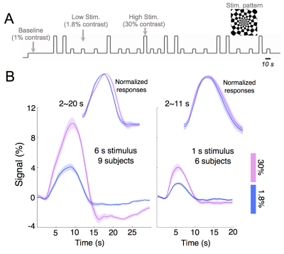 |
Probing the neuronal and vascular origins of task contrast-dependent hemodynamic response functions
Jingyuan E Chen1,2, Nina E Fultz1, Gary Glover3, Bruce R Rosen1,2, Jonathan R Polimeni1,2, and Laura D Lewis4
1Athinoula A. Martinos Center for Biomedical Imaging, Massachusetts General Hospital, Boston, MA, United States, 2Radiology, Harvard Medical School, Boston, MA, United States, 3Radiology, Stanford University, Stanford, CA, United States, 4Biomedical Engineering, Boston University, Boston, MA, United States
In this study, we employed concurrent EEG/fMRI to investigate the neuronal and vascular mechanisms driving task-contrast modulation of HRF shapes. Our results demonstrated that HRFs vary as a function of task contrast levels. Briefly, HRFs elicited by high-contrast stimuli exhibited delayed time-to-peaks and stronger post-stimulus undershoots that likely arose from neuronal origins, and wider full-width-at-half-maximums that were possibly driven by vascular changes.
|
|
1108.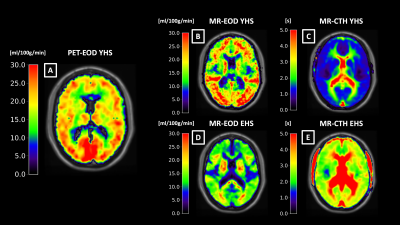 |
Effective oxygen diffusivity mapping with multiparametric quantitative BOLD and pCASL: Comparison between healthy young and elderly subjects
Jan Kufer1, Jens Goettler1,2,3, Samira Epp1, Mikkel Bo Hansen4, Claus Zimmer1, Kim Mouridsen4, Fahmeed Hyder2, Christine Preibisch1,5, and Stephan Kaczmarz1,2
1School of Medicine, Department of Neuroradiology, Technical University of Munich, Munich, Germany, 2MRRC, Yale University, New Haven, CT, United States, 3School of Medicine, Department of Radiology, Technical University of Munich, Munich, Germany, 4Institute of Clinical Medicine, Aarhus University, Aarhus, Denmark, 5School of Medicine, Clinic of Neurology, Technical University of Munich, Munich, Germany The effective oxygen diffusivity (EOD) of the capillary bed has gained increasing interest as a promising biomarker providing additional information on microvascular integrity. To overcome limitations in the applicability of existing and relatively complex EOD mapping techniques, we proposed a novel more easily applicable MR-based approach. We measured EOD in 16 young and 30 elderly healthy subjects. Our measurements of EOD by MRI in young subjects yielded comparably good results in comparison with PET-data as a reference. Furthermore, we found EOD reductions in elderly healthy subjects with concomitant capillary transit-time heterogeneity (CTH) increases, indicating disturbed capillary oxygen extraction ability. |
1109.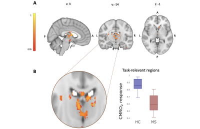 |
Voxel-wise CMRO2 mapping reveals focally-reduced task-related oxygen consumption in multiple sclerosis
Eleonora Patitucci1, Rachael C Stickland 2, Hannah L Chandler1, Michael Germuska1, Catherine Foster1, Sharmila Khot1, Neeraj Saxena1, Valentina Tomassini1,3, and Richard G Wise1,3
1CUBRIC - Cardiff University Brain Research Imaging Centre -Psychology, Cardiff University, Cardiff, United Kingdom, 2Department of Physical Therapy and Human Movement Sciences, Northwestern University, Chicago, IL, United States, 3Institute for Advanced Biomedical Technologies (ITAB), Department of Neurosciences, Imaging and Clinical Sciences, University of Chieti-Pescara “G. d’Annunzio”, Chieti, Italy
Calibrated fMRI can map the rate of cerebral oxygen consumption of the human brain, offering an important indicator of energy dysfunction in neurodegenerative and neuroinflammatory diseases. Previous studies investigated oxygen metabolism at rest or in response to tasks within BOLD signal defined region of interests (ROIs). Here, we investigate on a voxel-by-voxel basis the oxygen metabolic activity in patients with multiple sclerosis during the execution of a task. We show the feasibility of mapping task-induced CMRO2 changes, demonstrating reduced oxygen consumption in the basal ganglia in MS patients that was not otherwise evident from BOLD or CBF signals.
|
|
1110.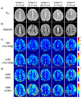 |
Venular Cerebral Blood Volume (vCBV) Mapping Using Fourier-Transform Based Velocity-Selective Pulse Trains
Wenbo Li1,2, Peter van Zijl1,2, and Qin Qin1,2
1Department of Radiology, Johns Hopkins University School of Medicine, Baltimore, MD, United States, 2F.M. Kirby Research Center for Functional Brain Imaging, Kennedy Krieger Institute, Baltimore, MD, United States
A new method is proposed to quantify the venular cerebral blood volume (vCBV) by using Fourier-transform based velocity-selective inversion (FT-VSI) to null the arterial blood signal while using Fourier-transform based velocity-selective saturation (FT-VSS) to suppress the tissue signal. Compared to previous schemes, the proposed method potentially has higher SNR and is more robust to tissue signal fluctuations attributed to system instabilities and physiological motion. The contamination of cerebrospinal fluid (CSF) signal is also corrected for by taking an extra image at a second echo with long TE. Using this method, vCBV of five volunteers were measured at 3T.
|
|
 |
1111.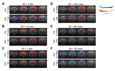 |
Increased negative BOLD responses along the rat visual pathway with short inter-stimulus intervals
Rita Gil1, Francisca F. Fernandes1, and Noam Shemesh1
1Champalimaud Neuroscience Programme, Champalimaud Centre for the Unknown, Lisbon, Portugal
We investigated BOLD responses along the rat visual pathway via inter-stimulus-intervals (ISI) and stimulus pulse width (PW) modulation. PWs did not impact negative BOLD responses (NBRs) while shortening ISI resulted in very large increases in NBRs. Visual cortex (VC) NBRs at short ISIs were accompanied by decreased positive BOLD responses (PBRs) in lateral geniculate nucleus of the thalamus (LGN) and superior colliculus (SC). At the shortest ISI (30ms) NBRs were observed in SC. Along with reported reduced visual evoked potentials amplitude at short ISIs, our findings suggest decreased net excitability as a source for negative BOLD responses in this scenario.
|
1112.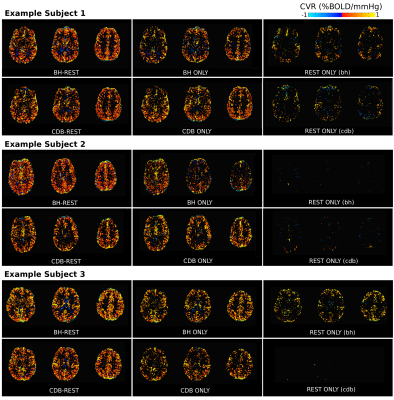 |
Short breathing tasks at the start of a resting state scan: feasible measures of cerebrovascular reactivity
Rachael Stickland1, Apoorva Ayyagari1, Kristina Zvolanek1, and Molly Bright1
Video Permission Withheld
1Northwestern University, Chicago, IL, United States
Cerebrovascular reactivity (CVR), the blood flow response to a vasodilatory stimulus, is changed in many pathologies. CVR can be estimated without gas challenges by performing breathing tasks or by analyzing natural CO2 fluctuations at rest. We added two short breathing tasks (hypercapnic: breath hold, hypocapnia: cued deep breathing) to the start of two resting state fMRI scans. When using all the data, or just the breathing segments, adequate CVR maps could be estimated; this was not the case when just using the resting portions. This paradigm can provide an estimate of CVR, and help improve analysis of resting state data.
|
|
1113.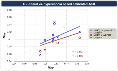 |
Comparison of calibrated fMRI with calibration factor M determined by hypercapnia vs. gas-free R2'
Stephan Kaczmarz1,2, Jan Kufer1, Lena Schmitzer1, Jens Göttler1,2,3, Mario Eduardo Archila Melendez1, Andreas Hock4, Christian Sorg1, Claus Zimmer1, Fahmeed Hyder2, and Christine Preibisch1,5
1School of Medicine, Department of Neuroradiology, Technical University of Munich, Munich, Germany, 2MRRC, Yale University, New Haven, CT, United States, 3School of Medicine, Department of Radiology, Technical University of Munich, Munich, Germany, 4Philips Healthcare, Hamburg, Germany, 5School of Medicine, Clinic of Neurology, Technical University of Munich, Munich, Germany Calibrated-fMRI is highly promising to quantify human brain function via mapping changes of cerebral metabolic rate of oxygen. While the R2’-based approach is easily applicable, systematic differences to the well-established hypercapnia-calibration have been reported. We present data from an ongoing study in seven healthy young subjects correlating calibration factors M from R2' vs. hypercapnia. We hypothesized better correlation after methodological improvements in R2'-mapping and pseudo-continuous arterial spin labeling (pCASL). Our results confirmed this hypothesis, with good correlations between both fMRI-calibrations. However, we found potentially confounding hypercapnia effects on pCASL. Thus, our results suggest benefits of gas-free R2’-calibration for future applications. |
|
1114.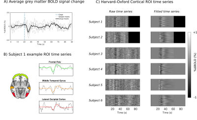 |
Probing dynamic cerebral autoregulation with BOLD fMRI using a thigh cuff challenge
Joseph R Whittaker1, Jessica Steventon1, Marcello Venzi1, and Kevin Murphy1
1CUBRIC, School of Physics and Astronomy, Cardiff University, Cardiff, United Kingdom
The Thigh Cuff Challenge (TCC) technique is a promising method for assessing dynamic cerebral autoregulation with fMRI. A TCC fMRI experiment was performed in order to better understand the BOLD fMRI signal changes associated with autoregulation. We demonstrate that TCC event-locked cortical fMRI signal changes are widespread across cortical grey matter, with varying response shape both within and between subjects. The TCC BOLD response is on average ~0.3%, which we estimate on a voxel-wise basis using a novel informed basis set, which provides a proof-of-concept demonstrating the potential of TCC and fMRI to probe cerebrovascular function.
|
|
|
1115.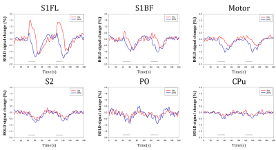 |
Effect of inhibitory neural activities to BOLD fMRI
Hyun Seok Moon1,2, Haiyan Jiang1, Won Beom Jung1,2, JungMi Lee1, Gunsoo Kim1, and Seong-Gi Kim1,2
1Center for Neuroscience Imaging Research (CNIR), Institute for Basic Science (IBS), Suwon, Korea, Republic of, 2Department of Biomedical Engineering, Sungkyunkwan University, Suwon, Korea, Republic of
BOLD fMRI combined with optogenetics allows for brain-wide neural network studies. Most studies have focused on activity of excitatory neurons, which is presumably to contribute BOLD fMRI dominantly. However, fMRI response evoked by inhibitory neural activities is unknown. Here, we investigated 15.2T BOLD response of optogenetically stimulated GABAergic neural activation, and verified the results with electrophysiology.
|
|
1116.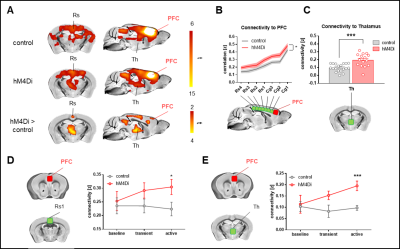 |
Paradoxical fMRI overconnectivity upon neural silencing of fronto-cortical activity
Carola Canella1,2, Federico Rocchi1,2, Shahryar Noei3, Daniel Gutierrez-Barragan1, Ludovico Coletta1, Elizabeth de Guzman1, Alberto Galbusera1, Massimo Pasqualetti4, Giuliano Iurilli5, Stefano Panzeri3, and Alessandro Gozzi1
1Functional Neuroimaging Laboratory, Istituto Italiano di Tecnologia, Rovereto, Italy, 2Center for Mind and Brain Sciences, University of Trento, Rovereto, Italy, 3Neuronal Computational Laboratory, Istituto Italiano di Tecnologia, Rovereto, Italy, 4Biology Department, University of Pisa, Pisa, Italy, 5Systems Neurobiology Laboratory, Istituto Italiano di Tecnologia, Rovereto, Italy
Neuroimaging measurements of functional connectivity are commonly interpreted as an index of reciprocal interareal communication. However, direct testing of this hypothesis has been lacking. Using chemogenetics, electrophysiology and resting-state fMRI in the mouse, we show that acute and chronic silencing of the prefrontal cortex result in paradoxical rsfMRI overconnectivity of the mouse default mode network (DMN) and increased delta activity, an effect relayed to wider cortical territories by polymodal thalamic areas. Our results challenge prevailing interpretations of functional connectivity and implicate a critical contribution of sub-cortical rhythm generators to the establishment of large-scale functional coupling.
|
|
1117.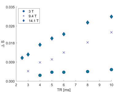 |
Towards intravascular BOLD signal characterization in balanced SSFP experiments of human blood at high to ultra-high fields
Marlon Pérez-Rodas1,2, Hildegard Schulz1, Rolf Pohmann1, Klaus Scheffler1,3, and Rahel Heule1
1High-Field MR Center, Max Planck Institute for Biological Cybernetics, Tübingen, Germany, 2Graduate Training Centre of Neuroscience, IMPRS for Cognitive and Systems Neuroscience, University of Tübingen, Tübingen, Germany, 3Department of Biomedical Magnetic Resonance, University of Tübingen, Tübingen, Germany
To fully understand the neurovascular fingerprint observed in BOLD experiments, extravascular and intravascular contributions have to be identified separately. Balanced steady-state free precession (bSSFP) imaging has demonstrated the ability for distortion-free fMRI with high microvascular sensitivity. However, the underlying intravascular contribution to BOLD bSSFP is not yet entirely known as literature R2 relaxation rates do not reflect the apparent diffusion-related R2 decrease in blood with shorter bSSFP refocusing intervals (TRs). This work thus focuses on characterizing the oxygen sensitivity of bSSFP in blood samples at high to ultra-high fields by means of passband signal differences and intrinsic R2 estimation.
|
1118.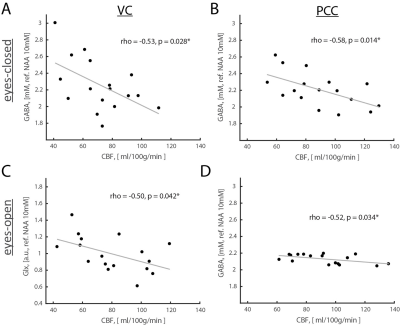 |
Metabolic basis of human brain network nodes in resting-states of eyes-closed and eyes-open
Yury Koush1, Robin A. de Graaf1, Peter Herman1, Douglas L. Rothman1, and Fahmeed Hyder1
1Yale University, New Haven, CT, United States
Resting-state fMRI studies are conducted with eyes-closed (EC) or eyes-open (EO) conditions. Given differences in spontaneous activity between EO and EC conditions, metabolic foundations of fMRI-derived networks, specifically activated (e.g., sensory network) and deactivated (e.g., default mode network, DMN) nodes, are poorly understood. We assessed aerobic glycolysis and excitatory-inhibitory balance in healthy volunteers’ visual cortex (VC, a non-DMN node) and posterior cingulate cortex (PCC, a DMN node) using J-edited fMRS and calibrated fMRI. Functional changes between EO and EC conditions are regionally non-specific to aerobic glycolysis and flow-metabolism coupling, but neurovascular coupling in VC depends on EO and EC conditions.
|
 Watch the Video