
Oral - Power Pitch Session
Interventional
Session Topic: Interventional
Session Sub-Topic: MRI Safety & Intervention
Oral - Power Pitch
MR Safety
| Monday Parallel 4 Live Q&A | Monday, 10 August 2020, 13:45 - 14:30 UTC | Moderators: Laleh Golestani Rad & Joseph Rispoli |
Session Number: PP-32
 |
0111.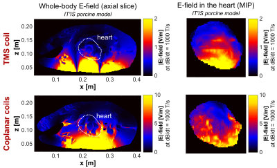 |
Feasibility of using transcranial magnetic stimulation devices to study magnetically induced cardiac stimulation in pigs
Valerie Klein1,2, Mathias Davids1,2,3, Christopher Nguyen2,3,4, Lothar R. Schad1, Lawrence L. Wald2,3,5, and Bastien Guérin2,3
1Computer Assisted Clinical Medicine, Medical Faculty Mannheim, Heidelberg University, Mannheim, Germany, 2A. A. Martinos Center for Biomedical Imaging, Department of Radiologoy, Massachusetts General Hospital, Charlestown, MA, United States, 3Harvard Medical School, Boston, MA, United States, 4Cardiovascular Research Center, Cardiology Division, Massachusetts General Hospital, Charlestown, MA, United States, 5Harvard-MIT Division of Health Sciences and Technology, Cambridge, MA, United States
This work evaluates the potential of porcine cardiac stimulation (CS) studies using transcranial magnetic stimulation (TMS) devices with the aim of determining appropriate safety limits for MRI gradients. We investigated the electric fields induced in electromagnetic porcine models and found that typical TMS coils may not generate fields strong enough for CS. Larger coplanar coils, however, may be suitable for CS studies. In addition to these investigations, we created a porcine model from MRI Dixon and cardiac CINE measurements. The use of such custom models of the animal under experimentation will facilitate the comparison between measured and simulated CS thresholds.
|
|
0112.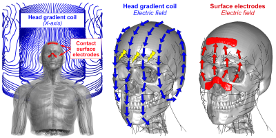 |
Mitigating Peripheral Nerve Stimulations for MRI Gradient Coils using Surface Electrodes
Mathias Davids1,2,3, Bastien Guerin1,2, and Lawrence L Wald1,2,4
1A.A. Martinos Center for Biomedical Imaging, Massachusetts General Hospital, Dept. of Radiology, Charlestown, MA, United States, 2Harvard Medical School, Boston, MA, United States, 3Computer Assisted Clinical Medicine, Medical Faculty Mannheim, Heidelberg University, Mannheim, Germany, 4Harvard-MIT Health Sciences and Technology, Cambridge, MA, United States
Peripheral Nerve Stimulation is becoming an important limitation for state-of-the-art head gradients, which despite higher PNS thresholds, are also operated at higher slew-rate and have limited degrees-of-freedom for FOV design mitigation strategies. We introduce a new mitigation approach, which uses contact surface electrodes driven simultaneously with the gradient coils to cancel the E-field induced by switching of the coil, thus increasing its PNS thresholds. We simulated the capability of four sets of electrodes placed in different areas of the face and found an up to 56% PNS reduction for the analyzed X-axis of a commercial head gradient coil.
|
0113.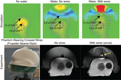 |
“Propeller Beanie” Passive Antennas to Alleviate Dark Bands in Transcranial MR-Guided Focused Ultrasound
Xinqiang Yan1,2, Steven Allen3, and William A. Grissom1,2,4
1Department of Radiology, Vanderbilt University Medical Center, Nashville, TN, United States, 2Institute of Imaging Science, Vanderbilt University, Nashville, TN, United States, 3Department of Biomedical Engineering, University of Virginia, Charlottesville, VA, United States, 4Department of Biomedical Engineering, Vanderbilt University, Nashville, TN, United States
Transcranial MR-guided focused ultrasound (tcMRgFUS) neurosurgery is a non-invasive treatment for essential tremor and many emerging applications. In the FDA-approved Insightec tcMRgFUS system, however, RF reflections inside the transducer create a curved dark band in brain images that runs through midbrain locations that are targeted for essential tremor, and signal is reduced at least 25% everywhere in the brain, which limits the set of scans that can be performed during treatment. This work proposes a simpler solution that alleviates the problem, which is to place a passive reflecting antenna or resonator above the patient’s head, with a “propeller-beanie” crossed-wire shape.
|
|
 |
0114.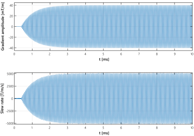 |
No substantial peripheral nerve stimulation beyond 5000T/m/s when driving a head gradient coil at 20kHz
Jolanda M Spijkerman1, Edwin Versteeg2, Dennis WJ Klomp2, David G Norris1,3, and Jeroen CW Siero2,4
1Donders Institute for Brain, Cognition and Behavior, Radboud University Nijmegen, Nijmegen, Netherlands, 2Department of Radiology, University Medical Center Utrecht, Utrecht, Netherlands, 3Erwin L. Hahn Institute for Magnetic Resonance Imaging, University of Duisburg-Essen, Essen, Germany, 4Spinoza Center for Neuroimaging Amsterdam, Amsterdam, Netherlands
In this work the PNS threshold of a recently introduced gradient head coil operating at 20kHz was determined in four healthy subjects. The gradient slew rate was increased to 5000T/m/s by varying the amplitude between 7.5–40mT/m, the maximal gradient strength currently available. One subject reported no PNS, two subjects reported mild PNS at the highest gradient amplitudes of 36.25, 37.5 and 40mT/m, and one subject reported mild sensations during 31.25mT/m, but was uncertain whether this was PNS. In conclusion, application of the coil at 20kHz is currently restricted by the available gradient strength, rather than PNS.
|
|
0115.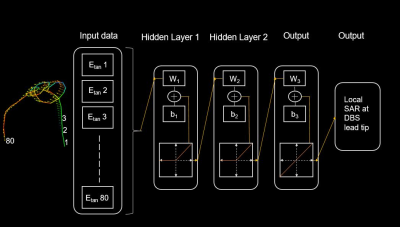 |
Prediction of subject-specific local SAR in patients with deep brain stimulation leads using artificial neural networks
Jasmine Vu1,2, Bach Nguyen2, Justin Baraboo1,2, Joshua Rosenow3, Julie Pilitsis4, and Laleh Golestanirad1,2
1Biomedical Engineering, Northwestern University, Chicago, IL, United States, 2Radiology, Northwestern University, Chicago, IL, United States, 3Northwestern Medicine, Chicago, IL, United States, 4Neurosurgery, Albany Medical Center, Albany, NY, United States
Patients with deep brain stimulation (DBS) implants can significantly benefit from MRI; however, their access is limited due to safety concerns associated with RF heating of implants. RF heating depends significantly on the trajectory of an implanted lead, but there is a lack of surgical guidelines about positioning the extracranial portion of the leads, resulting in substantial patient-to-patient variation in DBS lead trajectories. Thus, quick and reliable patient-specific assessment of RF heating is highly desirable. Here we present an artificial neural network (ANN) model that demonstrates great potential in predicting local SAR at the tips of the DBS leads.
|
0116.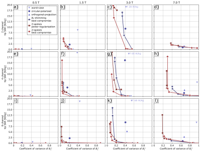 |
On the benefit of pTx for implant safety - a multiparameter simulation study
Johannes Petzold1, Sebastian Schmitter1, Bernd Ittermann1, and Frank Seifert1
1Physikalisch-Technische Bundesanstalt (PTB), Braunschweig and Berlin, Germany
FDTD simulations were used to generate the electromagnetic fields of a human voxel model with a generic implant in a pTx coil. B1+ homogeneity and SAR at the implant tip were systematically investigated for 4 field strength, 4 pTx channel counts, 3 implant positions and 6 excitation strategies. Simple and unsurprising conclusions can be drawn for 0.5 T (pTx is not necessary), 7 T (nothing goes without pTx), and on the channel count (the higher, the better). For the clinically most relevant field strength 1.5 T and 3 T, a much more complex pattern emerges.
|
|
|
0117.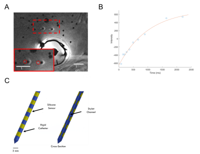 |
A Pilot Clinical Study of Quantitative Silicone Oxygen Sensors in Cervical Cancer
Gregory James Ekchian1, Junichi Tokuda2, Jahanara Freedman1, Hannah Harens1, Robert Cormack3, Larissa Lee3, and Michael Cima1,4
1Koch Institute for Integrative Cancer Research, Massachusetts Institute of Technology, Cambridge, MA, United States, 2Radiology, Brigham and Women's Hospital, Boston, MA, United States, 3Radiation Oncology, Brigham and Women's Hospital, Boston, MA, United States, 4Materials Science and Engineering, Massachusetts Institute of Technology, Cambridge, MA, United States
We report on the first-in-human evaluation of a class of silicone oxygen sensors capable of both high sensitivity and repeated and long-term monitoring of tumor oxygen levels. We are evaluating the use of this sensor in patients receiving high dose rate brachytherapy for cervical cancer. This sensor is a direct and quantitative measurement of tumor oxygen. Low oxygen regions of tumors are more resistant to many common forms of treatment. Understanding tumor oxygen levels can enable personalized radiation and chemotherapy treatments to overcome resistance and improve outcomes for patients.
|
|
0118.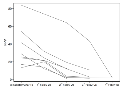 |
To assess and follow-up the mpMRI and prostate volumetric changes after whole prostate MR-guided transurethral prostate ultrasound ablation.
Afshin Azadikhah1, Holden Wu2, Melina Hosseiny2, and Steven S Raman2
1Radiology, University of California, Los Angeles, Los Angeles, CA, United States, 2University of California, Los Angeles, Los Angeles, CA, United States
To evaluate the changes of 3 Tesla (3T) mpMRI and PSA parameters before and during multiple time points after whole gland prostate cancer (PCa) treatment using MRI-guided directional transurethral ultrasound ablation (TULSA). Patients were treated and followed-up for 1, 3, 6 and 12-month in retrospective, cohort, a trial study from October 2017 to February 2019. The mean ADC value, T2W, NPV, PSAD, and prostate volume were significantly decreased after 1 to 12-month follow-up with significant differences. MRI-guided TULSA uses a minimally-invasive transurethral approach, and this appears to be an effective method especially in patients with localized, organ-confined prostate cancer.
|
0119.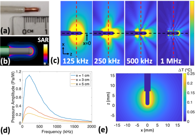 |
RF Power Deposition and Temperature Rise for Thermo-Acoustic Ultrasound Signal Generation from Lead Tips in MRI
Neerav Dixit1, John Pauly1, and Greig Scott1
1Electrical Engineering, Stanford University, Stanford, CA, United States Using the pressure signals resulting from RF energy absorption, thermo-acoustic ultrasound (TAUS) enables detection of excessive local SAR at the lead tips of implanted devices, which causes RF-induced lead tip heating in MRI. Interleaving TAUS acquisitions with MR sequences may also allow for real-time lead tip temperature tracking during MRI. However, generating TAUS signals requires some RF energy deposition and heating at lead tips. Here, we analyze the amount of RF power at lead tips and the associated lead tip temperature rise needed to generate the TAUS signal. |
|
|
0120.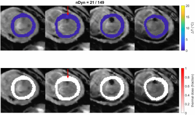 |
Active tracking based cardiac triggering of MR thermometry in an animal model
Ronald Mooiweer1, Rainer Schneider2, Radhouene Neji1,3, Rahul K Mukherjee1, Steven Williams1, Li Huang1, Valéry Ozenne4, Pierre Bour4, Jason Stroup5, Tom Lloyd5, Pierre Jaïs4, Bruno Quesson4, Mark O'Neill1, Tobias
Schaeffter1,6, Reza Razavi1, and Sébastien Roujol1
1Biomedical Engineering, King's College London, London, United Kingdom, 2Siemens Healthcare GmbH, Erlangen, Germany, 3MR Research Collaborations, Siemens Healthcare Limited, Frimley, United Kingdom, 4IHU-Liryc, Pessac, France, 5Imricor Medical Systems, Burnsville, MN, United States, 6Physikalisch-Technische Bundesanstalt, Berlin, Germany
MR thermometry can offer real-time temperature information during RF ablation in the heart. As ECG-triggering can be unreliable in these situations, cardiac triggering based on the position of the ablation catheter could provide an alternative. Active tracking was used to continuously measure the position of microcoils inside the catheter. Cardiac triggers were determined after respiratory motion filtering. Temperature stability over time was below 2.5 ˚C.
|
0121.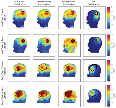 |
RF Power Deposition Optimization Algorithms for Thermal MR Targeting Human Brain Tumors
Eva Oberacker1, Andre Kuehne2, Cecilia Diesch1, Thomas Wilhelm Eigentler1, Jacek Nadobny3, Pirus Ghadjar3, Peter Wust3, and Thoralf Niendorf1,2,4
1Berlin Ultrahigh Field Facility (B.U.F.F.), Max Delbrück Center for Molecular Medicine in the Helmholtz Association, Berlin, Germany, 2MRI.TOOLS GmbH, Berlin, Germany, 3Clinic for Radiation Oncology, Charité Universitätsmedizin, Berlin, Germany, 4Experimental and Clinical Research Center (ECRC), joint cooperation between the Charité Medical Faculty and the Max-Delbrück-Center for Molecular Medicine in the Helmholtz Association, Berlin, Germany
Ultrahigh field (UHF) MR employs higher radio frequencies (RF) than conventional MR and has unique potential to provide focal temperature manipulation and high resolution imaging (ThermalMR). The advantage of integrated therapy monitoring allows the consideration of thermal interventions in brain tumor treatments. Optimization algorithms used to confine the RF power deposition to the target volume (TV) are under constant revision. This work compares three in-house developed optimization algorithms with the focus on power delivery to the target volume as well as sparing of the healthy tissue with a more commonly available approach.
|
|
0122.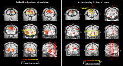 |
MR-guided neuromodulation of visual networks in Rhesus Monkey at a 3T system
Xiaojing Long1, Yangzi Qiao1, Teng Ma1, Weibao Qiu1, Chao Zou1, Jo Lee1, Yang Liu1, Changjun Tie1, Ye Li1, Lijuan Zhang1, Qiang He2, Xin Liu1, and Hairong Zheng1
Video Permission Withheld
1Shenzhen Institutes of Advanced Technology, Chinese Academy of Sciences, Shenzhen, China, 2Shanghai United Imaging Healthcare Co., Ltd., Shanghai, China
In this work, we applied BOLD fMRI in Rhesus monkey on a 3T MR system and investigated the functional effects induced by transcranial ultrasound stimulation (TUS) in both the target spot (the primary visual cortex) and the remote interconnected brain regions. We found that TUS can evoke BOLD reaction not only on the region-specific region but also the interconnected areas in the monkey brain. Additionally, our results demonstrated that the temporal features of BOLD time courses of TUS on the primary visual cortex and those of real visual stimulation have no significant difference in the regions of primary visual pathway.
|
|
0123.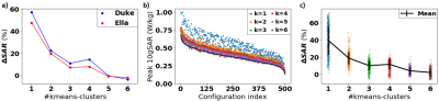 |
Required number of tissue compartments for electromagnetic safety simulation of the head: personalized RF safety for 7T pTx
Matthijs H.S. de Buck1, Peter Jezzard1, and Aaron T. Hess2
1Wellcome Centre for Integrative Neuroimaging, FMRIB Division, Nuffield Department of Clinical Neurosciences, University of Oxford, Oxford, United Kingdom, 2Oxford Centre for Clinical Magnetic Resonance Research, Department of Cardiovascular Medicine, University of Oxford, Oxford, United Kingdom
Personalized electromagnetic simulation models can be generated by segmenting MR-images. However, it is unclear how many tissue types are required for accurate 7T head models. Here, a clustering approach is used to determine the error in the simulated pTx SAR for models with different numbers of tissue types (clusters). Models consisting of only four different tissue types plus air were found to consistently generate low errors for human body-models of different ages and genders. Using the proposed method, it should be possible to operate scanners closer to the true SAR limits due to improved estimations of the actual patient-specific SAR.
|
|
0124.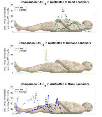 |
Specific Absorption Rate (SAR) Comparison in the Conventional and Open MRI Systems Utilizing an Anatomical Human Computational Model
Kyoko Fujimoto1, Tayeb A Zaidi1, David Lampman2, Joshua W Guag1, Hideta Habara3, and Sunder S Rajan1
1Center for Devices and Radiological Health, US Food and Drug Administration, Silver Spring, MD, United States, 2Hitach Healthcare Americas, Twinsburg, OH, United States, 3Healthcare Business Unit, Hitachi, Ltd., Tokyo, Japan
There is an increasing use of open-bore vertical Magnetic Resonance (MR) systems which consist of two planar radio-frequency (RF) coils. These planar coils generate different electric field distributions compared to that of the conventional cylindrical coils. A recent study showed that RF-induced heating of a neuromodulation device was much lower in the open-bore system. However, imaging landmarks other than the brain have not been evaluated. In this study, we examined the differences in RF exposure using computational modeling and compared specific absorption rate in an anatomical human model at a 1.2T open-bore system with a 1.5T conventional system.
|
 Watch the Video