
SMRT Live
President's Award, JAK, Research & Clinical Abstract Award 1st, 2nd, 3rd Q&A
Session Sub-Topic: President's Award, Jak, Research & Clinical Abstract Award 1st ,2nd, 3rd Q&A
SMRT Ed Session
| SMRT Parallel Room 11 Live Q&A | Monday, 10 August 2020, 23:20 - 23:59 UTC | Moderators: |
| Winner: John A. Koveleski Award for Professional Development | ||
 |
S7.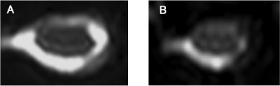 |
FA Value After 1 Week Decompressive Surgery is a Prognostic Factor in Patients with CSM
Takumi Yokohama1, Motoyuki Iwasaki2, Daisuke Oura1, Sho Furuya3, Yoshimasa Niiya2, and Tomoyuki Okuaki4
1Radiology, Otaru General Hospital, Otaru, Hokkaido, Japan, 2Neurosurgery, Otaru General Hospital, Otaru, Hokkaido, Japan, 3Nuclear of Medicine, Hokkaido University, Sapporo, Hokkaido, Japan, 4Radiology, Philips Healthcare, Tokyo, Japan
We evaluated the perioperative FA change in CSM patients using ZOOM DTI. As a result, the FA value after decompression surgery showed the proper state of the damaged cord and had a significant positive relationship with the patient’s outcome. The postsurgical FA value can become an accurate prognostic factor. Besides, the presurgical FA value did not have any relationships with the patient’s outcome, the reason why that a masking effect as an “Aligned Fibers Effect” due to strongly stenosis even using fine resolution DTI.
|
| Winner: SMRT President's Award | ||
 |
S119.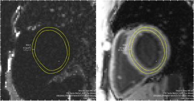 |
Using Novel Fat Water Separation Sequence to Quantify Intramyocardial Fat-Fraction
Xin Dong1,2, Olumide Awelewa1,2, Graham Galloway1, Viral Chikani3, Rob Robergs2, and Arnold Ng3
1Translational Research Institute, Woolloongabba, Australia, 2Queensland University of Technology, Brisbane, Australia, 3Princess Alexandra Hospital, Woolloongabba, Australia
VARPRO is a novel fat water separation sequence that shows promise for myocardial lipid characterisation. Two questions were addressed in this study: 1. Does VARPRO provide the same myocardial fat-fraction result compared to established magnetic resonance spectroscopy (MRS)? 2. Is the myocardial fat-fraction obtained by VARPRO reproducible? The results suggest the two methods are strongly associated but agreed poorly. The VARPRO estimation is proportionally biased, however, the bias can be easily accounted for using a linear equation. The temporal reproducibility of VARPRO is satisfactory. VARPRO could be a potential, alternative modality to MRS in myocardial lipid quantification.
|
| Winner: 1st Place, Clinical Abstract Award | ||
 |
S34.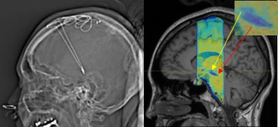 |
Utility of Susceptibility Map-Weighted Imaging for Enhanced Visual Identification of Subthalamic Nucleus at 3T: A Feasibility Study
Weiling Lee1, Peik Yen Teh1, Hartono Septian2,3, Nicolas Kon Kam King4, Prakash Kumar M 2, Jongho Lee5, and Ling Ling Chan1,3
1Division of Radiological Sciences, Singapore General Hospital, Singapore, Singapore, 2Department of Neurology, National Neuroscience Institute, Singapore, Singapore, 3Duke-NUS Graduate Medical School, Singapore, Singapore, 4Department of Neurosurgery, National Neuroscience Institute, Singapore, Singapore, 5Department of Electrical & Computer Engineering, Seoul National University, Seoul, Republic of Korea High resolution susceptibility map-weighted images (SMWI) has recently been developed to improve visualization of the nigrosome-1 region of the substantia nigra (SN) on 3T imaging. SMWI utilizes susceptibility weighting mask derived from quantitative susceptibility mapping (QSM) to enhance the contrast for the SWI magnitude images. In this study, we explore the utility of SMWI to enhance visual identification of the subthalamic nucleus (STN) at 3T, which may be useful to aid in neuro-navigational procedures requiring precise and accurate identification of the STN such as deep brain stimulation (DBS) surgery. |
| Winner: 1st Place, Research Abstract Award | ||
 |
S68.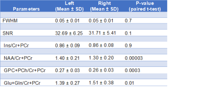 |
Metabolite concentrations in the parietal white matter: Is there a difference between left and right hemispheres in young children?
Petronella Samuels1,2, Kirsten Donald3,4, Catherine Wedderburn3,4,5, Ernesta Meintjes1,2, and Frances Robertson1,2
1Cape Universities Body Imaging Centre, University of Cape Town, Cape Town, South Africa, 2Department of Human Biology, Faculty of Health Sciences, University of Cape Town, Cape Town, South Africa, University of Cape Town, Cape Town, South Africa, 3Department of Paediatrics and Child Health, Red Cross War Memorial Children’s Hospital, University of Cape Town, SA, University of Cape Town, Cape Town, South Africa, 4Neuroscience Institute, University of Cape Town, Cape Town, South Africa, 5Department of Clinical Research, London School of Hygiene & Tropical Medicine,, London, United Kingdom
We retrospectively analyzed 1HMRS data from right and left parietal white matter in a study of 2-year-old children, aiming to determine whether there was a difference between right and left hemispheres in metabolite ratios to creatine. We found concentration ratios of NAA and choline to be higher on the left and Glu+Gln higher on the right. Correlation between right and left was highest for myo-inositol (r= 0.81), but fairly low for NAA (r = 0.63) and Glu+Gln (r = 0.42). Consideration should be given to hemisphere selection when planning 1HMRS studies in young children.
|
| Winner: 2nd Place, Clinical Abstract Award | ||
 |
S114.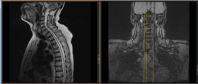 |
Implementation of Safety Checklist and Protocols for scanning patients with whole body conditional DBS implants
Nancy Talbot1 and Tanya Wah Kan1
1Joint Department of Medical Imaging, Princess Margaret Cancer Center, University Health Network, Toronto, ON, Canada
Implementing protocols for patients with deep brain neurostimulators (DBS) that have whole body conditions can be challenging. To obtain sequences that meet the B1+rms RF deposition requirements may require modifications image quality and coverage. T1 weighted imaging can be achieved utilizing gradient echo pulse sequences, however these do not suffice for T2 weighted imaging. For T2 weighted turbo spin echo sequences multiple paramter changes are made including echo train length, slice thickness and gap, matrix, averages, flip angle and gradient operation modes. The implementation of a checklists assists the technologists in ensure conditions are met, and patient safety is ensured.
|
| Winner: 2nd Place, Research Abstract Award | ||
 |
S108.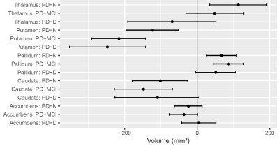 |
Tracking basal ganglia volume in Parkinson’s disease over 10 years with MRI
Nickolas J Palmer1,2, Tracy R Melzer3,4, Jenny H Sim1,5, Mustafa M Almuqbel2,3,4, Daniel J Myall4, Michael R MacAskill3,4, Reza Shoorangiz4,6, Ross J Keenan2,4, John C Dalrymple-Alford3,4, and Tim J Anderson3,4
1University of Auckland, Auckland, New Zealand, 2Pacific Radiology Group, Christchurch, New Zealand, 3University of Otago, Christchurch, New Zealand, 4The New Zealand Brain Research Institution, Christchurch, New Zealand, 5Monash University, Melbourne, Australia, 6The Department of Electrical and Computer Engineering, University of Canterbury, Christchurch, New Zealand
Basal ganglia degeneration is a hallmark of Parkinson’s disease. However, invivo tracking of these structures has not been fully explored longitudinally.This longitudinal study investigates changes in the volume of basal ganglia nuclei in PD over time, with correlation of these changes in measures of cognition and laterality of motor impairment. At cross-section, the PD group exhibited volume changes in the basal ganglia, but no evidence of a relationship with laterality of symptom onset. Over time, the PD group showed accelerated atrophy relative to controls in the thalamus, caudate, and putamen.
|
| Winner: 3rd Place, Clinical Abstract Award | ||
 |
S30.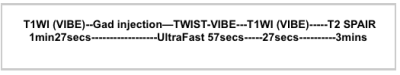 |
Abbreviated breast MRI: How have we achieved it?
Angela Agostinelli1
1Radiology, Royal Melbourne Hospital, Melbourne, Australia
An abbreviated breast MRI protocol has been developed utilising an ultrafast view sharing technique.
|
| Winner: 3rd Place, Research Abstract Award | ||
 |
S80.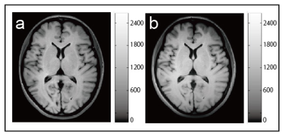 |
Development of the brain phantom for T1- and T2-weighted image showing image contrast and construction similar to those of in vivo MRI
Kousaku Saotome1
1Center for Evolutionary Cognitive Sciences,, University of Tokyo, Tokyo, Japan
We developed the brain phantom for T1-weighted image (T1WI) showing image contrast and construction similar to those of in vivo MRI by using 3D printer. This phantom was produced according to a novel method using a partial volume effect in a slice plane. As our results, we can see that the T1WI of in vivo and the phantom were in very good agreement. Furthermore, the correlation coefficient (R2) between both T1WIs was 0.941 (p < 0.001). Our phantoms might be powerful tools to use for experiments that need ethically unacceptable long scanning or systematic movements.
|
 Watch the Video