
SMRT Posters
SMRT Poster Tour Part 3
Session Sub-Topic: SMRT Poster Tour Part 3
SMRT Poster Presentations
| Winner: 1st Place, Clinical Focus Poster Award | ||
 |
S71.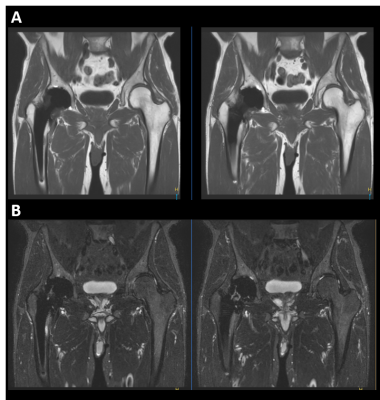 |
Optimization of Metal Artifact Reduction (O-MAR) Imaging
Dave Hitt1, Jonathan Chia1, Robert Lay1, Tom Lowe1, Michael Pawlak1, John Penatzer1, James Snicer1, Marcie Stopchinski1, Gregory Thomas1, Paul Worthington1, Kristen Williams1, and Brian Johnson2
1Philips Healthcare, Gainesville, FL, United States, 23545 SW 47th Ave, Philips Healthcare, Gainesville, FL, United States
Patients with MR Conditional orthopedic implants can be challenging to scan due to susceptibility artifact from metal implants. Use of metal artifact reduction (MARS) techniques can help reduce signal loss from metal implants, however, are not usually sufficient to allow for detailed peri-prosthetic tissue visualization. Metal-induced field inhomogeneities cannot be accurately modeled, which is why O-MAR (MARS+VAT) can improve in-plane distortions by utilizing a more robust spatial encoding approach. Here we present the conceptual background behind the O-MAR technique and work done to provide guidance for optimized metal reduction imaging protocols.
|
| Winner: 3rd Place, Clinical Focus Poster Award | ||
 |
S74.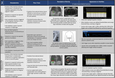 |
Respiratory Pacing – A Low-Tech Solution to a Universal Struggle?
Ortman Jason1, Jennifer Wagner2, Bharath Ambale1, Yoko Kato3, Jaclyn Sesso3, Yoshimori Kassai4, Larry Kasuboski2, and Joao Lima3
1Radiology, Johns Hopkins University, Baltimore, MD, United States, 2Canon Medical Research Unit, Mayfield VIllage, OH, United States, 3Cardiology, Johns Hopkins University, Baltimore, MD, United States, 4Canon Medical Systems, Otawara-Shi, Tochigi, Japan
For respiratory triggered and gated acquisitions, variance in the natural respiratory cycle often complicates workflow, extending scan times and reducing image quality. This abstract discusses the hypothesis that by providing continuous auditory guidance, or "respiratory pacing", the efficacy of respiratory triggered and gated sequences may be improved. The importance of mimicking natural respiratory patterns was quickly noted, as were key differences in the success of respiratory pacing when applied to different acquisitions. Other interesting observations included an observable increase in diaphragmatic travel when respiratory pacing was applied and the extension of scan lengths due to diaphragmatic fatigue over time.
|
| S77. | GRAPPA vs SMS Fast Brain MRI Compared to ACR Specifications
Mark J Burton1, Robert Thomen1, Talissa A. Altes1, Carlos Leiva Salinas1, Nicholas Tustison2, and Joseph P Cousins1
1Radiology, University of Missouri Hospital, Columbia, MO, United States, 2Radiology, University of Virginia, Charlottesville, VA, United States
This work was to assess the trade-off between scan time and image quality afforded by acceleration techniques GRAPPA and simultaneous multi-slice (SMS). We imaged an ACR phantom using standard clinical T1, T2, and FLAIR sequences. Then, without altering any other scan parameters, we increased GRAPPA and SMS acceleration factors (2, 4, and 6). All GRAPPA and SMS accelerated scans passed the ACR resolution criteria. Both GRAPPA and SMS passed low-contrast criteria at acceleration factor 2, but factors of 4 and 6 did not. GRAPPA and SMS gave nearly identical similarity indices and SNR at all acceleration factors.
|
|
S78.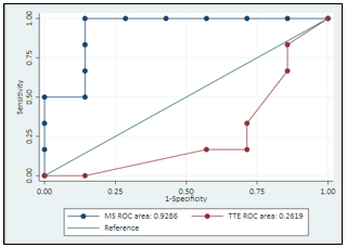 |
Evaluation of an ultra-fast 4D MRI sequence for the diagnosis of breast lesions
Belinda Lokaj1, Karen Kinkel2, Jean-Noel Hyacinthe3, Jérôme Schmid3, and Céline Gaignot3
1Master of Science in Health Sciences, HES-SO /UNIL, Lausanne, Switzerland, Lausanne, Switzerland, 2Institut de radiologie, Clinique des Grangettes, Chêne-Bougeries, Geneva, Switzerland, Geneva, Switzerland, 3Geneva School of Health Sciences, HES-SO // University of Applied Sciences and Arts of Western Switzerland, Geneva, Switzerland
Breast MRI is promoted as a tool for breast cancer screening because of its high sensitivity. Possibility of differentiating between benign and malign lesions has been studied using lesion enhancement parameters on DCE, thus helping to reduce examination time. This study was designed to verify sensitivity and specificity for lesion characterization. Time to Enhancement and Maximum Slope of the enhancement curve obtained from an ultrafast sequence were evaluated. Preliminary results tend to confirm that performing breast lesions screening using ultrafast-DCE is a valuable alternative to the standard MRI Breast protocol, possibly opening diagnostic perspectives to a wider population of women.
|
|
S86.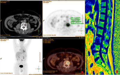 |
Fat Fraction may be used as a radiographic evaluation index for vertebral bone metastasis:a report of 4 cases
Hui Hao1, Jia Yin Tong1, Xiao Cheng Wei2, Jian Xin Guo1, and Jian Yang1
1Department of Radiology, The first affiliated hospital of xi 'an jiaotong university, Xi'an, China, 2MR Research China, GE Healthcare, BeiJing, China
Bone metastasis is a common complication of advanced malignant tumors with severe simptoms. Common imaging modalities for bone metastasis diagnosis include X-ray, CT,MRI, SPECT, and PET. PET is the golden standard, but the nature of radiation, metabolic risk and high price have limited PET from widely clinical use. In this work, we presented 4 cases of bone metastasis which had had a difference in MRI derived fat fraction between bone metastatic vertebrae and normal non-metastatic vertebrae. MR results were also coincided with PET results. MRI derived fat fraction may be used as an imaging evaluation index for bone metastasis.
|
|
S87.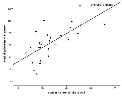 |
Analysis on breast tumor movement using MRIs scanned in prone and supine positions
chuan bing wang1 and da peng li2
1Department of Radiology, the First Affiliated Hospital of Nanjing Medical University (Jiangsu Province Hospital), Nanjing, China, 2the First Affiliated Hospital of Nanjing Medical University (Jiangsu Province Hospital), nan jing, China
The aim of this study is to evaluate the volume change of breast tumor, various kinds ofspatial movementin nipple and the bottom center of the sternum origin, and distance from tumor center to the chest wall from prone to supine MRIs, and then to predict more accuracy tumor positions for surgical planning in the operation room.
|
|
S91.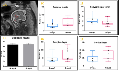 |
Lower risk of hearing loss without sacrificing image quality in fetal MR imaging: a feasibility study using acoustic reduction technique
Le Cao1, Xiang Liu1,2, and Jianxin Guo1
1The First Affiliated Hospital of Xi’an Jiaotong University, Xi’an, Shaanxi,, China, 2The First Affiliated Hospital of Xi’an Jiaotong University, Xi’an, Shaanxi, China
3.0T MR scanner can achieve superior image quality depicting fetal anatomic details over 1.5T, but may poses higher risk of adverse impact on fetal auditory development due to its intrinsically higher acoustic noise level. This comparative study investigated the value of acoustic noise reduction technique in fetal exam. The result shows the technique can acquire equivalent quality images in 3.0T scanner, meanwhile decrease hearing loss risk in fetal head examinations compared with the conventional method.
|
|
S97.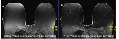 |
A technical approach to reduce inhomogeneities and image distortion in PET/MRI imaging using mMR Breast coil
Pradeep Singh Negi1,2,3, Shashi Bhushan Mehta1,2, Amarnath Jena1,2, and Sangeeta Bhushan Taneja1
1Molecular Imaging & Nuclear Medicine, Indraprastha Apollo Hospitals, New Delhi, India, 2Physics, Vivekananda Global University, Jaipur, India, 3PET SUITE, House of Diagnostics, New Delhi, India
Inhomogeneity in fat suppression and image distortion pose problem in Breast MRI specially in the diffusion and dynamic fat suppressed images using mMR breast coil. The purpose of this work to improve fat-superstation and minimize image distortion in the breast imaging before applying the MRI sequence specially in Dynamic fat-supressed Contrast and Diffusion images. 330 breast cancer patients have been taken for this study. Volume shim was applied in all patients and water peak shift corrected manually when required. Our results show reduction in image distortion in diffusion and improving in fat-supressed MRI images.
|
|
| Winner: 3rd Place, Research Focus Poster Award | ||
 |
S98.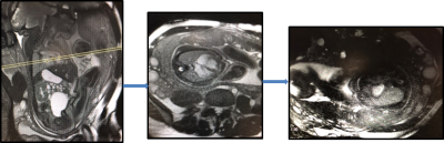 |
Fetal Cardiac Imaging Utilizing Retrospectively Gated 2D Radial MRI
Kristina Mary Pelkola1, Onur Afacan1, Tess E. Wallace2, Jason A. Ferro1, Carol M. Backman1, Davide Piccini3,4,5, Tobias Kober3,4,6, Carol E. Barnewolt1, Susan A. Connolly1, Judy A. Estroff2, and Simon K. Warfield1
1Radiology, Boston Children's Hospital, Boston, MA, United States, 2Boston Children's Hospital, Boston, MA, United States, 3Advanced Clinical Imaging Technology, Siemens Healthcare, Lausanne, Switzerland, 4Radiology, Lausanne University Hospital and University of Lausanne, Lausanne, Switzerland, 5LTS5, Ecole Polytechnique Federale de Lausanne (EPFL), Lausanne, Switzerland, 6LTS5, École Polytechnique Fédérale de Lausanne (EPFL), Lausanne, Switzerland
Fetal MRI is a rapidly growing field that facilitates diagnosis and treatment providing improved maternal and fetal care. New imaging strategies are needed to improve the assessment of cardiac structure and function. We assessed an interactive fetal cardiac prototype sequence enabling the fetal heart to be imaged in real-time, while prescribing the imaging plane to fetal movement. Once positioned to the long axis, a sequence of 2D radial readouts are acquired, enabling inline reconstruction of beating heart images with a frame rate between 0.5s and 1s. This new capability allowed assessment the anatomy and function of the fetal heart.
|
S99.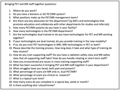 |
Operating a Hybrid PET/MR Scanner: Challenges and Opportunities
Dawn Holley1, Kim Halbert1, and Mehdi Khalighi1
1Radiology, Stanford University, Stanford, CA, United States
Simultaneous PET/MR imaging is still a relatively new dual modality. As such, unique challenges are commonly faced across departments. What were once commonly separate, Nuclear Medicine and Radiology departments now need to work together to create new workflows, protocols and management systems. Questions commonly arise regarding how these two modalities and departments can best come together for management success. A survey of 19 questions was distributed to 12 different PET/MR departments in order to gain an understanding of what challenges are often faced and how to resolve them, as well as any commonalities across successful departments.
|
|
S101.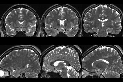 |
A comparison of 2D High resolution PROPELLER with 3D Fast spin echo imaging on multi-planar reconstruction
Ho Beom Lee1 and Kwan Woo Choi2
1Asan Medical Center, Seoul, Republic of Korea, 2Wonkwang Health Science University, Iksan, Republic of Korea
2D High resolution PROPELLER technique was compared to 3D Fast spin echo imaging on axial and reformatted (sag and cor) images.
|
|
S109.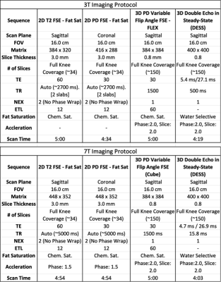 |
7T Knee MRI: Clinical Benefits and Challenges
Kevin Epperson1, Karla Epperson1, Garry Gold1, and Feliks Kogan1
1Radiology, Stanford University School of Medicine, Stanford, CA, United States
Diagnostic MRI scanning of the knee at 3T offers multiple contrasts and definition of anatomical structures, presenting pathology, and allows quantitative performance methods. Ultra high field (7T) systems offer improved resolution and SNR for visualization of small morphological changes and translation of quantitative methods presents challenges in field uniformity and shading artifacts which limit the robustness and clinical potential of these systems.In this work we implement a new method to overcome field inhomogeneity errors and assess the potential clinical benefits of knee imaging at 7T.
|
|
S115.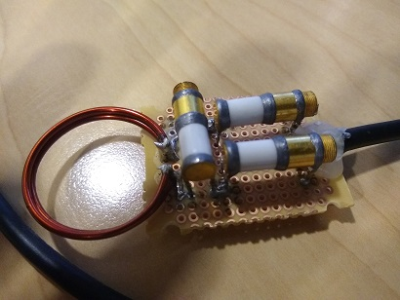 |
Building an MRI RF Coil for the Technologist
Rene Jack Roy1
1UVA, charlottesville, VA, United States
The complexity and costs of MRI RF coil design seems out of reach for most non-physicists in the medical research field. To facilitate learning more about coil design and functionality, we have attempted to build a simple loop coil that can be used in small animal research.
|
|
| S116. | An exploration of MRI patient safety decision making processes using ‘social media’ as a data resource
Renee Mineo1, Claire Mulcahy1, Shawna Farquharson1, and Alan Connely2,3
1MRI, The Florey Institute of Neuroscience and Mental Health, Heidelberg, Australia, 2Imaging, The Florey Institute of Neuroscience and Mental Health, Heidelberg, Australia, 3Imaging, The Florey Department of Neuroscience and Mental Health, University of Melbourne, Melbourne, Victoria, Australia, Melbourne, Australia
This study reviewed the engagement of MR Radiographers & Technologists with a social media platform seeking professional guidance with MR safety decision making processes regarding implants or devices. From June 2016 to August 2018 100 discussion items were reviewed and categorized. Our study identified that the ‘consensus to scan or not to scan’, was not completely dependent on the presence of detailed information about the implant or device.
|
|
 View the Poster
 Watch the Video