
Sunrise Session
CMR for Cardiac Function Beyond Ejection Fraction:
Relationships Between Function & Myocardial Remodeling
Session Topic: Educational Q&A: CV Sunrise
Session Sub-Topic: CMR for Cardiac Function Beyond Ejection Fraction: Relationships Between Function & Myocardial Remodeling
Sunrise Session
ORGANIZERS: Dana Peters, Peng Hu
| Wednesday Parallel 1 Live Q&A | Wednesday, 12 August 2020, 13:45 - 14:30 UTC | Moderators: Sebastien Roujol |
Skill Level: Basic to Advanced
Session Number: S-Th-02
Overview
This sunrise course will provide an overview of technologies and clinical applications of CMR for evaluating function of the heart, beyond LV EF, including systolic function and diastolic and atrial contraction phases. This overview will include evaluation of phasic volumes but will also describe newer technologies such as evaluation of strain, with tagging and feature-tracking, fibrosis, and flow, and how these relate to tissue characterization and myocardial remodeling.
Target Audience
- Physicists and engineers who wish to obtain a basic understanding of the clinical need for imaging cardiac function, which include quantification of volumes and ejection fractions, but also measurement of phasic volumes, strain and flow in the ventricles, atria, and aorta;
- Physicists and clinicians who wish to acquire an understanding of current CMR tools that are available for evaluating cardiac function, including systolic and diastolic function; and
- Physicists and clinicians who wish to obtain a current understanding of how cardiac structure and function are related.
Educational Objectives
As a result of attending this course, participants should be able to:
- Describe which functional measurements are useful based on clinical needs;
- Define the basic principles of myocardial strain imaging, feature-tracking, flow imaging and tissue characterization techniques; and
- Explain the basic natural history of cardiac remodeling that leads to reduced diastolic function.
 |
Imaging Myocardial Fibrosis
Michael Jerosch-Herold
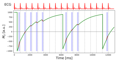
Mapping of the magnetization relaxation parameters such as T1 in the myocardium has transformed cardiac magnetic resonance (CMR) over the last decade into a highly versatile imaging modality for myocardial tissue characterization. T1 mapping rendered it possible to detect diffuse myocardial fibrosis that is not visible by late-gadolinium enhancement. Initially, this was accomplished by estimating the extracellular volume fraction for gadolinium contrast as a biomarker that correlates with the accumulation of connective tissue in the myocardial interstitium. Since then, significant advances have also been made in the use of native imaging markers (i.e. not requiring gadolinium contrast).
|
|
| Myocardial Tissue Remodeling: Fibrosis & Function
Jeanette Schulz-Menger
|
 Watch the Video