Riccardo Pascuzzo1, Alexandra L. Young2,3, Neil P. Oxtoby2, Janis Blevins4, Gianmarco Castelli1, Pierluigi Gambetti5, Brian S. Appleby4, Daniel C. Alexander2, and Alberto Bizzi1
1Neuroradiology Unit, Fondazione IRCCS Istituto Neurologico Carlo Besta, Milan, Italy, 2Centre for Medical Image Computing, Department of Computer Science, University College London, London, United Kingdom, 3Department of Neuroimaging, Institute of Psychiatry, Psychology and Neuroscience, King′s College London, London, United Kingdom, 4National Prion Disease Pathology Surveillance Center, Case Western Reserve University, School of Medicine, Cleveland, OH, United States, 5Department of Pathology, Case Western Reserve University, School of Medicine, Cleveland, OH, United States
1Neuroradiology Unit, Fondazione IRCCS Istituto Neurologico Carlo Besta, Milan, Italy, 2Centre for Medical Image Computing, Department of Computer Science, University College London, London, United Kingdom, 3Department of Neuroimaging, Institute of Psychiatry, Psychology and Neuroscience, King′s College London, London, United Kingdom, 4National Prion Disease Pathology Surveillance Center, Case Western Reserve University, School of Medicine, Cleveland, OH, United States, 5Department of Pathology, Case Western Reserve University, School of Medicine, Cleveland, OH, United States
A novel unsupervised machine-learning
technique found 5 clusters of patients with sporadic Creutzfeldt-Jakob disease
(sCJD), each having a distinct pattern of diffusion-weighted MRI abnormality progression. Clusters
were significantly associated to sCJD strains and phenotypes.
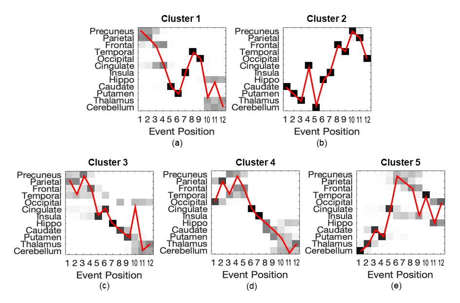
Figure 1. Orderings
of the 5 clusters identified by SuStaIn in the whole sCJD population. In each
subplot, the event position (x axis) is the disease stage (from 1 to 12) at
which the brain region (on the y axis) becomes abnormal. The darker the square,
the higher the probability (0=white; 1=black) that the corresponding region on
the y axis has become abnormal at the stage indicated on the x axis. The red
line of each subplot indicates (from left to right) the direction of disease
progression.
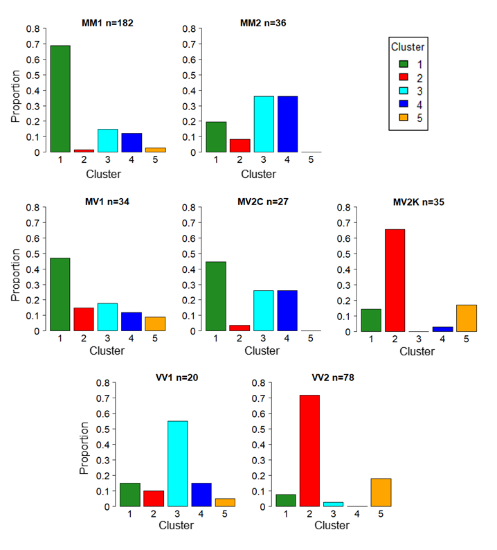
Figure 3. Proportion
of patients (y axis) in each sCJD subtype assigned to the 5 clusters (x axis)
identified by SuStaIn. Total number of patients for each sCJD subtype are
indicated in the title of each subplot.