Xiao Cui1, Xianshun Yuan1, Hui Gu1, Mo Wang2, Yin Dong1, Ximing Wang1, and Xiang Feng3
1Department of Radiology, Shandong Provincial Hospital Affiliated to Shandong University, Jinan, China, 2Department of Vascular Surgery, Shandong Provincial Hospital Affiliated to Shandong University, Jinan, China, 3MR Scientific Marketing, Diagnosis Imaging, Siemens Healthcare Ltd, Beijing, China
1Department of Radiology, Shandong Provincial Hospital Affiliated to Shandong University, Jinan, China, 2Department of Vascular Surgery, Shandong Provincial Hospital Affiliated to Shandong University, Jinan, China, 3MR Scientific Marketing, Diagnosis Imaging, Siemens Healthcare Ltd, Beijing, China
This
study supports the value of HR-MRI in non-invasive diagnosis of young adults due to cervical artery dissections, especially for the patients in the sub-acute stroke, by comparing the diagnostic values between HR-MRI and CTA.
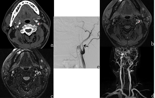
Fig 1. 39-year-old man with headache for 10 days
diagnosed as left carotid artery dissection by DSA eventually. (a) Axial CTA
image showed eccentric low
density (red star). (b, c) Axial T1-weighted and T2-weighted MRI images showed
crescent-shaped high signal caused by a mural hematoma
surrounding the lumen (red star) and intimal flap (white arrow) clearly. (d)
Reconstruction contrast-enhanced MR angiography image showed tapered occlusive
lumen of the left carotid artery (white arrow). (e) DSA image demonstrated the
dissection (black arrow).
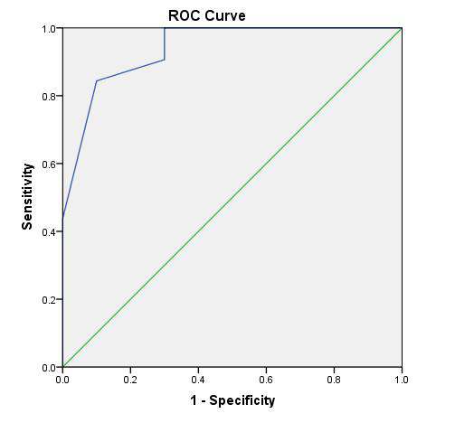
Fig 2. ROC curves
(a) ROC curve for diagnosis of HR-MRI (blue full
line, AUC = 0.94 [95% CI,
0.86–0.97]). Diagonal line represents line of reference.