Ryan McNaughton1, Mina Botros2, Ning Hua2, Xin Zhang1, and Hernan Jara2
1Mechanical Engineering, Boston University, Boston, MA, United States, 2Boston University Medical Center, Boston, MA, United States
1Mechanical Engineering, Boston University, Boston, MA, United States, 2Boston University Medical Center, Boston, MA, United States
This study presents a qMRI-based technique for repolarizing magnitude images obtained from the mixed-TSE pulse sequence. By isolating different tissue compartment via application of variable thresholds of PD, T2, and T1/T2 maps, a high contrast, correctly polarized T1 map can be calculated.
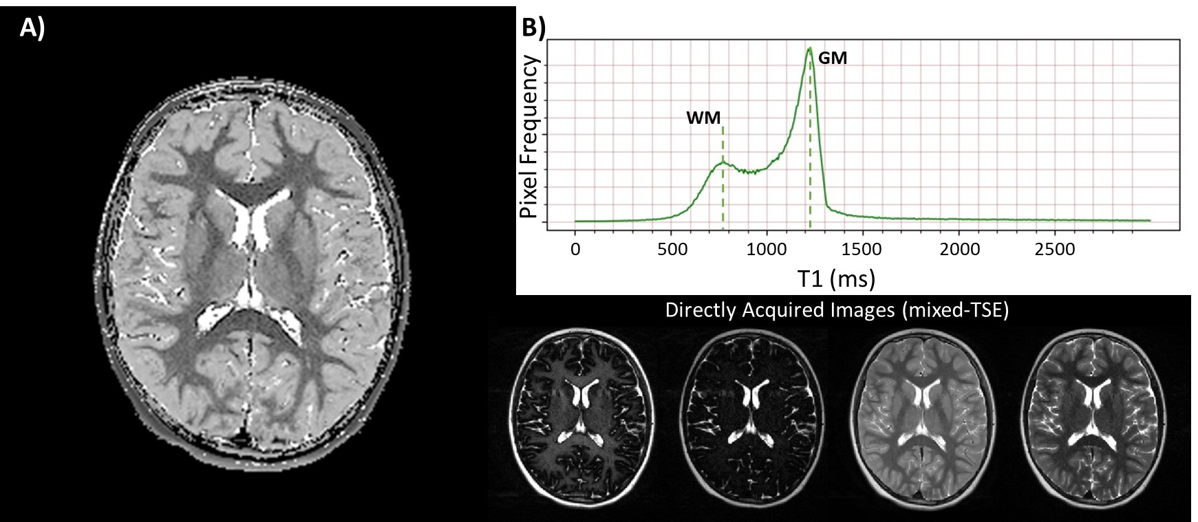
In vivo application of the qMRI-based T1
repolarization algorithm. A) Representative image of an extremely preterm born
subject. Strong contrast can be seen between all tissue types, including white
matter, grey matter, and cerebrospinal fluid. B) The repolarized T1
demonstrates a characteristic bimodal distribution, with clearly
distinguishable peaks for white matter and grey matter. Directly acquired
images from the mixed-TSE are shown for reference.
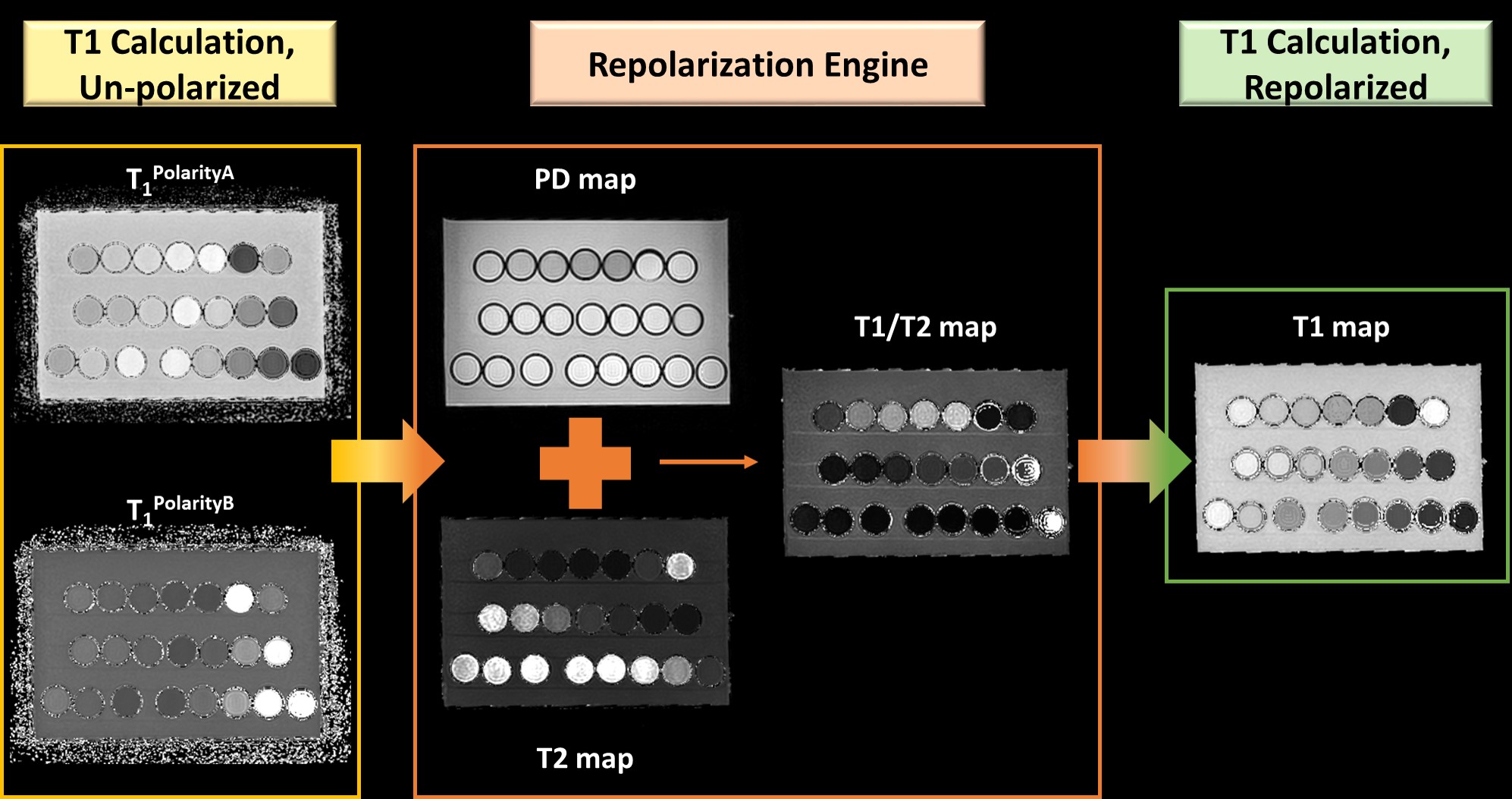
Process flow for the T1 repolarization, multispectral
quantitative MR algorithm, carried out on an agarose gel phantom. First, the two
polarities are calculated (Eq. 1) and account for the possible T1’s a voxel can
exhibit from a magnitude image. The two T1 polarity maps are read into a
qMRI-based repolarization engine along with maps for PD, T2, and T1/T2. Thresholds
for large PD and T2 values are applied to correctly polarize Polarity B T1 in
the final map, while a threshold for large T1/T2 map correctly polarizes polarity
A T1. The output of the engine is a correctly polarized T1 map.