Yukai Zou1,2, Wenbin Zhu3, Ho-Ching (Shawn) Yang1, Nicole L Vike4, Diana O Svaldi1, Trey E Shenk5, Victoria N Poole1,4, Gregory G Tamer, Jr.1, Larry J Leverenz6, Ulrike Dydak7, Eric A Nauman1,4,8, Thomas M Talavage1,5, and Joseph V Rispoli1,5
1Weldon School of Biomedical Engineering, Purdue University, West Lafayette, IN, United States, 2College of Veterinary Medicine, Purdue University, West Lafayette, IN, United States, 3Department of Statistics, Purdue University, West Lafayette, IN, United States, 4Department of Basic Medical Sciences, Purdue University, West Lafayette, IN, United States, 5School of Electrical and Computer Engineering, Purdue University, West Lafayette, IN, United States, 6Department of Health and Kinesiology, Purdue University, West Lafayette, IN, United States, 7School of Health Sciences, Purdue University, West Lafayette, IN, United States, 8School of Mechanical Engineering, Purdue University, West Lafayette, IN, United States
1Weldon School of Biomedical Engineering, Purdue University, West Lafayette, IN, United States, 2College of Veterinary Medicine, Purdue University, West Lafayette, IN, United States, 3Department of Statistics, Purdue University, West Lafayette, IN, United States, 4Department of Basic Medical Sciences, Purdue University, West Lafayette, IN, United States, 5School of Electrical and Computer Engineering, Purdue University, West Lafayette, IN, United States, 6Department of Health and Kinesiology, Purdue University, West Lafayette, IN, United States, 7School of Health Sciences, Purdue University, West Lafayette, IN, United States, 8School of Mechanical Engineering, Purdue University, West Lafayette, IN, United States
A population-specific brain atlas was developed and shown to better
characterize the neuroanatomy of the adolescent collision-sport
athletes, reduced biases introduced during spatial normalization, and exhibited
higher sensitivity in detecting regional FA differences.
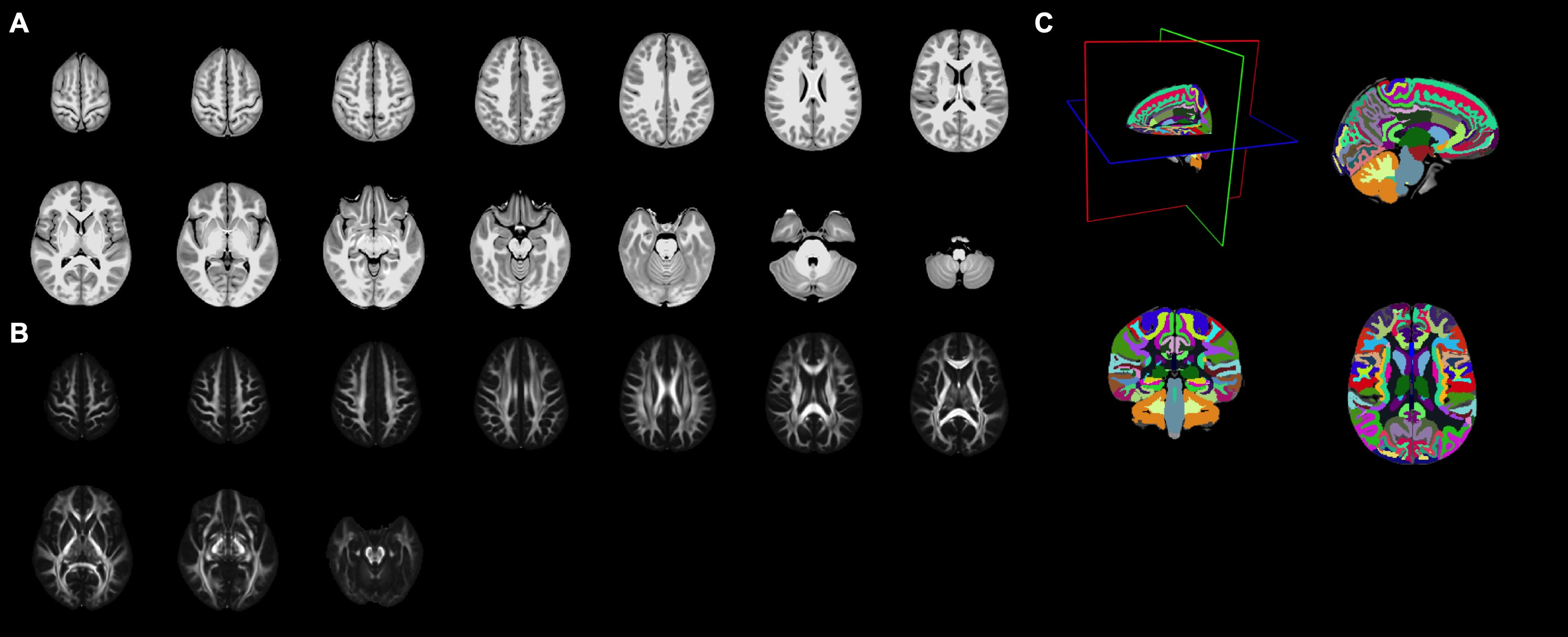
Figure 2. Illustrations of the population-specific brain atlas, including
the T1 (A) and DTI (B) templates in axial
views, and the semantic labels of cortical and white matter parcellations (C).
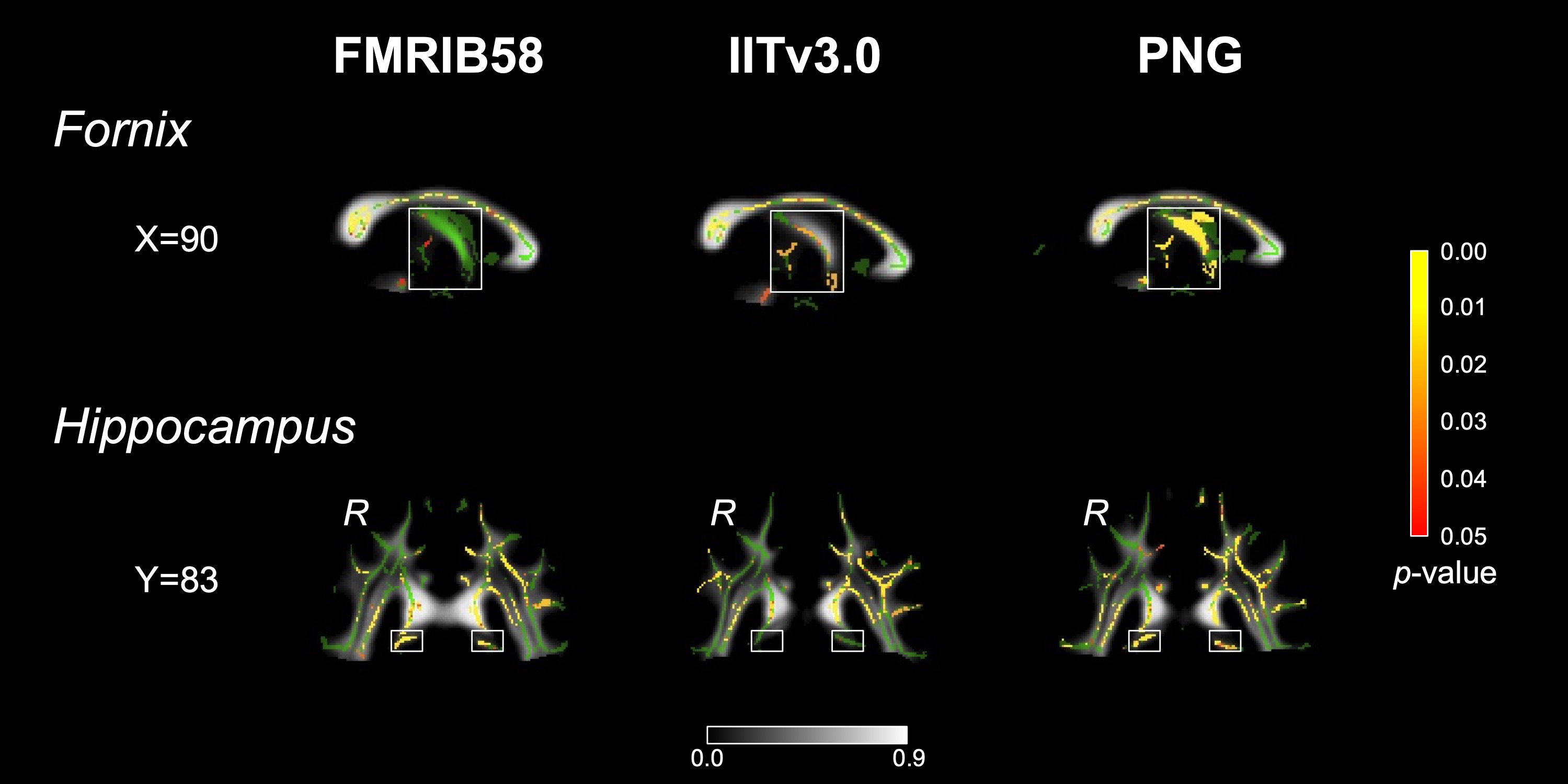
Figure 5. Illustrations of fornix and bilateral
hippocampi (in white box), in FMRIB58, IITv3.0, and PNG DTI templates. t-statistical maps (Red-yellow, p<0.05, FWE corrected) showing
decreased FA at In2 vs. Pre are overlaid on TBSS skeleton (green) and mean FA
image derived from each template respectively. R: right hemisphere.