Jaemin Shin1, Jungho Cha2, Jeffrey McGovern3, Patrick Quarterman1, Suchandrima Banerjee4, and Ki Sueng Choi2
1GE Healthcare, New York, NY, United States, 2Mount Sinai, New York, NY, United States, 3GE Healthcare, Waukesha, WI, United States, 4GE Healthcare, Menlo Park, CA, United States
1GE Healthcare, New York, NY, United States, 2Mount Sinai, New York, NY, United States, 3GE Healthcare, Waukesha, WI, United States, 4GE Healthcare, Menlo Park, CA, United States
This work does a quantitative evaluation of the effectiveness of a distortion correction method, clinically available at the scanner console, in diffusion MRI of the brain. Distortion correction reduced average residual distance error to 1.1 mm or less (average 76% reduction).
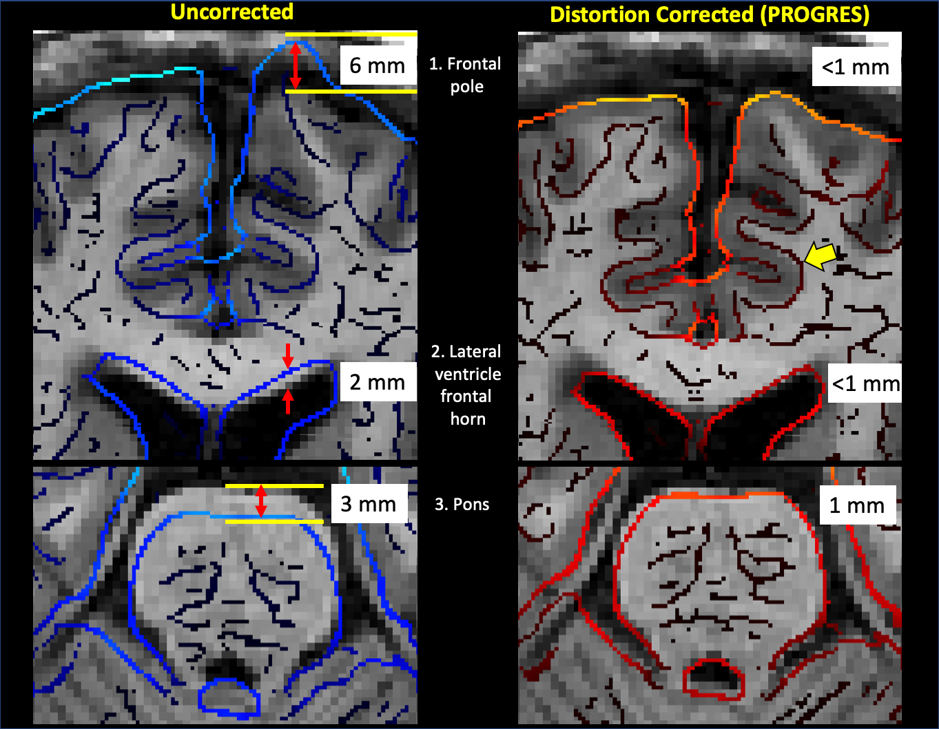
Figure 2. Illustration of geometric distortion in three anatomical landmarks (frontal pole, lateral ventricle, pons) from the same subject as Figure 1. Uncorrected DWI shows local residual distance error up to 6 mm and distortion correction reduced error to 1mm or less. Distortion correction noticeably improved match of the boundary between white matter and grey matter (yellow arrow).
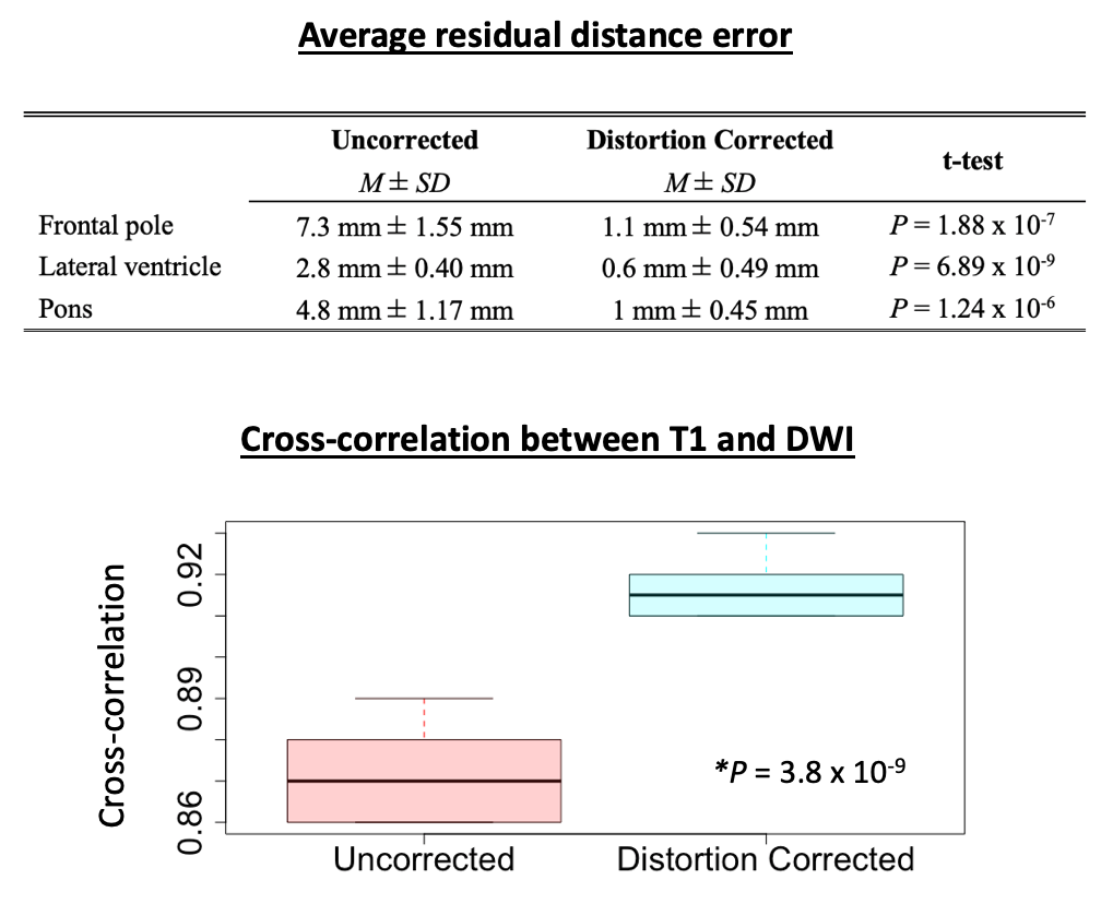
Figure 3. Average residual distance error (N=10) was significantly reduced after distortion correction in all of three regions of interests. Average cross-correlation coefficient between T1 and DWI, as a global similar measure, was significantly increased from 0.872 (uncorrected) to 0.916 (distortion) (P= 3.8 x 10⁻⁹).