Xin Li1, Yue Qin1, Shaoyu Wang2, Xiang Feng3, Yifan Qian1, Juan Tian1, Liyao Liu1, Yinhu Zhu1, Boyuan Jiang1, and Yanqiang Qiao1
1XI’AN DAXING HOSPITAL, ShaanXi, Xi’an, China, 2Siemens Healthcare Ltd., ShaanXi, Xi’an, China, 3Siemens Healthcare Ltd., Beijing, China
1XI’AN DAXING HOSPITAL, ShaanXi, Xi’an, China, 2Siemens Healthcare Ltd., ShaanXi, Xi’an, China, 3Siemens Healthcare Ltd., Beijing, China
Hypertension is known to be
a major risk factor for damage to target organs, including the brain. As people
grow older, iron accumulates, mainly in the form of hemosiderin, in several
brain regions and cell types. Hypertension may be associated with increased
content of heme and non-heme iron contents. The brain is one of the main target
organs affected by hypertension. Iron is the most abundant metal element in
human body, most of which are distributed in brain tissue in the form of non
heme iron. QSM is a potent imaging method that can be exploited in the clinic
to provide noninvasive, accurate, and precise measurements of iron for
diagnosing diseases and monitoring progression and treatment. This study
evaluated the feasibility of QSM in measurement of brain iron deposition between
hypertensive patients and health controls, and found increased iron
accumulation mainly in deep gray matter nucleus and CSF in hypertensive
patients. Indicate the role of excess brain iron in deep gray matter in
hypertension. This suggested iron may be a potential biomarker for further
understanding the pathophysiological mechanism of hypertension.
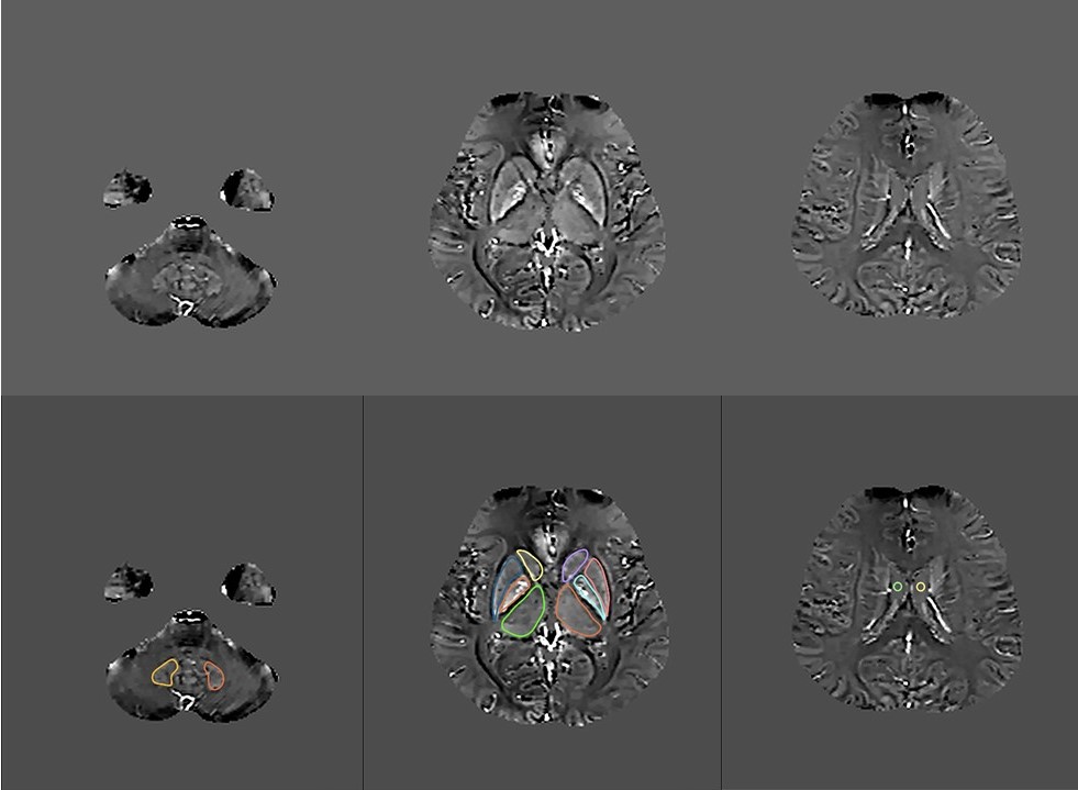
Fig. 1. This image
shows six selected regions of one QSM image slice from a 62-year-old subject.
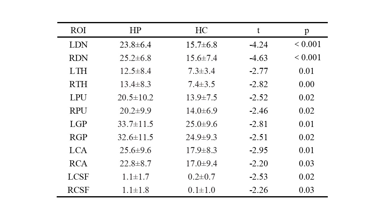
Table 1. Comparison of magnetic sensitivity of ROI between
hypertensive patients and healthy controls (p < 0.05 was considered statistically significant)