Alessandra Caporale1, Hyunyeol Lee1, Hui Lei2, Hengyi Rao3, Michael C Langham1, Alessandra A Caporale2, and Felix W Wehrli1
1Radiology, University of Pennsylvania Perelman School of Medicine, Philadelphia, PA, United States, 2Neurology, University of Pennsylvania Perelman School of Medicine, Philadelphia, PA, United States, 3Psychiatry, Division of Sleep and Chronobiology, University of Pennsylvania Perelman School of Medicine, Philadelphia, PA, United States
1Radiology, University of Pennsylvania Perelman School of Medicine, Philadelphia, PA, United States, 2Neurology, University of Pennsylvania Perelman School of Medicine, Philadelphia, PA, United States, 3Psychiatry, Division of Sleep and Chronobiology, University of Pennsylvania Perelman School of Medicine, Philadelphia, PA, United States
The cerebral metabolic
rate of oxygen (CMRO2) was measured with concurrent EEG in 9 healthy
volunteers, during wakefulness and non-REM sleep. CMRO2 decrease
during sleep was positively correlated with slow-wave activity increase when sleep
onset occurred.
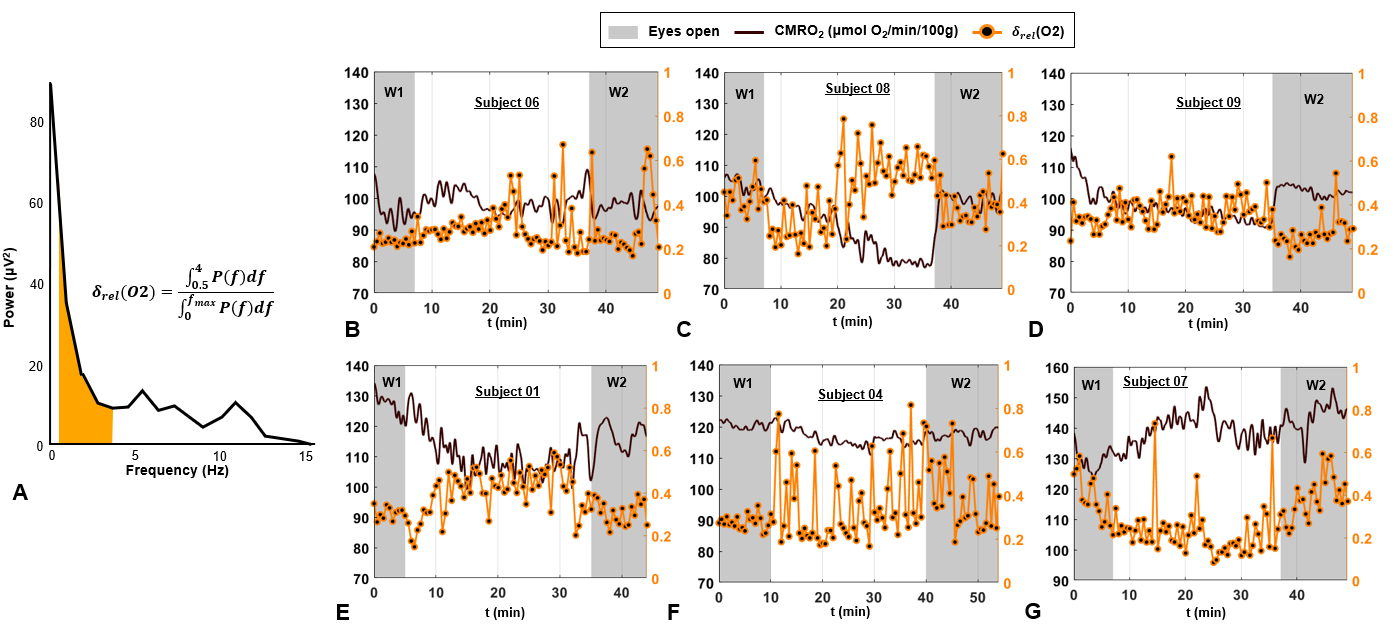
Figure 3. Concurrent EEG and rOxFlow during
wakefulness and sleep. A. Power
spectrum derived from EEG recordings of the channel O2 in a volunteer during
sleep (S01). The delta-band (0.5-4.0 Hz) is colored. Delta power ratio (δrel) was estimated as indicated (P, power; f,
frequency; fmax=30 Hz). B-G. δrel(O2), superimposed on CMRO2 time-course
for the three subjects of Figure 2,
and three additional subjects.
δrel(O2) is averaged every 30 seconds.
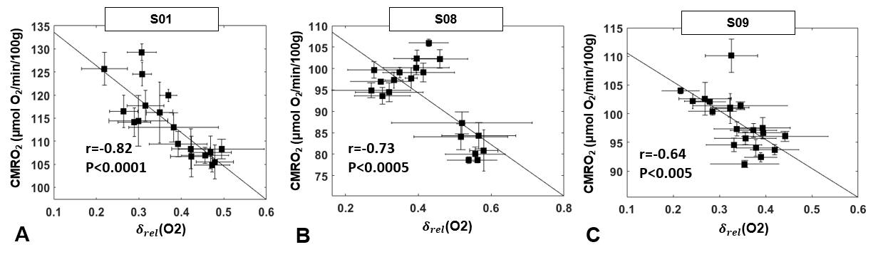
Figure 5. CMRO2 versus EEG delta power ratio. CMRO2 plotted as a function of δrel(O2)
for
three subjects for whom sleep duration greater than 5 minutes
was confirmed by EEG. A. S01, male, 40 years. B. S08, male, 36
years. C. S09, female, 24 years. Markers represent averages over 2.5-min time
windows, error bars represent standard deviations. Pearson’s correlation
coefficient r and the significance level P are indicated.