Masami Yoneyama1, Michinobu Nagao2, Yasuhiro Goto3, Shuo Zhang4, Isao Shiina3, Kazuo Kodaira3, Yutaka Hamatani3, and Marc Van Cauteren5
1Philips Japan, Tokyo, Japan, 2Department of Diagnostic Imaging and Nuclear Medicine, Tokyo Women’s Medical University, Tokyo, Japan, 3Department of Radiological Services,, Tokyo Women’s Medical University, Tokyo, Japan, 4Philips Healthcare, Hamburg, Germany, 5Philips Healthcare, Best, Netherlands
1Philips Japan, Tokyo, Japan, 2Department of Diagnostic Imaging and Nuclear Medicine, Tokyo Women’s Medical University, Tokyo, Japan, 3Department of Radiological Services,, Tokyo Women’s Medical University, Tokyo, Japan, 4Philips Healthcare, Hamburg, Germany, 5Philips Healthcare, Best, Netherlands
We demonstrated the feasibility of free-breathing whole-heart 3D T2-mapping with 2mm3 isotropic spatial resolution in the diastolic and systolic phases within a clinically acceptable scan time.
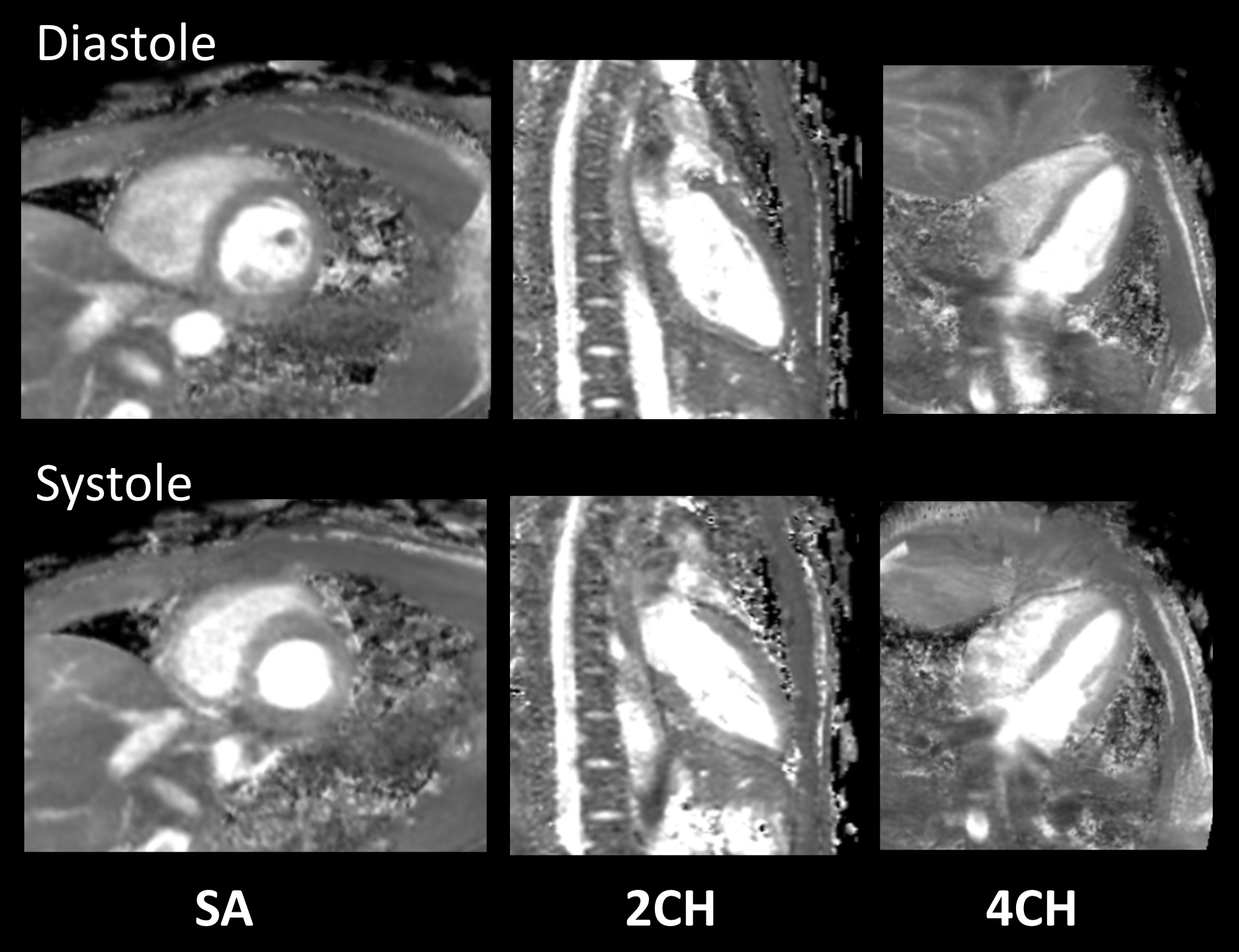
Figure 3. Representative short-axis (SA), two-chamber (2CH), and four-chamber (4CH) MPR images of 3D isotropic myocardial T2-mapping in diastole and systole.
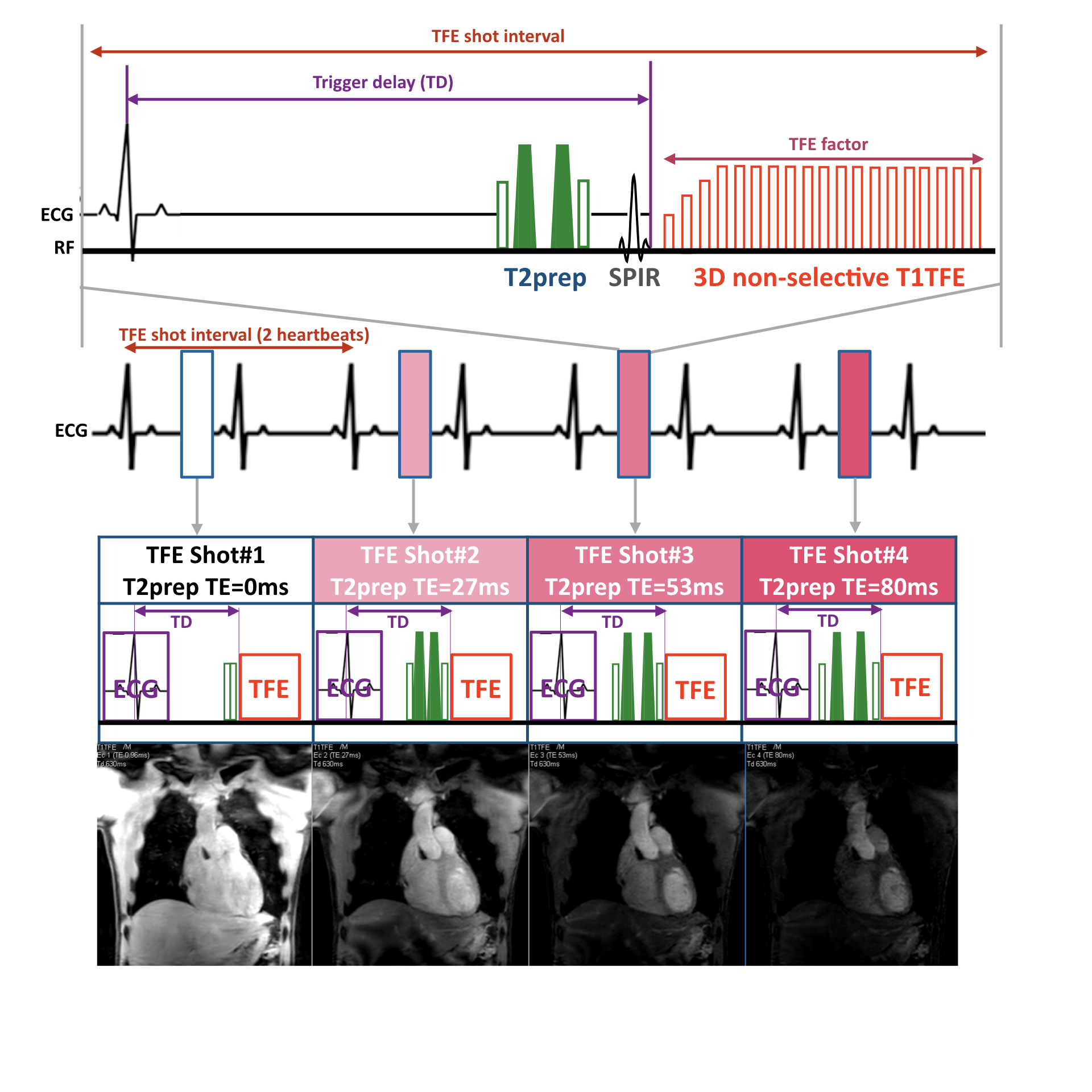
Figure 1. Scheme of the sequence for 3D isotropic myocardial T2-mapping.
T2-mapping was performed using a T2-prepared segmented gradient echo (turbo field-echo: TFE) sequence. 3D non-selective excitation pulses were also applied to shorten the TR/TE which leads to increase the TFE factor per one heartbeat. The numbers indicate the 4 images with different T2-preparation times (TE = 0, 27, 53 and 80ms). Each T2-weighted image was obtained with a navigator respiratory triggering and interleaved scanning, at both late diastole and systole timings separately.
