Giulio Ferrazzi1, Sarah McElroy1, Radhouene Neji1,2, Karl Kunze1,2, Muhummad Sohaib Nazir1, Peter Speier3, Daniel Stäb4, Christoph Forman3, Reza Razavi1, Amedeo Chiribiri1, and Sébastien Roujol1
1School of Biomedical Engineering and Imaging Sciences, King's College London, London, United Kingdom, 2MR Research Collaborations, Siemens Healthcare Limited, Frimley, United Kingdom, 3Cardiovascular MR predevelopment, Siemens Healthcare GmbH, Erlangen, Germany, 4MR Research Collaborations, Siemens Healthcare Limited, Melbourne, Australia
1School of Biomedical Engineering and Imaging Sciences, King's College London, London, United Kingdom, 2MR Research Collaborations, Siemens Healthcare Limited, Frimley, United Kingdom, 3Cardiovascular MR predevelopment, Siemens Healthcare GmbH, Erlangen, Germany, 4MR Research Collaborations, Siemens Healthcare Limited, Melbourne, Australia
We develop a SMS3 perfusion sequence which provides dual phase/contrast measurements at two different saturation delay times. In-vivo measurements demonstrate an increase of myocardial/defect contrast, and a decreased of blood/myocardium signal ratio when comparing long vs short delay.
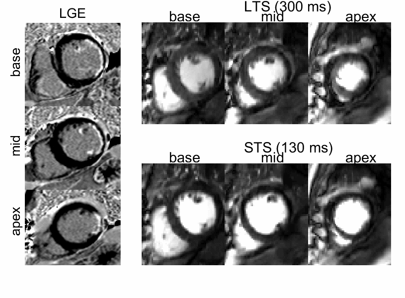
Figure3: LGE-positive patient. left) LGE
images at base, mid and apex. right) dual contrast perfusion data
acquired at short (left) and long (right) saturation delay times (TS).
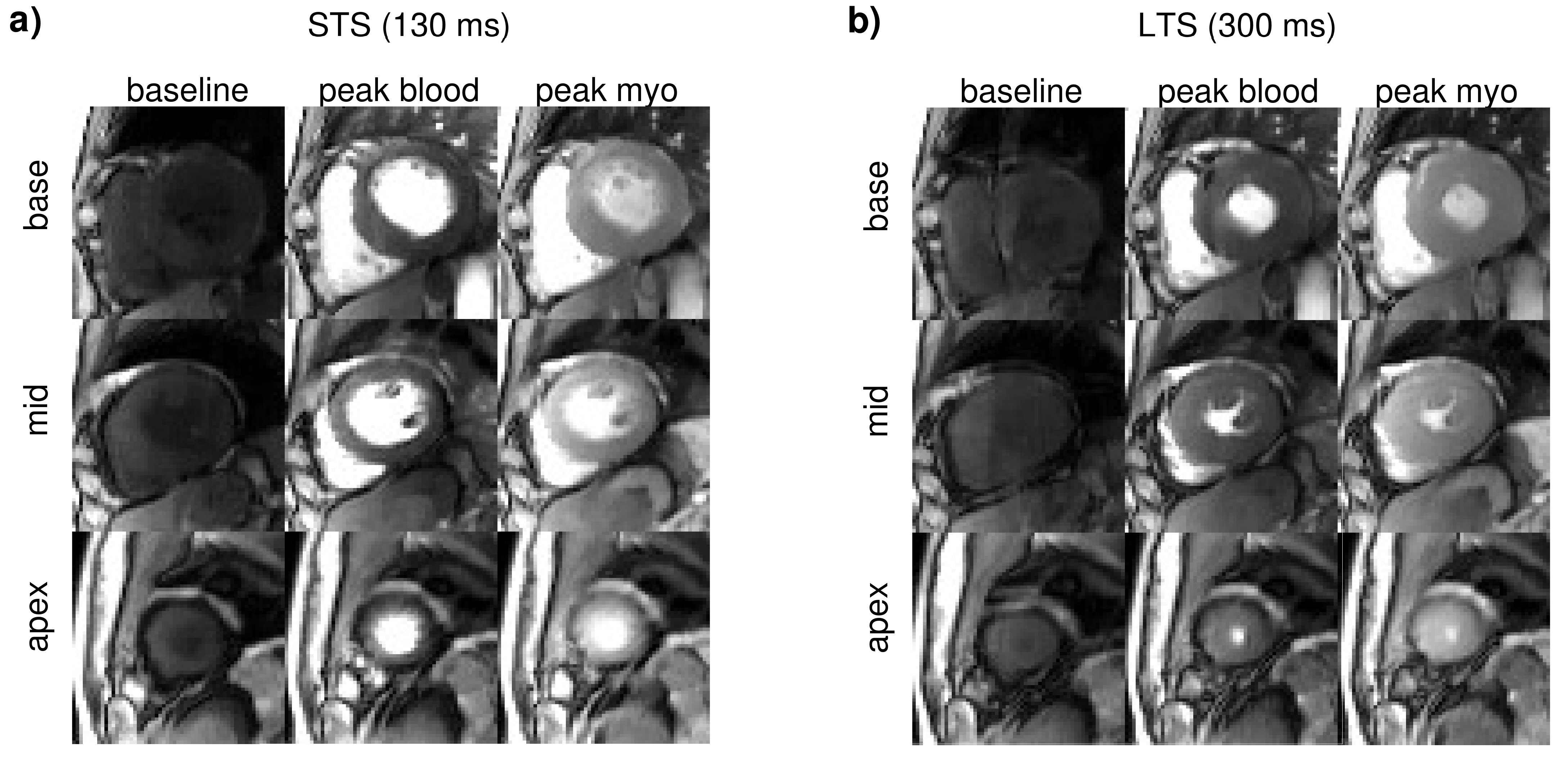
Figure3: In-vivo evaluation of the sequence in
5 patients. a) acquired data using saturation delay times (TS) of 130ms
(left) and b) 300ms (right) on one
exemplary subject without perfusion deficit. Top to bottom: the three
simultaneously excited slices (base to apex). Left to right: baseline frame,
peak blood and peak myocardium.