Zhengling Zeng1, Bin Sun1, Yunjing Xue1, Ning Jin2, and Jing An3
1Fujian Medical University union hospital, Fuzhou, China, 2Siemens Medical Solution, Chicago, IL, United States, 3Siemens Shenzhen Magnetic Resonance Ltd, Shenzhen, China
1Fujian Medical University union hospital, Fuzhou, China, 2Siemens Medical Solution, Chicago, IL, United States, 3Siemens Shenzhen Magnetic Resonance Ltd, Shenzhen, China
Patients with thoracic aorta atherosclerosis had lower circumferential WSS compared with that of non-plaque patients in planes 2-7, with lower mean and peak velocity in planes 5-7, respectively.Scanning time of Compressed Sensing 4D flow MRI was obviously less than that of 4D flow MRI.
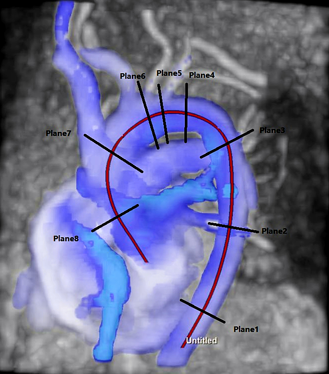
Figure1:Flow was evaluated in 8 planes at different levels of the thoracic aorta. Plane (P) 1 middle and lower segment of descending aorta, P2 mid-descending aorta, P3 end of the aortic arch, P4 plane immediately after left subclavian artery, P5 plane immediately after left common carotid artery, P6 plane immediately after tuncus brachiocephalicus, P7 proximal aortic arch, P8 proximal ascending aorta
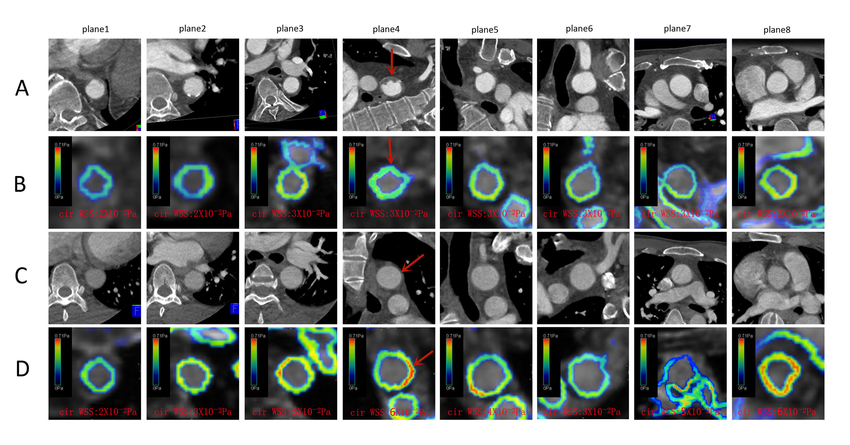
Figure2:A and B row:one plaque patient's CTA and cirWSS in 8 planes;C and D row:one non-plaque patient's CTA and cirWSS in 8 planes.The numbers below each picture was cirWSS values.B and D row:The color of D row was more inclined to warm tone than that of B row.And the values in D row were larger than B row(warm tone means high values in cirWSS).The patient's plaque was located at the fourth plane,and its cirWSS was lower than that of non-plaque patient visually(red arrow).