Bharath Ambale Venkatesh1, Jason Ortman2, Jaclyn Sesso2, Yoko Kato2, Elzbieta Chamera2, Jennifer Wagner3, Yoshimori Kassai4, and Joao Lima2
1Radiology, Johns Hopkins University, Baltimore, MD, United States, 2Johns Hopkins University, Baltimore, MD, United States, 3Canon Medical Research USA, Mayfield Village, OH, United States, 4Canon Medical Systems, Kanagawa, Japan
1Radiology, Johns Hopkins University, Baltimore, MD, United States, 2Johns Hopkins University, Baltimore, MD, United States, 3Canon Medical Research USA, Mayfield Village, OH, United States, 4Canon Medical Systems, Kanagawa, Japan
We have demonstrated the potential of non-contrast WB-MRA to
quantify atherosclerosis burden and Dixon imaging of muscle tissue to assess
underlying metabolic disease, as well as to monitor and quantify progression
towards frailty.
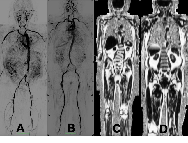
Figure 1. A Whole-body non-contrast MRA in frail
(A) and robust (B) individuals. Atheroma burden was 75% and 25%
respectively. Data was available in 90% of segments for evaluation. Whole body fat
percent maps in frail (C) and robust (D) individuals are also shown.
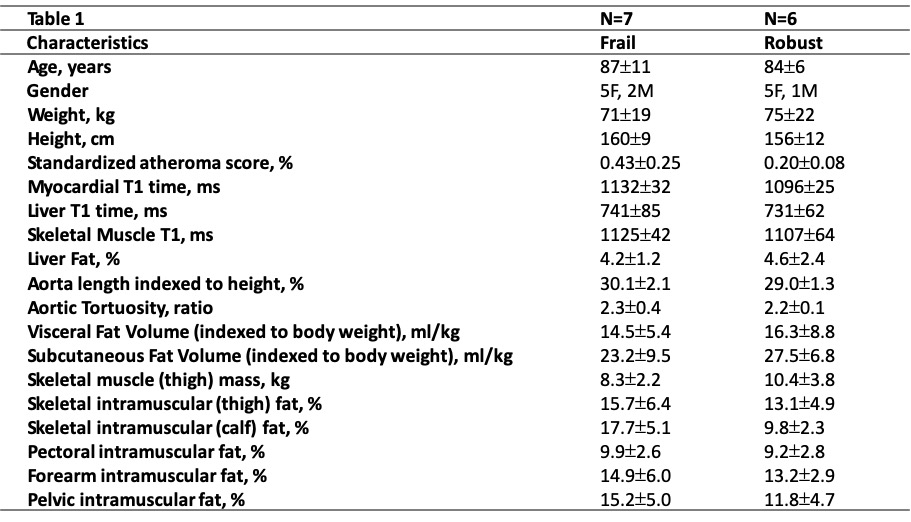
Table 1. The table below shows the participant
characteristics across all the participants in the study.