Ashitha Pathrose1, Hassan Haji-Valizadeh1,2, Roberto Sarnari1, James Carr1,2,3, and Daniel Kim1,2
1Radiology, Northwestern University, Chicago, IL, United States, 2Biomedical Engineering, Northwestern University, Chicago, IL, United States, 3Medicine, Northwestern University, Chicago, IL, United States
1Radiology, Northwestern University, Chicago, IL, United States, 2Biomedical Engineering, Northwestern University, Chicago, IL, United States, 3Medicine, Northwestern University, Chicago, IL, United States
CMR feature-tracking derived strain measurements from a 16-fold
accelerated, real-time cine had good agreement with measurements derived from
standard breath-hold cine. But, there can be underestimation which is dependent
on spatiotemporal resolution and CS regularization weights.
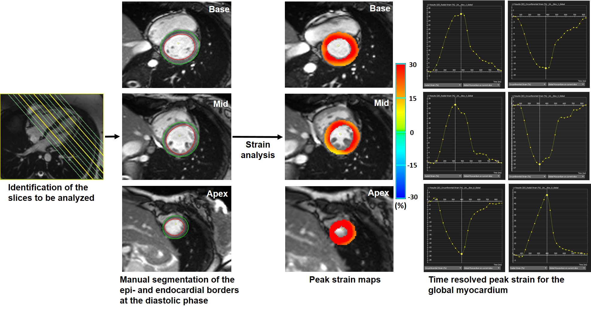
Figure
1: Image analysis workflow.
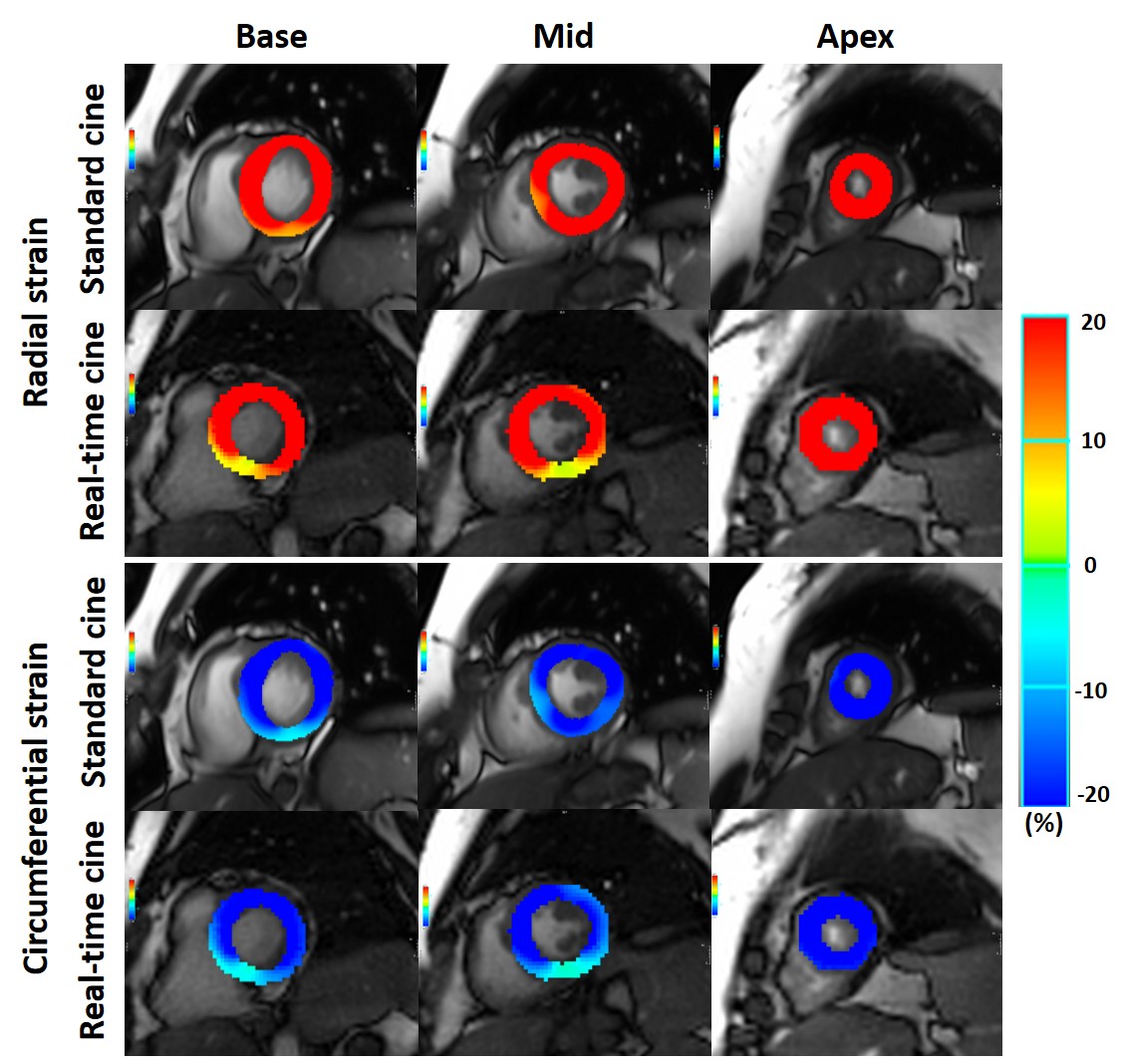
Figure
2: Comparison of CMR-FT derived radial and circumferential strain maps during peak
systole for the standard cine and real-time cine at the basal slice, mid-slice
and apical slice. Note that in the real-time cine, images have larger
tracking-windows when compared to the corresponding standard cine images.