El-Sayed H Ibrahim1, V Emre Arpinar1, L Tugan Muftuler1, Andrew Nencka1, and Kevin Koch1
1Medical College of Wisconsin, Milwaukee, WI, United States
1Medical College of Wisconsin, Milwaukee, WI, United States
UHF cardiac functional MRI can be improved by proper imaging
parameter optimization and using dielectric pads. This is expected to open the
door for more cardiac applications of UHF MRI and potential adoption in
clinical practice in the near future.
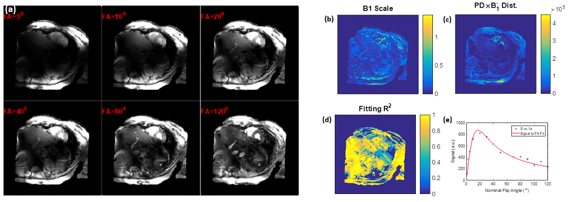
Figure 1. Estimation of the B1 transmission field in a volunteer scan. (a)
Gradient-echo magnitude images with different excitation flip angles. (b) B1
scale factor map. (c) Proton density distribution map. (d) Goodness-of-fit map.
(e) Example of signal intensity measurements at different flip angles (dots) and
the fitting curve (red line) for a certain pixel in the heart.
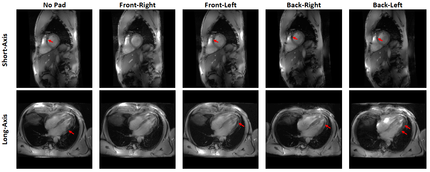
Figure 2. Short-axis and
long-axis slices acquired with dielectric pad placed at different locations
around the thorax area. Note how signal intensity drops appear at different
regions (arrows) based on pad location. In the case of this subject, placing
the pad at right anterior position resulted in most uniform signal intensity.